. 2018 Sep 17;8(1):13928.
doi: 10.1038/s41598-018-32303-7.

Attenuation of melanogenesis by Nymphaea nouchali (Burm. f) flower extract through the regulation of cAMP/CREB/MAPKs/MITF and proteasomal degradation of tyrosinase

Affiliations

Attenuation of melanogenesis by Nymphaea nouchali (Burm. f) flower extract through the regulation of cAMP/CREB/MAPKs/MITF and proteasomal degradation of tyrosinase

Md Badrul Alam et al. Sci Rep. .

Abstract

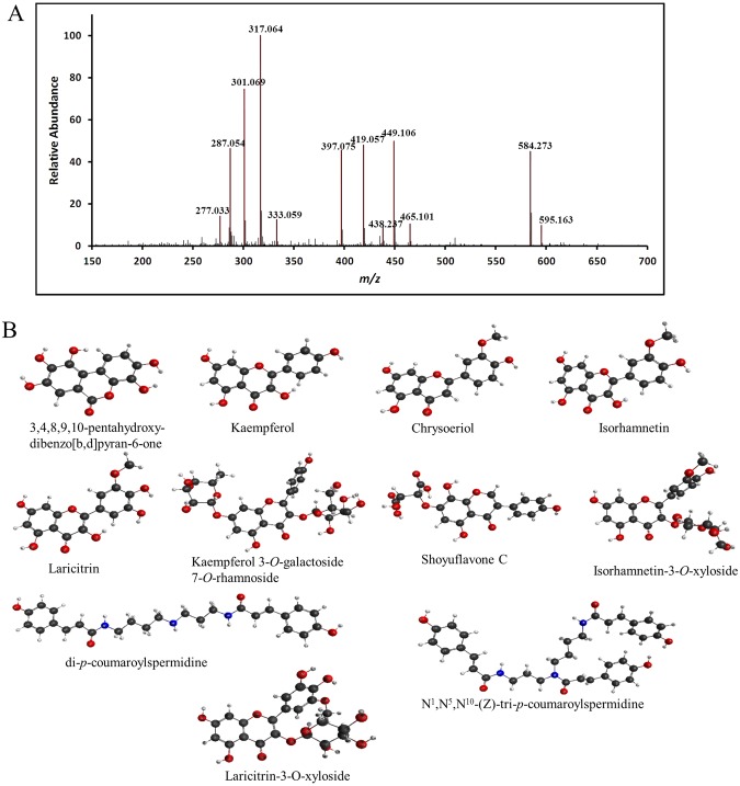
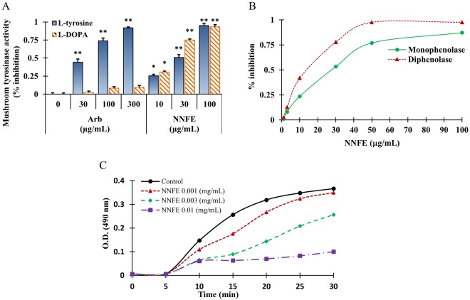
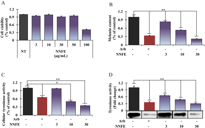
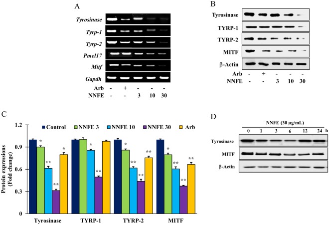
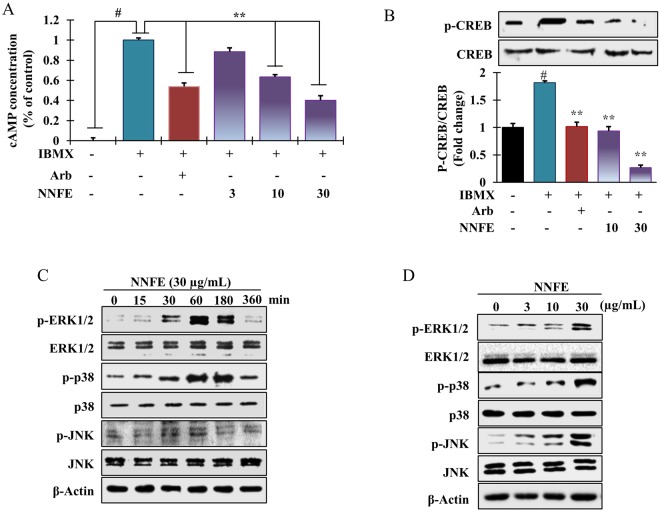
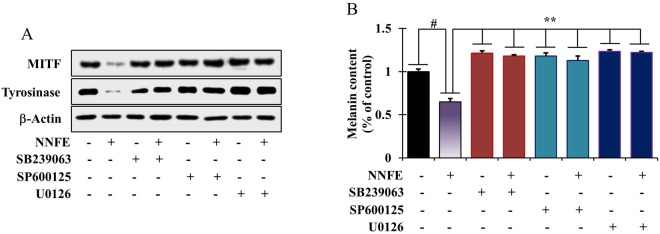
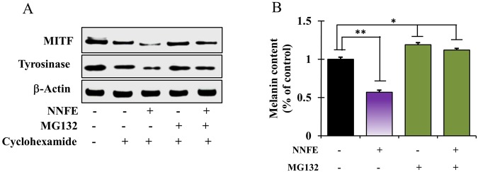
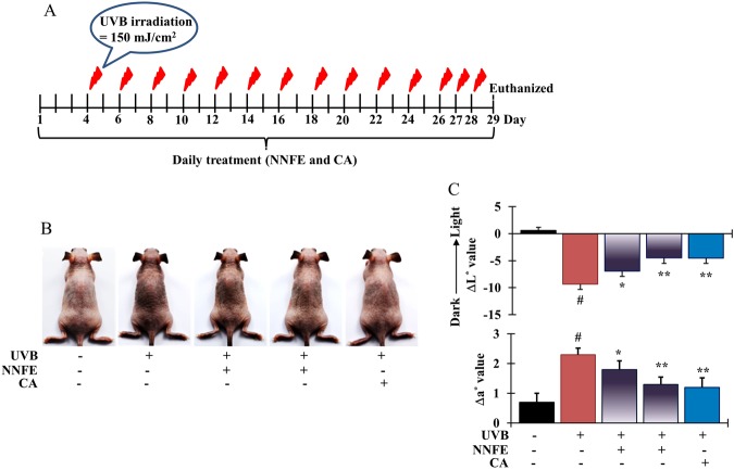
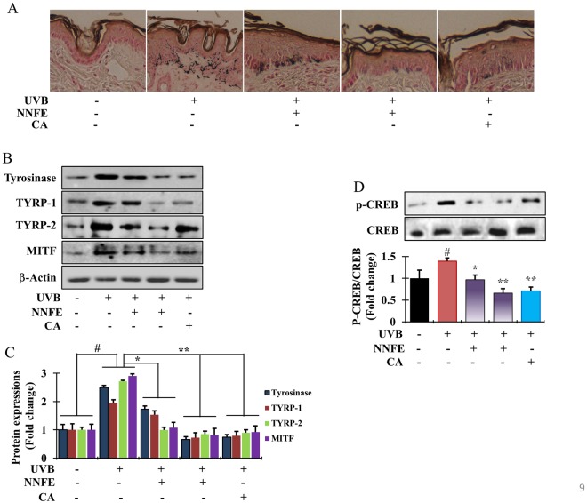
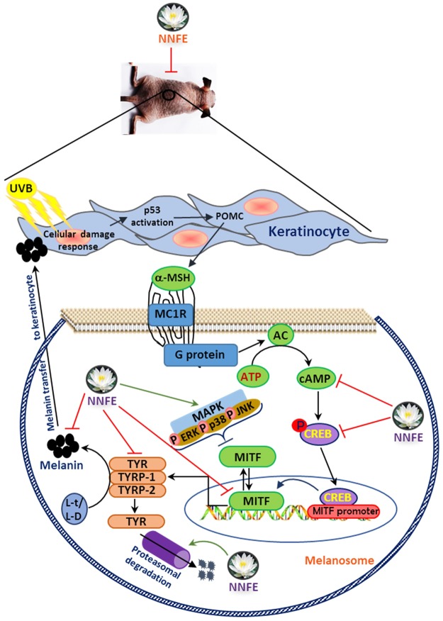
Medicinal plants have been used to treat diseases from time immemorial. We aimed to examine the efficacy of the ethyl acetate fraction of Nymphaea nouchali flower extract (NNFE) against melanogenesis process, and the underlying mechanisms in vitro and in vivo. Paper spray ionisation mass spectroscopy and (+) mode electrospray ionisation revealed the presence of seven flavonoids, two spermidine alkaloids, 3,4,8,9,10-pentahydroxy-dibenzo[b,d]pyran-6-one, and shoyuflavone C in NNFE. NNFE (100 µg/mL) significantly inhibited the monophenolase and diphenolase activities of mushroom tyrosinase at 94.90 ± 0.003% and 93.034 ± 0.003%, respectively. NNFE significantly suppressed cellular tyrosinase activity and melanin synthesis in vitro in melan-a cells and in vivo in HRM2 hairless mice. Furthermore, NNFE inhibited tyrosinase (TYR), tyrosinase-related protein (TYRP)-1, TYRP-2, and microphthalmia-associated transcription factor (MITF) expression, thereby blocking melanin synthesis. In particular, NNFE suppressed cAMP production with subsequent downregulation of CREB phosphorylation. Additionally, it stimulated MAP kinase phosphorylation (p38, JNK, and ERK1/2) and the proteasomal debasement pathway, leading to degradation of tyrosinase and MITF and the suppression of melanin production. Moreover, selective inhibitors of ERK1/2, JNK, and p38 attenuated NNFE inhibitory effects on melanogenesis, and MG-132 (a proteasome inhibitor) prevented the NNFE-induced decline in tyrosinase protein levels. In conclusion, these findings indicate that NNFE is a potential therapy for hyperpigmentation.


Conflict of interest statement

The authors declare no competing interests.

Figures

Figure 1
Figure 1
Representative paper spray ionization mass spectroscopy profile of N. nouchali flower extract. Paper spray ionisation mass spectrum (A) and structures of the identified compounds (B) are shown.
Figure 2
Figure 2
Inhibitory effects of NNFE on mushroom tyrosinase activity. (A) Different concentrations of NNFE or arbutin were incubated with mushroom tyrosinase. After incubation, the amount of dopachrome produced was determined spectrophotometrically at 490 nm. (B) Effects of NNFE on the monophenolase and diphenolase activities of tyrosinase. Enzyme activity was tested in the presence of L-tyrosine and L-DOPA, as a substrate for monophenolase and diphenolase activities, respectively. (c) Effects of NNFE on the monophenolase activity of tyrosinase. Enzyme activity was tested in the presence of L-tyrosine, as a substrate Results are presented as the means ± SDs of three experiments. *p < 0.05, **p < 0.01, versus the non-treated controls, Student’s t-test. Arb, arbutin; NNFE, ethyl acetate fraction of N. nouchali flower extract.
Figure 3
Figure 3
Effects of NNFE on melanogenesis in melan-a cells. Cells were cultured with NNFE (3–30 μg/mL) for 3 days. (A) cytotoxicity, (B) melanin content, (C) intracellular tyrosinase, and (D) tyrosinase activity by zymography were measured as described in the Materials and Methods. Experiments were performed in triplicate, and the results are expressed as the means ± SDs. *p < 0.05, **p < 0.01, Student’s t-test. NT, no treatment; Arb, arbutin; NNFE, ethyl acetate fraction of N. nouchali flower extract.
Figure 4
Figure 4
Effects of NNFE on the expression of melanogenesis-related mRNA and proteins in melan-a cells. (A) Cells (5 × 105 cells/mL) were cultured for 24 h, and the medium was then replaced with fresh medium containing the indicated concentrations of NNFE or arbutin for 24 h. mRNA was extracted using TRIzol, and mRNA expression was determined by RT-PCR. (B) Cells (1 × 105 cells/mL) were cultured for 24 h, and the medium was replaced with fresh medium containing the indicated concentrations of NNFE or arbutin for 3 days. Total cell lysates were extracted and assayed by western blotting using antibodies against tyrosinase, TYRP-1, TYRP-2, and MITF. Equal protein loading was confirmed using β-actin. (C) Statistical analysis of the band intensities of tyrosinase, TYRP-1, TYRP-2, and MITF obtained by western blot analysis. *p < 0.05, **p < 0.01, versus the non-treated controls, Student’s t-test. (D) Total cell lysates were extracted and assayed by western blotting using antibodies against tyrosinase, and MITF. Equal protein loadings were confirmed using β-actin.
Figure 5
Figure 5
Effects of NNFE treatment on the intracellular cAMP concentration and melanogenesis-related signalling protein expression in melan-a cells. (A) Cells were treated with the indicated concentrations of NNFE for the indicated times. Intracellular cAMP levels were measured using a cAMP ELISA kit. Results are representative of three independent experiments. #p < 0.05, versus the non-treated controls; **p < 0.01, versus the IBMX-treated controls, Student’s t-test. Western blot analysis showing the changes in p-CREB, CREB, p-ERK1/2, ERK1/2, p-p38, p38, p-JNK, and JNK expression in melan-a cells treated with (B and D) the indicated concentrations, (C) at the indicated times. Arb, arbutin; NNFE, ethyl acetate fraction of N. nouchali flower extract.
Figure 6
Figure 6
Effects of NNFE on MAP kinase signalling in melan-a cells. (A) Melan-a cells were co-treated with NNFE and selective inhibitors of ERK (U0126), JNK (SP600125), or p38 (SB239063). MITF and tyrosinase levels were determined by western blot analysis, and (B) melanin content was also determined. Measurements were made in triplicate, and results are expressed as the means ± SDs. #p < 0.05, versus the non-treated controls; **p < 0.01, versus the IBMX-treated controls, Student’s t-test. NNFE, ethyl acetate fraction of N. nouchali flower extract.
Figure 7
Figure 7
Effects of NNFE on the proteasomal degradation of tyrosinase in melan-a cells. (A) Cells (3 × 105 cells/mL) were pre-treated with 25 μg/mL cycloheximide (a protein synthesis inhibitor) for 1 h. Separately, cells were pre-treated with 10 μM MG-132 (a proteasomal inhibitor) for 1 h. Cycloheximide and MG-132-pretreated cells were then treated with NNFE for 6 h. Whole cell lysates were subjected to western blot analysis using anti-tyrosinase antibodies. Equal protein loadings were confirmed using β-actin antibodies. (B) Melanin contents were determined in triplicate, and results are expressed as the means ± SDs. *p < 0.05, versus the non-treated controls, Student’s t-test. NNFE, ethyl acetate fraction of N. nouchali flower extract.
Figure 8
Figure 8
Effects of NNFE on pigmentation of mouse skin exposed to UVB. (A) HRM-2 melanin-possessing hairless mice were treated with the vehicle or NNFE on a designated site on the dorsal skin, according to the indicated schedules. (B) Representative photos show pigmentation differences in the dorsal skin among the tested animals. (C) The upper panel shows ΔL* values calculated as the average values after the final UVB exposure minus the average baseline values before treatment. The lower panel shows Δa* values, which were calculated as the average values after final UVB exposure minus the average baseline values before treatment. Results are presented as the means ± SDs. *p < 0.05, versus the non-treated controls, Student’s t-test. NNFE, ethyl acetate fraction of N. nouchali flower extract; CA, caffeic acid.
Figure 9
Figure 9
Effects of NNFE on pigmentation of mouse skin exposed to UVB. (A) Fontana-Masson staining of dorsal skin sections from the mice shown in (Fig. 8B) reveals differences in melanin content. (B) After completing UVB treatment, dorsal skin was excised, homogenised, and assayed by western blot using antibodies against tyrosinase, TYRP-1, TYRP-2, and MITF. Equal protein loading was confirmed using anti-β-actin. Arb, arbutin. (C) Quantification and statistical analysis of the band intensities of tyrosinase, TYRP-1, TYRP-2, and MITF obtained by western blot analysis. *p < 0.05, **p < 0.01, versus the non-treated controls, Student’s t-test. (D) Phosphorylation of CREB confirmed by western blot analysis. NNFE, ethyl acetate fraction of N. nouchali flower extract; CA, caffeic acid.
Figure 10
Figure 10
A proposed molecular mechanism of NNFE against melanogenesis process.

Similar articles

Cited by

References

    1. Miyamura Y, et al. Regulation of human skin pigmentation and responses to ultraviolet radiation. Pigment Cell Res. 2007;20:2–13. doi: 10.1111/j.1600-0749.2006.00358.x. - DOI - PubMed
    1. Hwang E, et al. A novel synthetic Piper amide derivative NED-180 inhibits hyperpigmentation by activating the PI3K and ERK pathways and by regulating Ca2+ influx via TRPM1 channels. Pigment cell melanoma Res. 2016;29:81–91. doi: 10.1111/pcmr.12430. - DOI - PubMed
    1. Hwang E, et al. Gallic Acid Regulates Skin Photoaging in UVB-exposed Fibroblast and Hairless Mice. Phytother. Res. 2014;28:1778–1788. doi: 10.1002/ptr.5198. - DOI - PubMed
    1. Kondo T, Hearing VJ. Update on the regulation of mammalian melanocyte function and skin pigmentation. Exp.t Rev. Dermatol. 2011;6:97–108. doi: 10.1586/edm.10.70. - DOI - PMC - PubMed
    1. Dong Y, et al. Nitric oxide enhances the sensitivity of alpaca melanocytes to respond to α-melanocyte-stimulating hormone by up-regulating melanocortin-1receptor. Biochem. Biophys. Res. Comm. 2010;396:849–853. doi: 10.1016/j.bbrc.2010.05.001. - DOI - PubMed

Publication types

MeSH terms