. 2018 Sep 17;8(1):13948.
doi: 10.1038/s41598-018-31887-4.

Kidney-resident macrophages promote a proangiogenic environment in the normal and chronically ischemic mouse kidney

Affiliations

Kidney-resident macrophages promote a proangiogenic environment in the normal and chronically ischemic mouse kidney

Amrutesh S Puranik et al. Sci Rep. .

Abstract

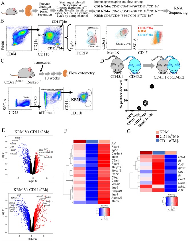
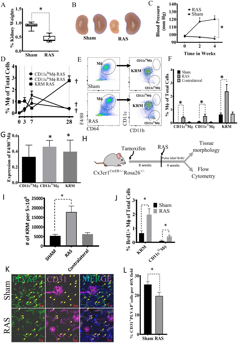
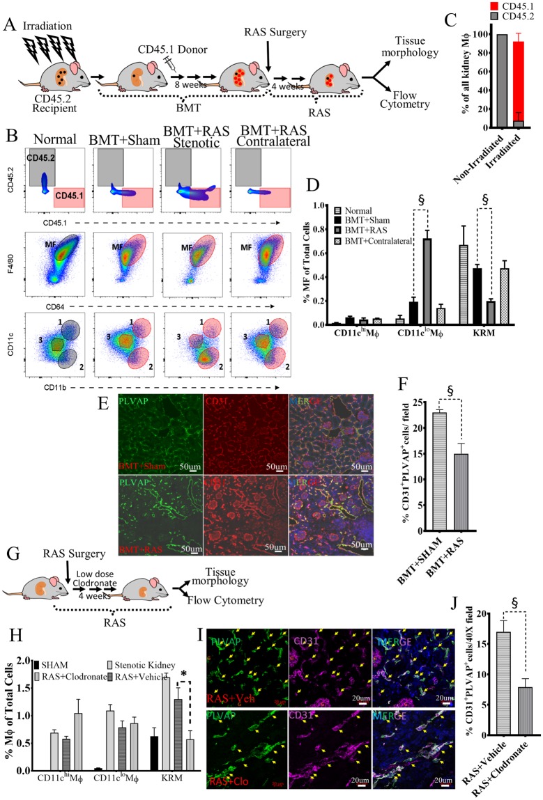
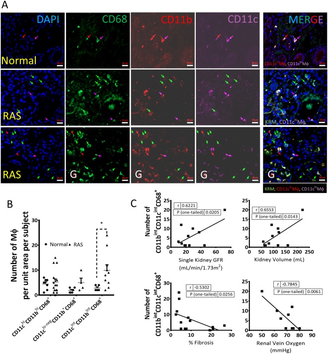
Renal artery stenosis (RAS) caused by narrowing of arteries is characterized by microvascular damage. Macrophages are implicated in repair and injury, but the specific populations responsible for these divergent roles have not been identified. Here, we characterized murine kidney F4/80+CD64+ macrophages in three transcriptionally unique populations. Using fate-mapping and parabiosis studies, we demonstrate that CD11b/cint are long-lived kidney-resident (KRM) while CD11chiMϕ, CD11cloMϕ are monocyte-derived macrophages. In a murine model of RAS, KRM self-renewed, while CD11chiMϕ and CD11cloMϕ increased significantly, which was associated with loss of peritubular capillaries. Replacing the native KRM with monocyte-derived KRM using liposomal clodronate and bone marrow transplantation followed by RAS, amplified loss of peritubular capillaries. To further elucidate the nature of interactions between KRM and peritubular endothelial cells, we performed RNA-sequencing on flow-sorted macrophages from Sham and RAS kidneys. KRM showed a prominent activation pattern in RAS with significant enrichment in reparative pathways, like angiogenesis and wound healing. In culture, KRM increased proliferation of renal peritubular endothelial cells implying direct pro-angiogenic properties. Human homologs of KRM identified as CD11bintCD11cintCD68+ increased in post-stenotic kidney biopsies from RAS patients compared to healthy human kidneys, and inversely correlated to kidney function. Thus, KRM may play protective roles in stenotic kidney injury through expansion and upregulation of pro-angiogenic pathways.


Conflict of interest statement

This work was partly funded by a research grant from Biogen. IL is a current Biogen employee, and JSD a former employee of Biogen (currently at Vertex Pharmaceuticals). Other author(s) have no competing interests.

Figures

Figure 1
Figure 1
Renal macrophages comprise of long-lived kidney-resident macrophages and monocyte-derived CD11chi and CD11clo macrophages. (A) Workflow of the experiment. Mouse kidneys were enzyme-digested, percoll separated and stained for lineage and macrophage markers. After removing the lineage positive cells, three populations of macrophages were identified and flow sorted in the RNA lysis buffer and subjected to transcriptional profiling by RNA-sequencing. (B) Live, LineagenegCD45+ were gated as F4/80+CD64+/lo macrophages while non-macrophage population is CD45+11b/cnegCD64negF4/80neg. We classified kidney macrophages as CD11chiMϕ (CD11bhiCD11chi), CD11chiMϕ (CD11bhiCD11clo-neg), and Kidney-resident macrophages (KRM) (CD11cIntCD11bInt). Overlay of CD11chiMϕ (red), CD11cloMϕ (blue) and KRM (orange) gated on Ly6c vs FCRIV, Cx3cr1 vs MerTK, and SSA vs CD45. KRM are Ly6cFcrIV+MerTK+Cx3cr1+MHCII+CD45int while the non-KRM CD11cloMϕ are FcrIV+MerTK+Cx3cr1+MHCII+Ly6chiCD45int-hi and CD11chiMϕ are FcrIV+MerTK+Cx3cr1+MHCII+Ly6cloCD45hi. (C) Fate-mapping studies using Cx3cr1CreER+/−Rosa26+/− mice demonstrates >80% of tdTomato+ cells gated as KRM. Live, LineagenegCD45+ were gated as F4/80+tdTomato+ that were then gated as CD11b vs CD11c to identify KRM. (D) CD45.1 and CD45.2 congenic mice analyzed on 10 weeks of parabiosis. Histograms represents percentage of partner-derived cells in the kidney. n = 4 parabionts. Bars indicate mean value ± SEM. Symbols represent individual mice. (E-G) Transcriptional differences detected by RNA-Seq. (E) Comparison of gene expression between KRM and CD11chiMϕ (above), CD11cloMϕ (below) displayed as volcano plots of individual genes, where fold-change between populations is plotted on the x-axis and significance on the y-axis. Genes upregulated >2-fold are colored in red and genes downregulated >2-fold in blue. 1257 genes are differentially expressed between KRM and CD11chiMϕ, 649 are up- and 608 down-regulated; and 2674 genes are differentially expressed between KRM and CD11chiMϕ, 1386 are up- and 1288 down-regulated. (F) Selected genes reflecting tissue resident status and upregulated in KRM, and (G) inflammatory genes downregulated in KRM, are presented as heatmaps with hierarchical clustering. Mean values per macrophage populations are shown. The z-score-based color-scale shows gene expression standard deviations below (blue) or above (red) the population mean. Data is representative of n = 4 independent experiments with at least n = 3 mice per group (KRM, n = 4, CD11chiMϕ, n = 3, CD11cloMϕ, n = 3; DEGs: fold change >2, P < 0.05).Kidney image cropped and adopted from openclipart.org (https://openclipart.org/detail/28929/kidneyreins) and mouse images adopted from (https://openclipart.org/detail/174870/mouse and https://openclipart.org/detail/17558/simple-cartoon-mouse).
Figure 2
Figure 2
KRM self-renew while monocyte-derived macrophages expand in RAS kidneys and this increase correlates with loss of peri-tubular endothelial cells. (A) The ratio of stenotic relative to the contralateral kidney size falls after a RAS surgery (n = 20 sham and RAS mice). (B) Representative pictures show reduced size of stenotic (left) and increased size of contralateral (right) Sham and RAS kidneys. (C) Systolic blood pressure in RAS and Sham mice at weeks 0, 2, and 4. (D) CD11chiMϕ, CD11cloMϕ and KRM expand gradually in RAS kidneys, (n = 4–6/time point). (E, F) At day 28, RAS significantly increases stenotic kidney CD11chiMϕ, CD11cloMϕ and KRM compared to their respective Sham or contralateral kidney macrophages (n = 10–14). (G) Macrophage Expression (determined by resolution metric, see Supplemental Methods) significantly increases in CD11cloMϕ and KRM (n = 10–14). (H) Experimental Scheme for Fate-mapping of KRM Cx3cr1CreER+/-Rosa26+/− mice. Tamoxifen was injected and after 8 weeks mice undergo RAS surgery. Mice were injected BrdU for 4 weeks and then euthanized. (I) Numbers of tdTomato + ve KRM in Cx3cr1CreER+/− mice significantly increase with RAS compared to Sham (n = 5 each). (J) BrdU-positive KRM and CD11chiMϕ in RAS and Sham, indicating proliferation. (K, L) Immunofluorescence quantification of peri-tubular endothelial cells by co-staining for CD31 and PLVAP in Sham and RAS. P < 0.05 vs Day-0 of the same group; *P < 0.01 vs Sham. Kidney pictures taken by AP.
Figure 3
Figure 3
KRM repopulate from donor bone marrow (BM) cells but do not sustain in stenotic kidneys resulting in loss of peritubular capillaries. (A) Experimental Schema for BM transplantation (BMT) studies. (BD) Reconstitution by donor-derived (CD45.1) BM in BMT + Sham, BMT + RAS stenotic and BMT + RAS contralateral kidneys (Top Row); Native (black tinted) and donor-derived macrophages (pink tinted) (Middle Row); Donor-derived KRM population decreased in BMT + RAS (Bottom Row). (E) Immunostaining of PLVAP and CD31 showing peri-tubular endothelial cells in representative images of BMT + Sham and BMT + RAS kidney sections. Images acquired on Zeiss confocal at 40X and stitched together to show a larger area. Note CD31 (red) stains peri-glomerular cells while PLVAP (green) is specific to peri-tubular endothelial cells. (F) Quantifying PLVAP+CD31+ cells showing significant reduction in BMT + RAS Vs BMT + Sham. (G) Experimental Schema for administration of liposomal clodronate at low-doses. (H) Comparing percent of all three macrophages in the stenotic kidneys of Sham, RAS, RAS + Vehicle and RAS + Clodronate mice. Note significant reduction in KRM in RAS + Clodronate group. (I) Immunostaining of PLVAP and CD31 showing peri-tubular endothelial cells in representative images of RAS + Vehicle and RAS + Clodronate kidney sections. (J) Quantifying PLVAP+CD31+ cells. Significant loss of peritubular endothelial cells seen after administration of clodronate. n = 6 mice/group; *P ≤ 0.01 vs Sham; §P < 0.05 vs BMT + Sham , RAS + Vehicle; ¥P < 0.01 vs RAS. Mouse images adopted from Openclipart.org https://openclipart.org/detail/174870/mouse and https://openclipart.org/detail/28929/kidneyreins.
Figure 4
Figure 4
Transcriptional profiling of Kidney-resident macrophages (KRM) demonstrates upregulation of Angiogenesis and Wound healing pathways in stenotic kidneys. (A) Ischemia-associated gene expression changes in RAS compared to Sham kidneys in CD11chiMϕ, CD11cloMϕ and KRM, respectively, displayed as volcano plots of individual genes, where fold-change between populations is plotted on the x-axis and significance on the y-axis. Genes upregulated >2-fold are in red, and genes downregulated >2-fold in blue. In RAS-CD11chiMϕ, 934 genes show changes in expression, with 766 up- and 168 down-regulated; while in RAS- CD11cloMϕ, 307 genes change, 241 up- and 66 down-regulated; and KRMs display the greatest changes in stenotic injury, with 3162 DEGs; 1506 are up- and 1656 down-regulated. (B) Enrichment analysis of biological process ontology in CD11chiMϕ, CD11cloMϕ and KRM. Top upregulated (top, red) and downregulated (bottom, blue) pathways in macrophage populations isolated from stenotic compared to Sham kidneys (pathway enrichment P < 0.05). (C) Contributions of different macrophage populations to injury response shown as log2 fold-change in expression of CD11chiMϕ, CD11cloMϕ and KRM. Each dot represents a gene that is up (red) or downregulated (blue) in RAS > 2-fold compared to sham control. (D) Gene signatures that are upregulated (angiogenesis, wound healing, and inflammation) or downregulated (interferon signature) in KRM in injury presented as heat-maps with hierarchical clustering. Mean values per Mϕ populations are shown. The z-score-based color-scale shows gene expression standard deviations below (blue) or above (red) the population mean. (E) Expression of genes involved in angiogenesis, and anti-inflammatory response presented as fold change over respective Sham and validated by RT-qPCR for individual macrophage samples from Sham and RAS mice. RAS KRM n = 3, RAS CD11chiMϕ, n = 3 and RAS CD11cloMϕ, n = 3; *P < 0.01 RAS-KRM vs RAS-CD11chiMϕ and *P < 0.01 RAS-KRM vs RAS-CD11cloMϕ. #P < 0.01 RAS vs respective Sham.
Figure 5
Figure 5
KRM promote proliferation of PLVAP+CD31+ renal peri-tubular endothelial cells and inhibit TGF-β-induced expression of Col1a in vitro. (A) Co-culture of RAS-KRM with PLVAP+CD31+ endothelial cells. EdU incorporation between BM macrophages (Mϕ), RAS-KRM and Sham-KRM compared to FOXO1-inhibitor AS1842856. (B) CellTrace Far Red dye dilution assay of PLVAP+CD31+ cells in a contact co-culture system. (C) TGF-β induces dose-dependent expression of Col1a in murine embryonic fibroblasts (MEF) derived from Col1-GFP mice (Lane1–3, Left and right graph). The increase in Col1a was inhibited by UO126 (MEK inhibitor) and LY2109761 (TGF-β receptor I and II dual inhibitors) (Left). Co-incubation of Sham-KRM (Lane5) and RAS-KRM (Lane6) with MEF (GFP) significantly inhibited the increase in Col1a1-GFP (Right), while BMϕ (Lane7) and CD11chi/loMϕ (Mϕ1,2) (Lane8) had no effect. BMϕ = bone marrow macrophages; Mϕ1,2 are (n = 5 technical replicates and n = 3 biological replicates per sample); (D) Representative images demonstrating contact co-culture of Col1a1-GFP MEFs (green) with (bottom) and without (top) KRM stained with anti-mouse CD64-AF594 (red) *P < 0.01 vs MEF untreated control; §P < 0.01 by One-sample T-test; P < 0.05 vs Control.
Figure 6
Figure 6
CD11cIntCD11bIntCD68+ macrophage numbers increase in stenotic human kidneys and directly correlate with better kidney function markers. (A) Representative images (40 X ) showing DAPI, CD68-AF488 (green), CD11b-AF594 (red), CD11c-AF647 (magenta), and merged (arrows); KRM-like cells were identified as CD11cIntCD11bIntCD68+ in healthy (row 1) and stenotic (rows 2–3) kidneys; G: Glomerulus. (B) CD11cIntCD11bIntCD68+ macrophage numbers are significantly higher in RAS compared to normal human kidneys. (C) The number of CD11cIntCD11bIntCD68+ correlated directly with GFR and kidney volume, and inversely with renal vein oxygen tension and degree of fibrosis measured by Trichrome staining *P < 0.05 vs Normal (n = 5–7 healthy human kidneys, n = 14 stenotic kidneys).

References

    1. Bohlke M, Barcellos FC. From the 1990s to CORAL (Cardiovascular Outcomes in Renal Atherosclerotic Lesions) trial results and beyond: does stenting have a role in ischemic nephropathy? Am J Kidney Dis. 2015;65:611–622. doi: 10.1053/j.ajkd.2014.11.026. - DOI - PubMed
    1. Lerman LO, Textor SC, Grande JP. Mechanisms of tissue injury in renal artery stenosis: ischemia and beyond. Prog Cardiovasc Dis. 2009;52:196–203. doi: 10.1016/j.pcad.2009.09.002. - DOI - PMC - PubMed
    1. Textor SC, Lerman L. Renovascular hypertension and ischemic nephropathy. American journal of hypertension. 2010;23:1159–1169. doi: 10.1038/ajh.2010.174. - DOI - PMC - PubMed
    1. Edeling M, Ragi G, Huang S, Pavenstadt H, Susztak K. Developmental signalling pathways in renal fibrosis: the roles of Notch, Wnt and Hedgehog. Nat Rev Nephrol. 2016;12:426–439. doi: 10.1038/nrneph.2016.54. - DOI - PMC - PubMed
    1. Gloviczki ML, et al. TGF expression and macrophage accumulation in atherosclerotic renal artery stenosis. Clin J Am Soc Nephrol. 2013;8:546–553. doi: 10.2215/CJN.06460612. - DOI - PMC - PubMed

Publication types