

Review
doi: 10.1038/s41556-018-0201-5.
Epub 2018 Sep 17.
Life, death and autophagy
Affiliations
- PMID: 30224761
- PMCID: PMC9721133
- DOI: 10.1038/s41556-018-0201-5
Item in Clipboard
Review
Life, death and autophagy
Nat Cell Biol.
2018 Oct.
Abstract
Autophagy influences cell survival through maintenance of cell bioenergetics and clearance of protein aggregates and damaged organelles. Several lines of evidence indicate that autophagy is a multifaceted regulator of cell death, but controversy exists over whether autophagy alone can drive cell death under physiologically relevant circumstances. Here, we review the role of autophagy in cell death and examine how autophagy interfaces with other forms of cell death including apoptosis and necrosis.
Figures
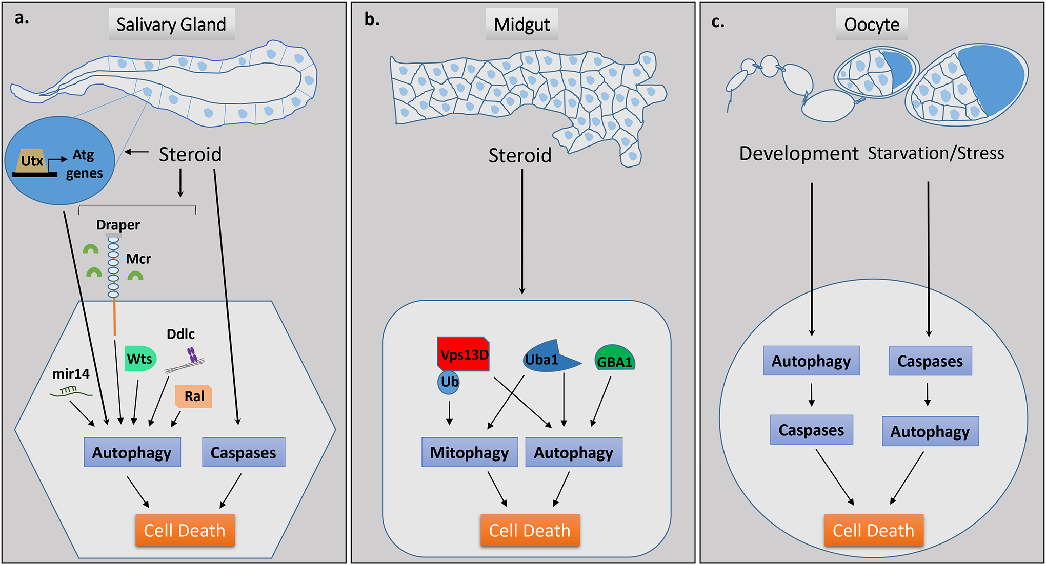
a, Drosophila developmental salivary gland cell death occurs in response to steroid signaling and is dependent upon both autophagy and caspases. Several context-specific regulators of autophagic cell death have been identified from screens performed during developmental salivary gland cell death, including Utx, mir-14, Draper/Mcr, Wts, Ddlc, and Ral. b, Drosophila developmental midgut regression and mitochondrial clearance through mitophagy occurs in response to steroid signaling, is dependent upon autophagy but not caspases, and involves factors including Vps13D, Uba1 and GBA1. c, Autophagy contributes to developmental cell death in the Drosophila oocyte through degradation of an anti-apoptotic factor and subsequent activation of caspases resulting in apoptosis. During starvation and/or stress, autophagic cell death occurs in the oocyte and this is dependent upon caspases.
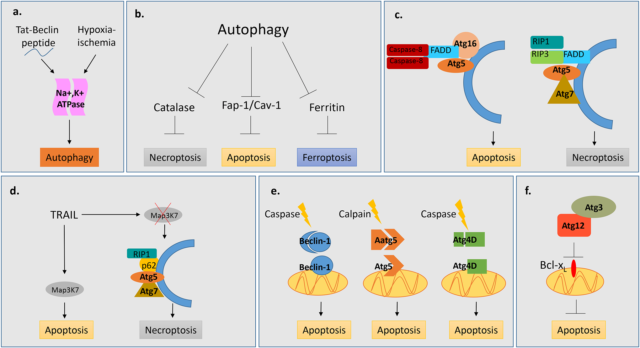
a, Tat-Beclin, hypoxia and ischemia can drive autophagic cell death which is dependent upon the Na+,K+ ATPase. b, Autophagy facilitates cell death by degrading anti-apoptotic (Fap-1/Cav-1), anti-ferroptotic (Ferritin) and anti-necroptotic (Catalase) factors. c, Components of the autophagic machinery serve as a scaffold for apoptotic and necroptotic machinery. Atg5 and Atg16 serve as a scaffold for FADD and Caspase-8 to promote apoptosis. Atg5 and Atg7 serve as a scaffold for necrosome components, RIP1, RIP3 and FADD to promote necroptosis. d, In response to TRAIL, autophagy acts as a switch between the apoptotic and necroptotic machinery by serving as a scaffold for the necrosome component RIP1 and p62 in the absence of Map3K7.. e, Cleavage of autophagy factors, Atg5, Beclin-1 and Atg4D results in products which localize to mitochondria and drive apoptotic cell death. f, Non-canonical conjugation of Atg12 with Atg3 drives intrinsic apoptosis through downstream repression of Bcl-xl.
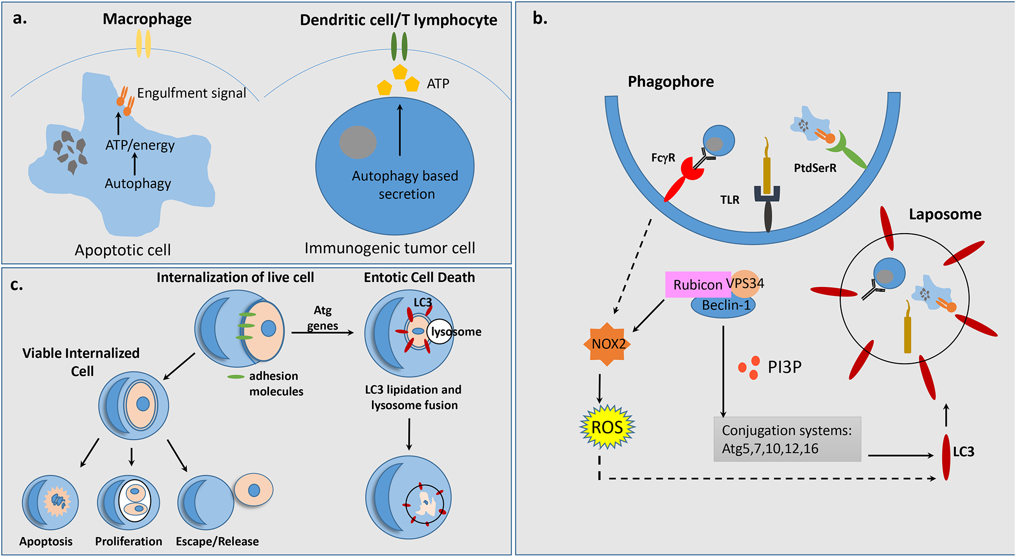
a, Autophagy regulates apoptotic cell clearance and immunogenic cell death by enabling the exposure of energy-dependent engulfment signals and release of ATP from dying cells and live tumor cells respectively, which are recognized and engulfed either by macrophages or dendritic cells/T lymphocytes. b, Autophagy facilitates cell death by acting within engulfing cells to promote LC3 Associated Phagocytosis (LAP) in response to FcγR, TLR or PtdSerR signalingLocalization of PI(3)P, results in sustained NOX2 and increased ROS as well as recruitment of downstream autophagy factors, Atg5, Atg7, Atg10, Atg12 and Atg16, allowing for LC3 lipidation of a single membrane vesicle, termed the laposome. c, Entosis is a sub-type of LAP which utilizes adhesion machinery to internalize live neighboring cells. The internalized cells often undergo entotic cell death through LAP involving LC3 lipidation and autophagosome-lysosome fusion. However, it is possible for internalized cells to undergo apoptotic cell death, proliferate within the host cell, or escape the host cell.
References
Publication types
MeSH terms
Substances
Grants and funding
LinkOut - more resources
Full Text Sources
Other Literature Sources
