. 2018 Sep 17;8(1):13929.
doi: 10.1038/s41598-018-31762-2.

Differential regulation of chloride homeostasis and GABAergic transmission in the thalamus

Affiliations

Differential regulation of chloride homeostasis and GABAergic transmission in the thalamus

Tobias Schmidt et al. Sci Rep. .

Abstract

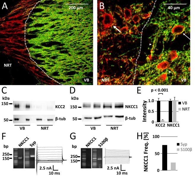
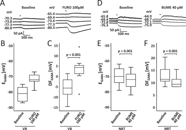
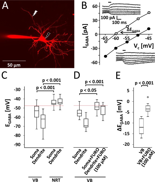
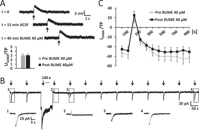
The thalamus is important for sensory integration with the ventrobasal thalamus (VB) as relay controlled by GABAergic projections from the nucleus reticularis thalami (NRT). Depending on the [Cl-]i primarily set by cation-chloride-cotransporters, GABA is inhibitory or excitatory. There is evidence that VB and NRT differ in terms of GABA action, with classical hyperpolarization in VB due to the expression of the Cl- extruder KCC2 and depolarizing/excitatory GABA action in the NRT, where KCC2 expression is low and Cl- accumulation by the Cl- inward transporter NKCC1 has been postulated. However, data on NKCC1 expression and functional analysis of both transporters are missing. We show that KCC2-mediated Cl- extrusion set the [Cl-]i in VB, while NKCC1 did not contribute substantially to Cl- accumulation and depolarizing GABA action in the NRT. The finding that NKCC1 did not play a major role in NRT neurons is of high relevance for ongoing studies on the therapeutic use of NKCC1 inhibitors trying to compensate for a disease-induced up-regulation of NKCC1 that has been described for various brain regions and disease states like epilepsy and chronic pain. These data suggest that NKCC1 inhibitors might have no major effect on healthy NRT neurons due to limited NKCC1 function.


Conflict of interest statement

The authors declare no competing interests.

Figures

Figure 1
Figure 1
Differential expression of KCC2 and NKCC1 in VB and NRT detected by immunostaining and Western blot analysis. (A) KCC2 (green) protein expression was high in VB, whereas it was barely detectable in the NRT. MAP2 was used as a somato-dendritic marker (red). The same distribution was also visible at the cellular level in the transition zone between VB and NRT (B), where MAP2-positive somata (thin arrow) and dendrites (arrow heads) in VB were labelled by KCC2 immunoreactivity (KCC2-IR), while KCC2-IR was not detectable around somata in the NRT (thick arrow). KCC2-positive dendrites in the NRT originated presumably from thalamo-cortical VB neurons. (C) KCC2 was present in immunoblots with samples of VB tissue, but not in NRT samples. β-tubulin (β-tub) was used as loading control. (D) NKCC1 was expressed ubiquitously in VB and NRT. (E) Quantification of KCC2- and NKCC1-immunoreactivity. Protein expression of KCC2 in NRT reached 9% of the level seen in VB (n = 7), whereas NKCC1 levels did not differ between VB and NRT (n = 6; paired Student’s t-test). (F) Agarose gel images showing NKCC1 transcript expression and respective current pattern of a NRT neuron after de- and hyperpolarization of the cell. Synaptophysin (Syp) served as positive control for neuronal mRNA detection. Expected product lengths were 159 bp for NKCC1 and 215 bp for synaptophysin. (G) Agarose gel and respective current pattern of a NRT astrocyte. S100β (product length: 186 bp) served as positive control for astrocytal mRNA detection. (H) NKCC1 mRNA was present in most NRT neurons (30 out of 40 Syp-positive cells) but to a lesser extent in NRT astrocytes (3 out of 13 S100β-positive cells).
Figure 2
Figure 2
NRT neurons exhibited more positive GABA reversal potentials than VB neurons. (A,B) Perforated patch recordings revealed KCC2-mediated transport in VB neurons. Bath application of the KCC2 inhibitor furosemide (FURO; in the presence of 10 µM bumetanide) shifted EGABA to more positive values (n = 13; paired Student’s t-test). (C) Accordingly, FURO shifted the GABA driving force (DFGABA) to more positive values. (D,E) NRT neurons exhibited native values of EGABA which were more positive than those of VB neurons and similar to VB neurons after FURO application. Bath application of the NKCC1 inhibitor bumetanide (BUME) induced a slight (although significant) shift to more negative EGABA values and reduced DFGABA (F) (n = 15; paired Student’s t-test). Sample traces (A,D) show representative current traces in response to command potential steps of single neurons before and after bath application of FURO or BUME, respectively (bar indicates GABA puff application).
Figure 3
Figure 3
KCC2-mediated Cl extrusion is present in VB, but not in NRT neurons. (A) Superimposed image stack of a VB neuron loaded with a defined Cl concentration and Alexa 594 as a fluorescent dye. GABA was locally uncaged at the indicated positions (flash). (B) Exemplary I-V-curves recorded from a VB neuron with subsequent GABA uncaging at the soma (filled circles; lower inlay) and at the dendrite in 50 µm distance from the soma (open circles; upper inlay). Currents were recorded at command potentials (VC) steps of +5 mV starting from −70 mV (inset: bar represents GABA photolysis). KCC2 shifted the imposed GABA reversal potential (EGABA; defined by the X-intercept) at the dendrite to more negative values compared to the soma by extruding Cl, since the slow diffusion of Cl from the pipette via the soma is not able to fully counteract KCC2-mediated Cl extrusion in dendrites. (C) Whole cell recordings revealed that imposed EGABA values of VB neurons at both soma and dendrite were more negative compared to NRT neurons (n = 17 and n = 15 at the soma, n = 13 and n = 7 at the dendrite, respectively; Student’s t-test). Moreover VB neurons exhibited gradients between soma and dendrite. In addition EGABA values of VB neurons are hyperpolarized from the calculated reversal potential of Cl (−47.5 mV; illustrated by the red line), which is determined by the pipette solution (20 mM Cl). The hyperpolarization compared to the NRT and to the calculated Cl reversal potential and somato-dendritic gradients of the VB together indicate a strong KCC2 activity. In the NRT, where KCC2 was not detectable, EGABA values at the soma as well as at the dendrite were close to calculated reversal potential and no somato-dendritic gradients were found. (D) In a new set of experiments bath application of the KCC2 inhibitor furosemide (FURO; in the presence of 10 µM bumetanide) shifted the imposed EGABA of VB neurons towards the calculated reversal potential at the soma and even more at the dendrite (n = 10 and n = 8 for control and FURO, respectively; Student’s t-test). Furthermore, FURO induced a significant decrease in somato-dendritic gradients (ΔEGABA) in VB neurons (n = 10 and n = 8, respectively; Student’s t-test) (E).
Figure 4
Figure 4
Inhibition of NKCC1 did not restrain neuronal Cl uptake in the NRT. (A) Example of a NRT neuron (VM = -91.4 mV) responding to local GABA puff application (indicated by bar). Under perforated patch conditions in current clamp, all recorded NRT neurons were depolarized from resting membrane potential (I = 0 pA). Application of the NKCC1 inhibitor bumetanide (BUME) did not alter the amplitude of the GABA response (UGABA/TP: voltage peak amplitude normalized to test pulse; p = 0.924 for n = 14; paired Student’s t-test). (B) In voltage clamp, depletion of intracellular Cl was achieved by application of 100 GABA puffs during hyperpolarization of the cell to −100 mV. GABA puffs were depolarizing, resulting in negative current responses. The first response to GABA immediately after the depletion was hyperpolarizing, thus the recorded current turned positive. Thereafter, GABA responses recovered to baseline level (magnification of boxed parts depicted in insets). GABA puffs are marked by arrows. (C) After a complete depletion protocol (Pre BUME) as shown in (B), bumetanide was bath applied for 20 min, before a second depletion protocol was performed in the same neuron (Post BUME). BUME application did neither significantly alter the time course nor the amplitude of the GABA responses, which suggests that NKCC1 is not responsible for Cl uptake in NRT neurons (repeated measures ANOVA: p = 0.407 and p = 0.331, respectively, for n = 6, see text for detailed description).

Similar articles

Cited by

References

    1. Pinault D. The thalamic reticular nucleus: Structure, function and concept. Brain Res. Rev. 2004;46:1–31. doi: 10.1016/j.brainresrev.2004.04.008. - DOI - PubMed
    1. Llinás RR, Ribary U, Jeanmonod D, Kronberg E, Mitra PP. Thalamocortical dysrhythmia: A neurological and neuropsychiatric syndrome characterized by magnetoencephalography. Proc. Natl. Acad. Sci. USA. 1999;96:15222–7. doi: 10.1073/pnas.96.26.15222. - DOI - PMC - PubMed
    1. Herd MB, Brown AR, Lambert JJ, Belelli D. Extrasynaptic GABAA Receptors Couple Presynaptic Activity to Postsynaptic Inhibition in the Somatosensory Thalamus. J. Neurosci. 2013;33:14850–14868. doi: 10.1523/JNEUROSCI.1174-13.2013. - DOI - PMC - PubMed
    1. Steriade M. Sleep, epilepsy and thalamic reticular inhibitory neurons. Trends Neurosci. 2005;28:317–24. doi: 10.1016/j.tins.2005.03.007. - DOI - PubMed
    1. Huguenard JR, McCormick DA. Thalamic synchrony and dynamic regulation of global forebrain oscillations. Trends Neurosci. 2007;30:350–6. doi: 10.1016/j.tins.2007.05.007. - DOI - PubMed

Publication types

LinkOut - more resources