. 2018 Aug 26:2018:2581031.
doi: 10.1155/2018/2581031. eCollection 2018.

SYKT Alleviates Doxorubicin-Induced Cardiotoxicity via Modulating ROS-Mediated p53 and MAPK Signal Pathways

Affiliations

SYKT Alleviates Doxorubicin-Induced Cardiotoxicity via Modulating ROS-Mediated p53 and MAPK Signal Pathways

Ting Chen et al. Evid Based Complement Alternat Med. .

Abstract

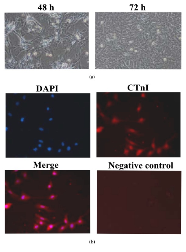
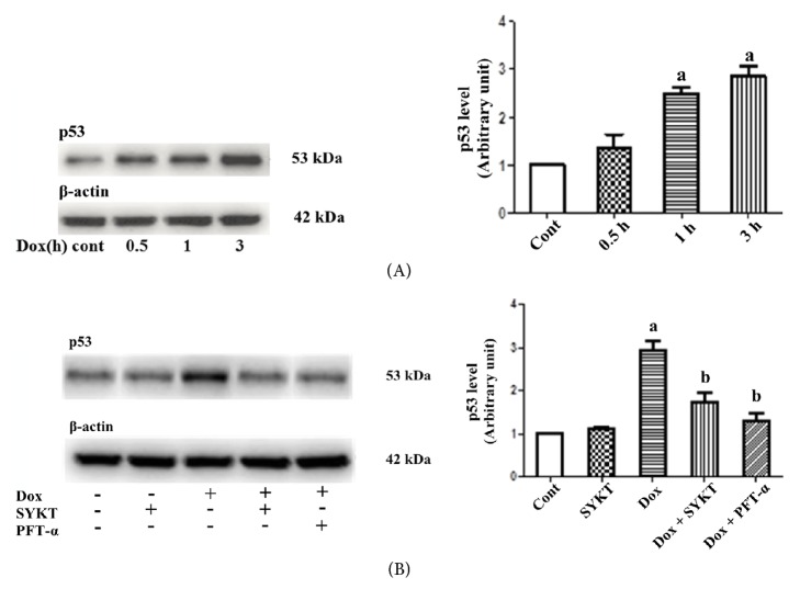
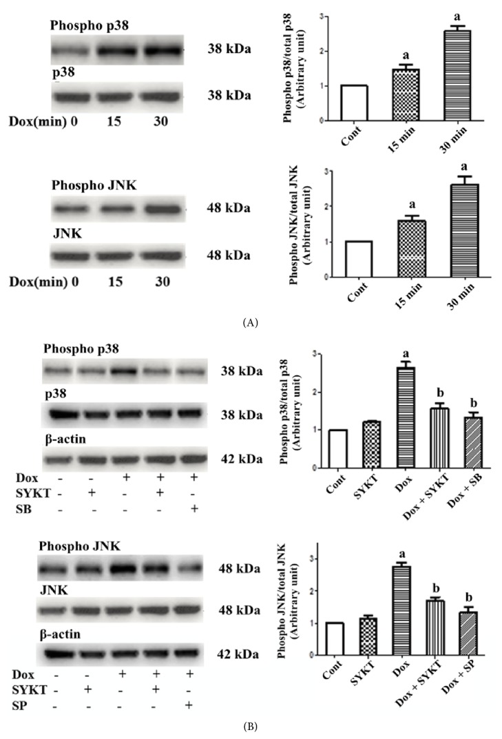
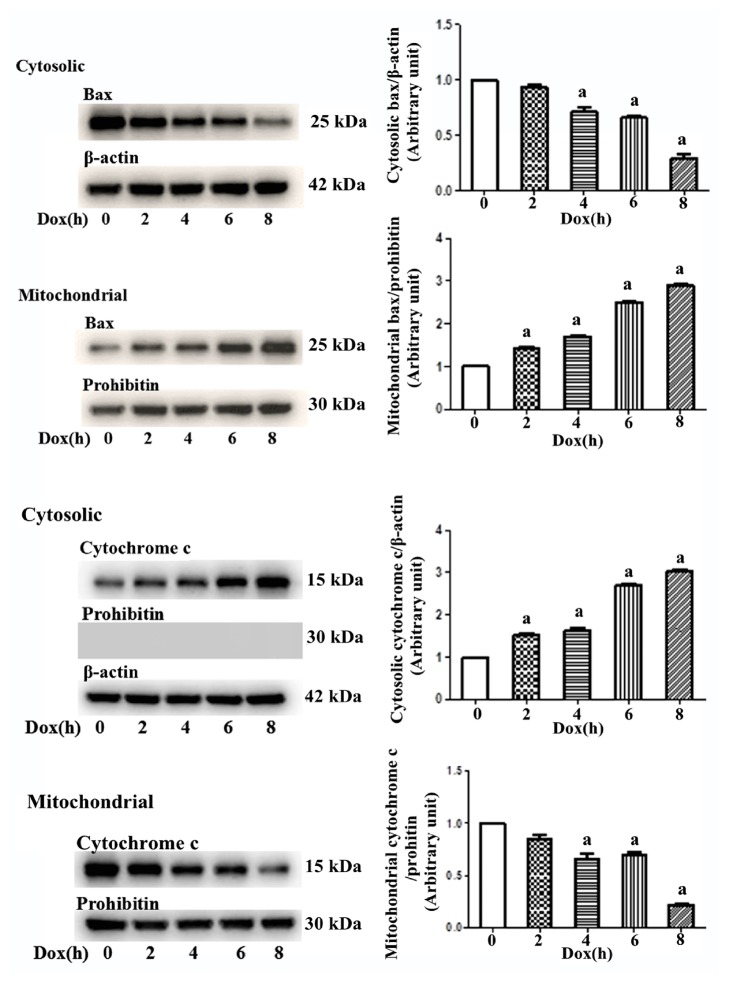
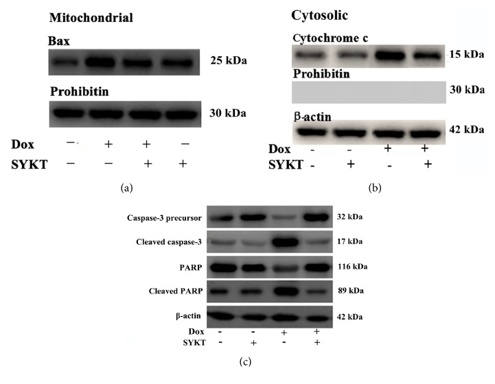
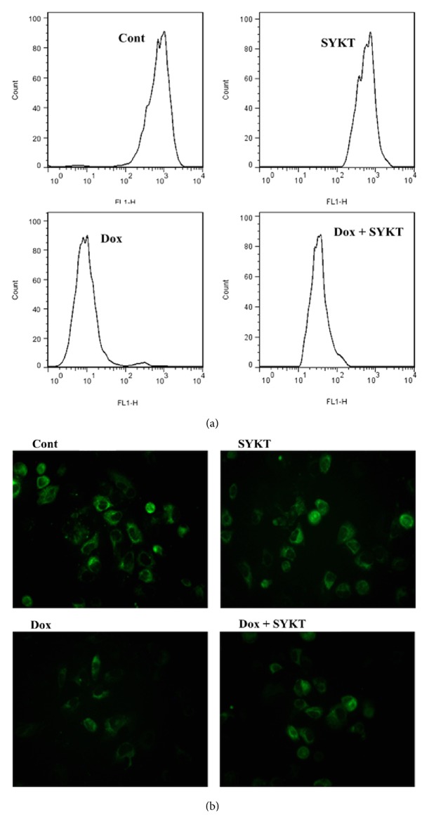
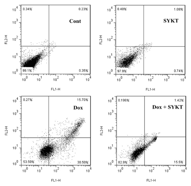
Backgrounds. Doxorubicin (DOX) is an effective therapeutic drug for malignant tumors; however, its clinical applications were limited by its side effects, especially the cardiotoxicity caused by ROS-mediated p53 and MAPK signal pathways' activation-induced cell apoptosis. Sanyang Xuedai mixture (SYKT) has been reported as an antioxidant agent and attenuated DOX-induced cardiotoxicity by targeting ROS-mediated apoptosis, but the mechanisms are still not fully delineated. Objective. This study aimed at investigating whether SYKT alleviated DOX-induced cardiotoxicity by inhibiting ROS-mediated apoptosis and elucidating the role of ROS-mediated p53 and MAPK signal pathways' activation in this process. Materials and Methods. Identification, separation, and culture of mouse primary cardiomyocytes. Cells were treated with DOX (1 μM), SYKT (30 mg/mL), or SYKT coupled with DOX. The p53 inhibitor Pifithrin-α (PFT-α), p38/MAPK inhibitor SB203583 (SB), and JNK inhibitor SP600125 (SP) were used as positive control. Western blot was employed to detected p53 and p38 as well as JNK expressions and the activation and translocation of Bax and cytochrome C. Flow cytometer (FCM) was used to detect the mitochondrial membrane potential and cell apoptosis. Results. After separation and culture, 95% of cells showed positive cTnI expression, which indicated that mouse primary cardiomyocytes were successfully identified in our research. DOX activated p53 and MAPK signal pathways in a time-dependent manner, which were inactivated by being cotreated with SYKT, PFT-α, or SB, respectively. DOX significantly decreased Bax and increased cytochrome c expressions in the cytoplasm, whereas Bax was upregulated and cytochrome c was downregulated in the mitochondria, which were reversed by SYKT treatment. Besides, DOX reduced mitochondria membrane potential (MMP) in cardiomyocytes compared to the control group; SYKT recovered its MMP and attenuated DOX-induced cardiomyocyte injury. Of note, DOX increased the expression levels of cleaved caspase-3 as well as poly ADP-ribose polymerase (PARP) and promoted cell apoptosis, which were also reversed by SYKT treatment. Discussion and Conclusions. Our results indicated that SYKT alleviated DOX-induced cardiotoxicity by inhibiting p53 and MAPK signal pathways' activation-mediated apoptosis, and it might serve as a potential therapeutic agent for DOX-induced cardiotoxicity.


Figures

Figure 1
Figure 1
Culture and identification of mouse primary cardiomyocytes. (a) After 48 h of incubation, adherent cardiomyocytes accounted for approximately 90% of the cells. The cells exhibited various morphologies, such as spindle and polygonal shapes. After 72 h of incubation, the cells fused together (10×). (b) Cardiomyocytes identification using cTnI immunofluorescent staining (10×).
Figure 2
Figure 2
SYKT inhibits DOX-induced p53 activation. (A) DOX induces p53 expression in a time-dependent manner. Cardiomyocytes were treated with DOX for the indicated periods, and the p53 levels were analyzed by immunoblotting. β-Actin served as a loading control. (B) Effects of DOX (1 μM for 3 h), SYKT (30 mg/ml), and PFT-α (40 μM) on p53 expression. ap < 0.05 versus control; bp < 0.05 versus DOX; n = 6.
Figure 3
Figure 3
SYKT inhibits DOX-induced MAPK activation. (A) DOX induces p38/JNK MAPK activation in a time-dependent manner. (B) DOX-mediated p38/JNK MAPK activation is inhibited by SB 203580/SP600125 and by SYKT. The cardiomyocytes were treated with SB 203580/SP600125 prior to DOX (1 μM for 30 min) addition. β-Actin was used as an internal control. ap < 0.05 versus control; bp < 0.05 versus DOX; n = 6.
Figure 4
Figure 4
Influence of DOX on cytoplasmic and mitochondrial Bax and cytochrome c expression is time-dependent. Mouse primary cardiomyocytes were treated with 1 μM DOX for 1-8 h. Over time, cytoplasmic Bax expression gradually decreased and cytochrome c expression gradually increased, whereas mitochondrial Bax expression gradually increased and cytochrome c expression gradually decreased. β-Actin was used as the internal control, and Prohibitin was used as a mitochondrial marker.
Figure 5
Figure 5
Effects of SYKT on DOX-induced cardiomyocyte apoptosis-related proteins at 8 h after DOX processing. (a) The Bax levels were analyzed in the mitochondrial extracts by immunoblotting. (b) Effects of DOX and SYKT on cytoplasmic cytochrome c release in cardiomyocytes. (c) Western blotting analysis of the effects of DOX, SYKT, and DOX+SYKT on caspase-3 activation and PARP cleavage in cardiomyocytes. β-Actin was used as the internal control, and Prohibitin was used as a mitochondrial marker.
Figure 6
Figure 6
Determination of the MMP of cardiomyocytes from each group (n = 6 rats/group) at 8 h after DOX processing. (a) Histograms of the MMP of cardiomyocytes detected by Rhodamine 123 staining by FCM. (b) Photos of Rhodamine 123 staining of MMP in all groups by CLSM (100×).
Figure 7
Figure 7
Percent distribution of apoptotic and necrotic cells from each group (n = 6 rats/group) at 8 h after DOX processing. The cell distribution was analyzed using Annexin V binding and PI uptake. The FITC and PI fluorescence was measured using a FCM with FL-1 and FL-2 filters, respectively.

Similar articles

Cited by

References

    1. Vadhan-Raj S., Patel S., Bueso-Ramos C., et al. Importance of predosing of recombinant human thrombopoietin to reduce chemotherapy-induced early thrombocytopenia. Journal of Clinical Oncology. 2003;21(16):3158–3167. doi: 10.1200/JCO.2003.08.003. - DOI - PubMed
    1. Carresi C., Musolino V., Gliozzi M., et al. Anti-oxidant effect of bergamot polyphenolic fraction counteracts doxorubicin-induced cardiomyopathy: Role of autophagy and c-kit pos CD45 neg CD31 neg cardiac stem cell activation. Journal of Molecular and Cellular Cardiology. 2018;119:10–18. doi: 10.1016/j.yjmcc.2018.04.007. - DOI - PubMed
    1. Wei X., Liu L., Guo X., Wang Y., Zhao J., Zhou S. Light-Activated ROS-Responsive Nanoplatform Codelivering Apatinib and Doxorubicin for Enhanced Chemo-Photodynamic Therapy of Multidrug-Resistant Tumors. ACS Applied Materials & Interfaces. 2018;10(21):17672–17684. doi: 10.1021/acsami.8b04163. - DOI - PubMed
    1. Zhang M., Harashima N., Moritani T., Huang W., Harada M. The roles of ROS and caspases in TRAIL-induced apoptosis and necroptosis in human pancreatic cancer cells. PLoS ONE. 2015;10(5) doi: 10.1371/journal.pone.0127386.e0127386 - DOI - PMC - PubMed
    1. Qin G., Wu L., Liu H., et al. Artesunate induces apoptosis via a ROS-independent and Bax-mediated intrinsic pathway in HepG2 cells. Experimental Cell Research. 2015;336(2):308–317. doi: 10.1016/j.yexcr.2015.07.004. - DOI - PubMed

LinkOut - more resources