

Unicameral bone cysts: Current concepts
- PMID: 30224948
- PMCID: PMC6138978
- DOI: 10.1016/j.amsu.2018.06.005
Unicameral bone cysts: Current concepts
Abstract
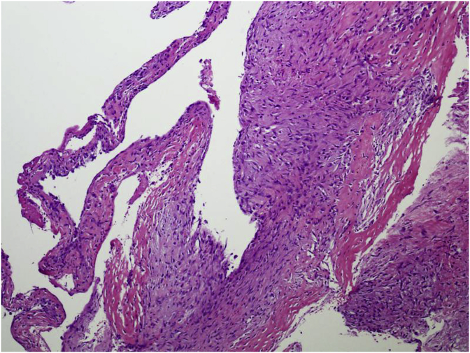
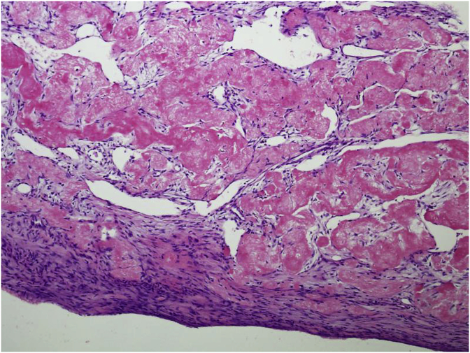
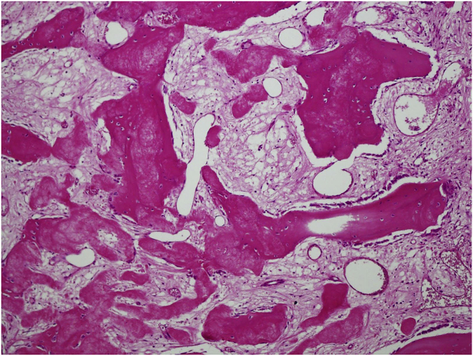
Unicameral bone cysts (UBC) or simple/solitary bone cysts are benign fluid filled cavities that enlarge over time, resulting in thinning of the bone. Usually these cysts are reported in the metaphyseal areas of long bones with open physes. 85% of UBCs occur almost exclusively in children and adolescents. UBCs are more aggressive in the first decade of life and correspondingly the recurrence rate for these patients is four times that for adolescents. The proximal humerus and femur account for almost 90% of these cases. UBCs are classified as active when they are within 1 cm of the physis and latent as they progress to a diaphyseal location. Differential diagnoses for UBC include aneurysmal bone cyst, fibrous dysplasia, enchondroma, and intraosseous ganglia. By the time of skeletal maturity most UBCs tend to resolve. Nonoperative treatment may be a viable option for many patients with small or symptomatic lesions. Interventions include steroid injection, open curettage and bone grafting, decompression and percutaneous injection of marrow or graft substitutes.
Keywords: Current concepts; Unicameral bone cyst.
Figures
References
-
- Wilkins R.M. Unicameral bone cysts. J. Am. Acad. Orthop. Surg. 2000;8(4):217–224. - PubMed
-
- Pretell-Mazzini J., Murphy R.F., Kushare I., Dormans J.P. Unicameral bone cysts: general characteristics and management controversies. J. Am. Acad. Orthop. Surg. 2014;22(5):295–303. - PubMed
-
- Biermann J.S. Common benign lesions of bone in children and adolescents. J. Pediatr. Orthop. 2002;22(2):268–273. - PubMed
-
- Boseker E., Bickel W., Dahlin D. A clinicopathologic study of simple unicameral bone cysts. Surg. Gynecol. Obstet. 1968;127(3):550–560. - PubMed
-
- Jaffe H.L., Lichtenstein L. Solitary unicameral bone cyst: with emphasis on the roentgen picture, the pathologic appearance and the pathogenesis. Arch. Surg. 1942;44(6):1004–1025.
Publication types
LinkOut - more resources
Full Text Sources
Other Literature Sources
