

doi: 10.1167/tvst.7.5.8.
eCollection 2018 Sep.
Translational Retinal Research and Therapies
Affiliations
- PMID: 30225158
- PMCID: PMC6138060
- DOI: 10.1167/tvst.7.5.8
Item in Clipboard
Translational Retinal Research and Therapies
Transl Vis Sci Technol.
.
Abstract
The following review summarizes the state of the art in representative aspects of gene therapy/translational medicine and evolves from a symposium held at the School of Veterinary Medicine, University of Pennsylvania on November 16, 2017 honoring Dr. Gustavo Aguirre, recipient of ARVO's 2017 Proctor Medal. Focusing on the retina, speakers highlighted current work on moving therapies for inherited retinal degenerative diseases from the laboratory bench to the clinic.
Keywords: Leber congenital amaurosis; X-linked Retinoschisis; animal models; retinitis pigmentosa.
Figures
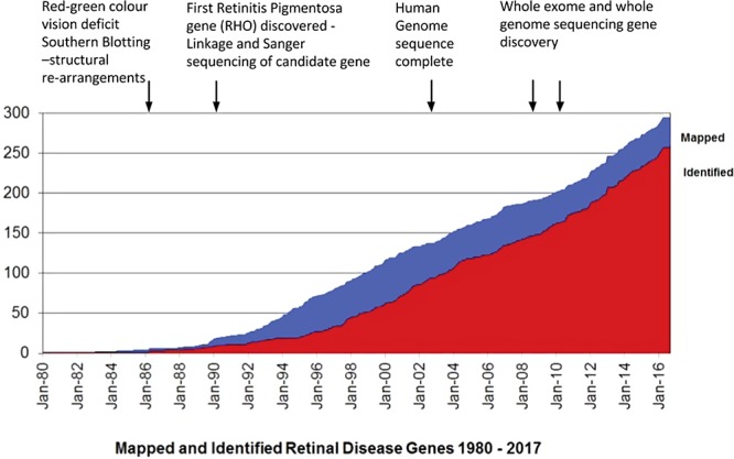
Mapped and identified retinal disease genes. Figure modified from RetNet (in the public domain, https://sph.uth.edu/retnet ) to highlight landmark time points in path to disease gene discovery.
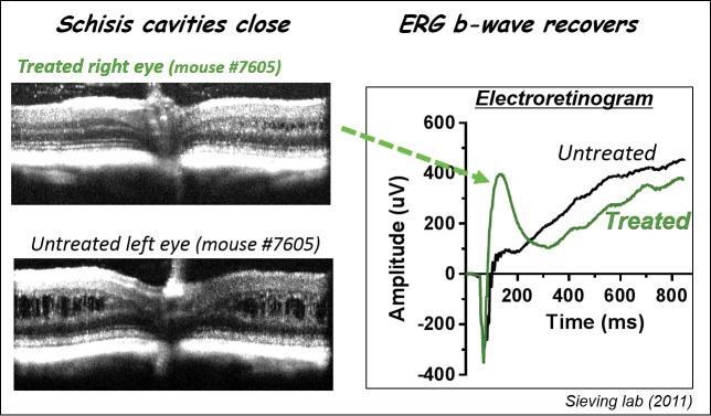
AAV-RS1 rescues structure and function in the XLRS mouse. scAAV-hRS1/IRBP- hRS1 injected intravitreally at 4 weeks of age and OCT and ERG evaluated 12 weeks later. Left panel demonstrates that schisis cavities close. Right panel demonstrates restoration of ERG b-wave (green arrow).
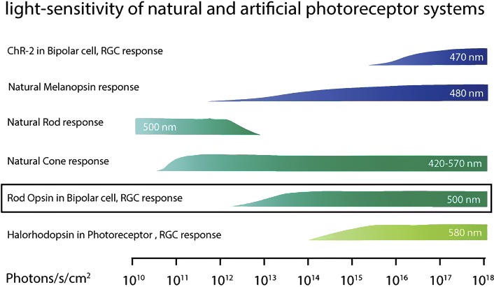
Light sensitivity of natural and artificial photoreceptor systems. Reprinted with permission from Gaub BM, Berry MH, Visel M, Holt A, Isacoff EY, Flannery JG. Optogenetic retinal gene therapy with the light-gated gpcr vertebrate rhodopsin. In: Boon C, Wijnholds J, eds. Retinal Gene Therapy. Methods in Molecular Biology. Humana Press: New York; 2018:177–189.
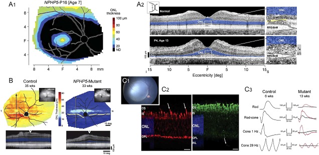
NPHP5 disease phenotypes and response to gene therapy. (A1) ONL thickness topography of NPHP5 patient shows the distribution of the detectable photoreceptors to be limited to a central ellipse corresponding to the cone-dominant region, and more peripherally corresponding to the rod-dominant region. Wide annular region shows no detectable photoreceptors (black). (A2) Cross-sectional OCT scans along the vertical meridian through the fovea, extending 15° into superior (S) and inferior (I) retina in a healthy subject (age 30 years; top) and NPHP5-LCA patient P4 (age 13 years). Magnified views of the central retina (right; white rectangles) with overlapping longitudinal reflectivity profiles (LRP). Photoreceptor nuclear layer (ONL) is highlighted in blue; ellipsoid zone is highlighted in yellow. OLM, outer limiting membrane; COS, cone outer segments; RPE/BrM, retinal pigment epithelium/Bruch's membrane. In the patients, there are wide hyperscattering bands (S+) and more narrow hyposcattering bands (S−) distal to the ONL (far right along image). Icon (upper left) is location of the scans on a retinal schematic. (B) Pseudocolor maps of ONL thickness topography (upper) in a 35-week-old control dog (left) and an NPHP5-mutant (right) at 33 weeks of age. Insets, near-infrared reflectance images. Arrows on the maps localize the reconstituted OCT scans (lower) along a superior–inferior meridian crossing the central visual streak at the gaps of the lines. ONL on reconstituted scans is highlighted in blue and location of the scan relative to the vertical arrows is shown with a white wedge. All eyes shown as equivalent right eyes and optic nerve, major blood vessels (black) and tapetum boundary (yellow) are overlaid for ease of orientation. T, temporal; N, nasal retina. (C1) Right eye of a 5.7-week-old vector-treated NPHP5 mutant with large subretinal bleb that encompasses the fovea-like central region. (C2) Six-week-old NPHP5 mutant dog retina showing that most cones lack an outer segment (red = hCAR labeling; arrows indicate present cone outer segments); rod outer segments (green = rod opsin labeling) form, but are abnormal and there is extensive rod opsin mislocalization to the ONL. (C3) Treatment at 5.7 weeks with AAV-cNPHP5 vector (red traces = treated eye; black traces = untreated fellow eye) results in robust recovery rod, mixed rod-cone and cone ERG responses by 13 weeks. Fig. A1 modified and reprinted with permission from Cideciyan AV, Rachel RA, Aleman TS, et al. Cone photoreceptors are the main targets for gene therapy of NPHP5 (IQCB1) or NPHP6 (CEP290) blindness: generation of an all-cone Nphp6 hypomorph mouse that mimics the human retinal ciliopathy. Hum Mol Genet 2011; 20:1411–1423. Figs. B, C2, and C3 modified and reprinted with permission from Downs LM, Scott EM, Cideciyan AV, et al. Overlap of abnormal photoreceptor development and progressive degeneration in Leber congenital amaurosis caused by NPHP5 mutation. Hum Mol Genet 2016;25:4211–4226.
References
-
- Dryja TP, McGee TL, Hahn LB, et al. Mutation within the rhodopsin gene in patients with autosomal dominant retinitis pigmentosa. N Eng J Med. 1990;323:1302–1307. - PubMed
-
- Zhang Q, Acland GM, Wu WX, et al. Different RPGR exon ORF15 mutations in Canids provide insights into photoreceptor cell degeneration. Hum Mol Genet. 2002;11:993–1003. - PubMed
-
- Fiorentino A, Fujinami K, Arno G, et al. Missense variants in the X-linked gene PRPS1 cause retinal degeneration in females. Hum Mutat. 2018;39:80–91. - PubMed
-
- Hardcastle AJ, Thiselton DL, Zito I, et al. Evidence for a new locus for X-linked retinitis pigmentosa (RP23) Invest Ophthalmol Vis Sci. 2000;41:2080–2086. - PubMed
Grants and funding
LinkOut - more resources
Full Text Sources
Other Literature Sources
