

Review
doi: 10.1007/s00259-018-4146-5.
Epub 2018 Sep 17.
Translational molecular imaging in exocrine pancreatic cancer
Affiliations
- PMID: 30225616
- PMCID: PMC6208802
- DOI: 10.1007/s00259-018-4146-5
Item in Clipboard
Review
Translational molecular imaging in exocrine pancreatic cancer
Eur J Nucl Med Mol Imaging.
2018 Dec.
Abstract
Effective treatment for pancreatic cancer remains challenging, particularly the treatment of pancreatic ductal adenocarcinoma (PDAC), which makes up more than 95% of all pancreatic cancers. Late diagnosis and failure of chemotherapy and radiotherapy are all too common, and many patients die soon after diagnosis. Here, we make the case for the increased use of molecular imaging in PDAC preclinical research and in patient management.
Keywords: Molecular imaging; PET; Pancreatic ductal adenocarcinoma; Preclinical developments; SPECT.
Conflict of interest statement
Conflicts of interest
None.
Figures

18F-FDG PET imaging for initial staging of PDAC in an 80-year-old woman. a The initial staging MR image shows a hypoenhancing mass in the pancreatic body (arrows) that has resulted in pancreatic ductal dilatation. b The PET/CT image shows corresponding FDG uptake in the mass (arrows). c, d Additionally, the fused PET/CT image (c) and PET-only image (d) show an 18F-FDG-avid peripancreatic node (arrows). Adapted from Yeh et al. [7]
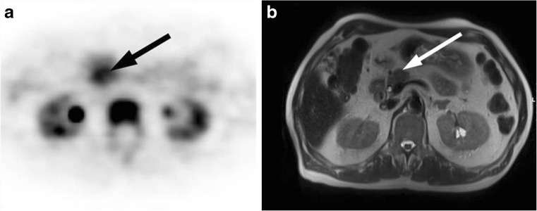
18F-FLT PET image in a 70-year-old patient with a 2-cm tumour in the pancreatic head [19]
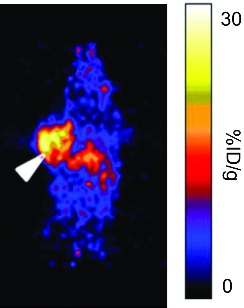
Coronal PET image of a mouse bearing a GRP78-positive BxPC-3 tumour xenograft 48 h after injection of the 64Cu-labelled anti-GRP78 antibody MAb159 [44]

MRI imaging in an orthotopic mouse model of pancreatic cancer 4 months after surgical implantation of the tumour showing the difference in signal between intravenously administered conventional contrast agent (free Magnevist) and the TfRscFv-Lip-Mag complex [50]

a Mesothelin in PDAC (protein atlas: 11/12 positive [56]). b
89Zr-MMOT0530 PET/CT image in a patient with PDAC shows the primary tumour (red oval), as well as uptake in healthy liver [55]

68Ga-avebehexin PET image (maximum-intensity projection, 60 min after injection) of a H2009-bearing SCID mouse. The tumour is indicated by the arrow. Blad. bladder, Kid. kidneys [63]

Coronal small-animal PET images of HT29 tumour-bearing immunodeficient mice injected with 68Ga-8, a radiolabelled neurotensin peptide analogue. The mouse on the right received a blocking dose of cold, unlabelled compound to saturate the receptor [71]

a Representative in-vivo image of a human primary pancreatic cancer tumour xenograft in a mouse after administration of a Cy5 fluorophore-labelled cathepsin E substrate [74]. b A Cy5.5-labelled cathepsin B-targeting DARPin is taken up in a 4T1 allograft murine breast tumour (red circle), but not in a healthy mammary fat pad (black circle) [75]

NIR fluorescence imaging in a mouse bearing subcutaneous PDAC tumours acquired 72 h after injection of CEA-targeting ssSM3E/800CW or F73/800CW [78]

a CEA expression in human PDAC tissue (protein atlas [56]) b SPECT/CT image in a 38-year-old patient acquired 24 h after injection of 111In-IMP288 (185 MBq, 25 μg) pretargeted with 75 mg of TF2 (1-day interval) [80]

PET, PET/CT, and near infrared imaging in an orthotopic PDAC mouse model using a 89Zr-labelled anti-CA19.9 antibody (LN lymph node, M metastasis, T tumour) [84]
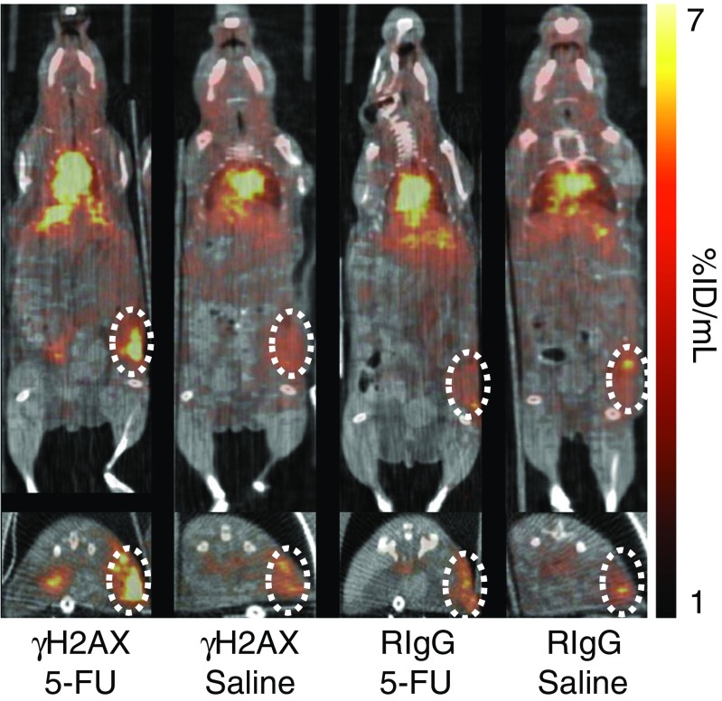
Monitoring 5-FU therapy with 89Zr-anti-γH2AX-TAT: PET/CT images show coronal (top) and transaxial (bottom) sections intersecting the centre of the allograft tumour (dotted circles) [13]. Tumour uptake of a nonspecific control antibody (RIgG) was not different between 5-FU-treated and vehicle-treated animals
Similar articles
-
Rare Solid Tumor of the Exocrine Pancreas: A Pictorial Review.Semin Ultrasound CT MR. 2019 Dec;40(6):483-499. doi: 10.1053/j.sult.2019.04.007. Epub 2019 Apr 25. Semin Ultrasound CT MR. 2019. PMID: 31806147 Review.
-
Detection of pancreatic ductal adenocarcinoma in mice by ultrasound imaging of thymocyte differentiation antigen 1.Gastroenterology. 2013 Oct;145(4):885-894.e3. doi: 10.1053/j.gastro.2013.06.011. Epub 2013 Jun 18. Gastroenterology. 2013. PMID: 23791701 Free PMC article.
-
Advances in Diagnostic and Intraoperative Molecular Imaging of Pancreatic Cancer.Pancreas. 2018 Jul;47(6):675-689. doi: 10.1097/MPA.0000000000001075. Pancreas. 2018. PMID: 29894417 Free PMC article. Review.
-
Utility of 18 fludeoxyglucose in preoperative positon-emission tomography-computed tomography (PET-CT) in the early diagnosis of exocrine pancreatic cancer: A study of 139 resected cases.Cir Esp. 2016 Nov;94(9):511-517. doi: 10.1016/j.ciresp.2016.07.007. Epub 2016 Oct 4. Cir Esp. 2016. PMID: 27712835 English, Spanish.
-
Contrast-enhanced ultrasound in the differential diagnosis of exocrine versus neuroendocrine pancreatic tumors.Pancreas. 2013 Jul;42(5):871-7. doi: 10.1097/MPA.0b013e31827a7b01. Pancreas. 2013. PMID: 23531999
Cited by
-
Non-invasive molecular imaging for precision diagnosis of metastatic lymph nodes: opportunities from preclinical to clinical applications.Eur J Nucl Med Mol Imaging. 2023 Mar;50(4):1111-1133. doi: 10.1007/s00259-022-06056-5. Epub 2022 Nov 29. Eur J Nucl Med Mol Imaging. 2023. PMID: 36443568 Review.
-
Radiolabeled cCPE Peptides for SPECT Imaging of Claudin-4 Overexpression in Pancreatic Cancer.J Nucl Med. 2020 Dec;61(12):1756-1763. doi: 10.2967/jnumed.120.243113. Epub 2020 May 15. J Nucl Med. 2020. PMID: 32414951 Free PMC article.
-
Diagnosis of Pancreatic Ductal Adenocarcinoma by Immuno-Positron Emission Tomography.J Clin Med. 2021 Mar 10;10(6):1151. doi: 10.3390/jcm10061151. J Clin Med. 2021. PMID: 33801810 Free PMC article. Review.
-
ImmunoPET/NIRF/Cerenkov multimodality imaging of ICAM-1 in pancreatic ductal adenocarcinoma.Eur J Nucl Med Mol Imaging. 2021 Aug;48(9):2737-2748. doi: 10.1007/s00259-021-05216-3. Epub 2021 Feb 3. Eur J Nucl Med Mol Imaging. 2021. PMID: 33537836 Free PMC article.
-
Preliminary study on SPECT/CT imaging of pancreatic cancer xenografts by targeting integrin α5 in pancreatic stellate cells.J Cancer. 2021 Jan 18;12(6):1729-1733. doi: 10.7150/jca.51190. eCollection 2021. J Cancer. 2021. PMID: 33613761 Free PMC article.
References
-
- Koopmans KP, Dierckx RA, Elsinga PH, Links TP, Kema IP, Fiebrich HB, et al. Other radiopharmaceuticals for imaging GEP-NET. In: Hubalewska-Dydejczyk A, Signore A, de Jong M, Dierckx RA, Buscombe J, Van de Wiele C, et al., editors. Somatostatin analogues: from research to clinical practice. Hoboken: Wiley; 2015.
-
- Cohen Joshua D., Li Lu, Wang Yuxuan, Thoburn Christopher, Afsari Bahman, Danilova Ludmila, Douville Christopher, Javed Ammar A., Wong Fay, Mattox Austin, Hruban Ralph H., Wolfgang Christopher L., Goggins Michael G., Dal Molin Marco, Wang Tian-Li, Roden Richard, Klein Alison P., Ptak Janine, Dobbyn Lisa, Schaefer Joy, Silliman Natalie, Popoli Maria, Vogelstein Joshua T., Browne James D., Schoen Robert E., Brand Randall E., Tie Jeanne, Gibbs Peter, Wong Hui-Li, Mansfield Aaron S., Jen Jin, Hanash Samir M., Falconi Massimo, Allen Peter J., Zhou Shibin, Bettegowda Chetan, Diaz Luis A., Tomasetti Cristian, Kinzler Kenneth W., Vogelstein Bert, Lennon Anne Marie, Papadopoulos Nickolas. Detection and localization of surgically resectable cancers with a multi-analyte blood test. Science. 2018;359(6378):926–930. doi: 10.1126/science.aar3247. - DOI - PMC - PubMed
Publication types
MeSH terms
Grants and funding
LinkOut - more resources
Full Text Sources
Other Literature Sources
Medical
