. 2018 Nov;40(5):2628-2636.
doi: 10.3892/or.2018.6703. Epub 2018 Sep 12.

Upregulation of macrophage migration inhibitory factor promotes tumor metastasis and correlates with poor prognosis of pancreatic ductal adenocarcinoma

Affiliations

Upregulation of macrophage migration inhibitory factor promotes tumor metastasis and correlates with poor prognosis of pancreatic ductal adenocarcinoma

Dong Wang et al. Oncol Rep. 2018 Nov.

Abstract

Macrophage migration inhibitory factor (MIF) is a pro‑inflammatory cytokine that serves important roles in cancer. MIF overexpression is frequently observed in numerous human cancer types, including pancreatic carcinoma. However, the prognostic value and function of MIF in pancreatic ductal adenocarcinoma (PDAC) have not been fully elucidated. In the present study, upregulation of MIF expression in PDAC tissue compared with adjacent normal tissue was observed. Furthermore, MIF overexpression was identified to be significantly associated with poor survival rates in patients with PDAC. Multivariate Cox regression analysis confirmed that MIF was an independent risk factor for poor survival. Functional analyses demonstrated that MIF knockdown significantly inhibited the proliferation and invasion of pancreatic cancer cells in vitro compared with control cells. IN addition, mechanistic investigations revealed that silencing MIF leads to inhibition of AKT serine/threonine kinase and extracellular‑signal‑regulated kinase activation, and suppression of cyclin D1 and matrix metalloproteinase‑2 expression, which may suppress tumor proliferation and invasion. These results highlight the importance of MIF overexpression in PDAC aggressiveness, and indicate that MIF may be a potential therapeutic target for pancreatic cancer.


Figures

Figure 1.
Figure 1.
Analysis of MIF expression in paired PDAC and adjacent non-tumor tissues. (A) Immunohistochemistry analysis of MIF expression in 85 human PDAC samples. A brown signal was considered positive. Images were captured at ×400 magnification. Scale bar, 50 µm. (B) Comparison of the MIF level in PDAC and adjacent non-tumor tissues in 85 human PDAC samples. Statistical analysis was performed by Wilcoxon matched-pairs signed-ranks test. (C) Reverse transcription-semi-quantitative polymerase chain reaction and immunoblotting analysis of MIF expression in PDAC. GAPDH was used as internal control. ***P<0.001. MIF, Macrophage migration inhibitory factor; PDAC, pancreatic ductal adenocarcinoma; T, PDAC tissue; N, adjacent noncancerous tissue.
Figure 2.
Figure 2.
Kaplan-Meier analysis for pancreatic ductal adenocarcinoma patient survival according to MIF level. MIF levels were analyzed by immunohistochemistry, and the median value of all 85 cases was chosen as the cut-off point for separating MIF low-expression tumors (n=42) from MIF high-expression cases (n=43). MIF, Macrophage migration inhibitory factor.
Figure 3.
Figure 3.
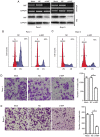
Silencing of MIF expression inhibits tumor invasion in vitro. (A) siRNA-mediated knockdown of MIF in PDAC cells. Control Panc-1 or Bxpc-3 cells (Mock) and Panc-1 or Bxpc-3 cells transfected with 50 nM MIF siRNA (si-MIF) or NC were subjected to RT-sqPCR or immunoblotting analysis. GAPDH was used as internal control. MIF knockdown caused accumulation of G1-population. (B) Panc-1 or (C) Bxpc-3 cells transfected with NC or si-MIF were treated with nocodazole (40 ng/ml) 32 h after transfection, and then cultured for an additional 16 h before harvest for FACS analysis. The percentage of the G1-population is indicated within each histogram. The results were reproduced in two independent experiments and representative data are shown. MIF knockdown inhibited PDAC (D) Panc-1 or (E) Bxpc-3 cell invasion. Control Panc-1 or Bxpc-3 cells (Mock) and Panc-1 or Bxpc-3 cells transfected with NC or si-MIF were added to Transwell chambers with Matrigel coating and incubated for 24 h, followed by staining with crystal violet. Scale bar, 100 µm. **P<0.01. MIF, Macrophage migration inhibitory factor; PDAC, pancreatic ductal adenocarcinoma; si/siRNA, small inteferring RNA; NC, negative control siRNA; wb, western blot; RT-sqPCR, reverse transcription-semi-quantitative polymerase chain reaction.
Figure 4.
Figure 4.
Silencing of MIF expression negatively regulates the AKT and ERK signaling pathways. Knockdown of MIF attenuated AKT and ERK activities in PDAC cells. Panc-1 or Bxpc-3 cells were transfected with NC or si-MIF and subjected to immunoblotting for (A) p-AKT and AKT; and (B) p-ERK1/2 and ERK1/2 expression. GAPDH was used as an internal control. Knockdown of MIF decreased the mRNA and protein levels of CCND1 and MMP-2. Panc-1 or Bxpc-3 cells transfected with si-MIF or NC were subjected to (C) reverse transcription- quantitative polymerase chain reaction or (D) immunoblotting analysis of CCND1 and MMP-2. For (A), (B), and (D), the intensity of each band was densitometrically quantified using Image J software and was normalized according to the value of NC group. Results were reproduced in three independent experiments and the cumulative data as well as the representative immunoblots are shown. ***P<0.001. MIF, Macrophage migration inhibitory factor; PDAC, pancreatic ductal adenocarcinoma; si/siRNA, small inteferring RNA; NC, negative control siRNA; CCND1, cyclin D1; MMP-2, matrix metalloproteinase-2; p- phosphorylated; AKT, AKT serine/threonine kinase; ERK, extracellular-signal-regulated kinase.

References

    1. Siegel RL, Miller KD, Jemal A. Cancer statistics, 2015. CA Cancer J Clin. 2015;65:5–29. doi: 10.3322/caac.21254. - DOI - PubMed
    1. Wolfgang CL, Herman JM, Laheru DA, Klein AP, Erdek MA, Fishman EK, Hruban RH. Recent progress in pancreatic cancer. CA Cancer J Clin. 2013;63:318–348. doi: 10.3322/caac.21190. - DOI - PMC - PubMed
    1. Liu SL, Friess H, Kleeff J, Ji ZL, Buchler MW. Surgical approaches for resection of pancreatic cancer: An overview. Hepatobiliary Pancreat Dis Int. 2002;1:118–125. - PubMed
    1. Ryan DP, Hong TS, Bardeesy N. Pancreatic adenocarcinoma. N Engl J Med. 2014;371:2140–2141. doi: 10.1056/NEJMra1404198. - DOI - PubMed
    1. Garcea G, Dennison AR, Steward WP, Berry DP. Role of inflammation in pancreatic carcinogenesis and the implications for future therapy. Pancreatology. 2005;5:514–529. doi: 10.1159/000087493. - DOI - PubMed

MeSH terms