. 2018 Nov;53(5):1836-1846.
doi: 10.3892/ijo.2018.4542. Epub 2018 Aug 28.

Inhibition of atypical protein kinase C‑ι effectively reduces the malignancy of prostate cancer cells by downregulating the NF-κB signaling cascade

Affiliations

Inhibition of atypical protein kinase C‑ι effectively reduces the malignancy of prostate cancer cells by downregulating the NF-κB signaling cascade

André H Apostolatos et al. Int J Oncol. 2018 Nov.

Abstract

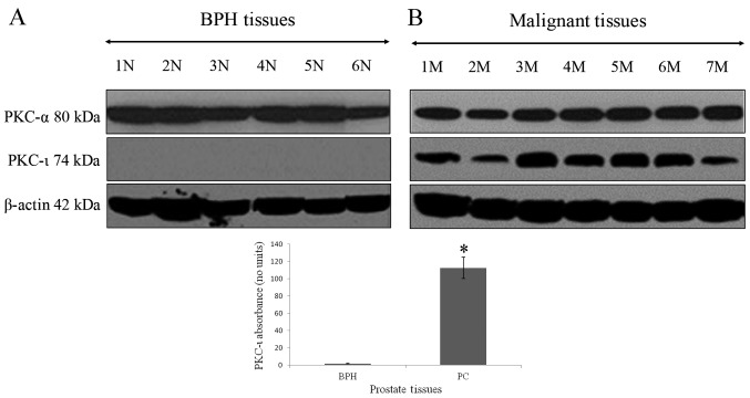
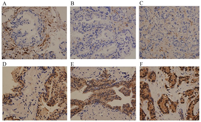
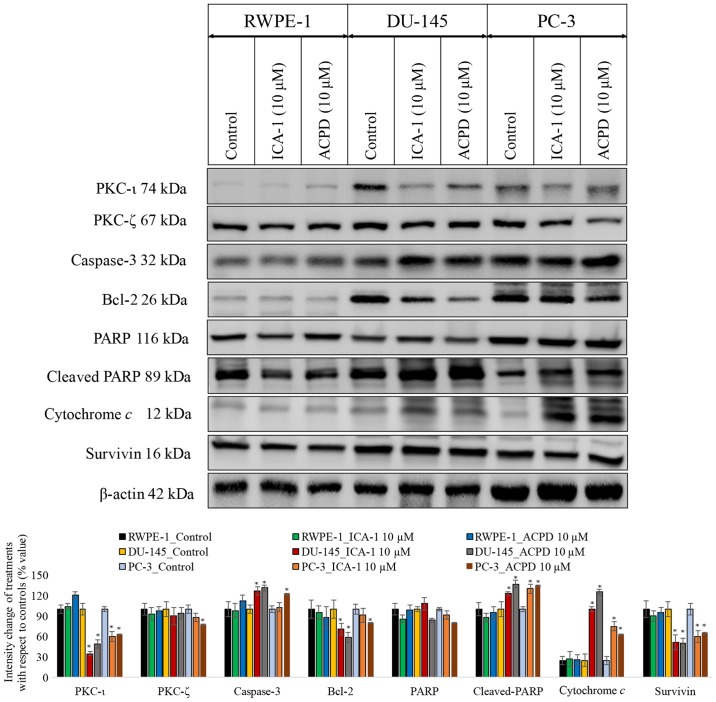
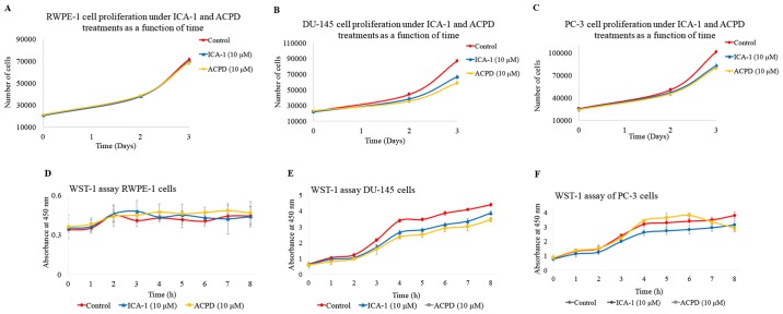
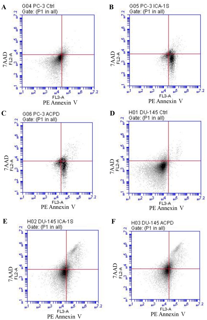
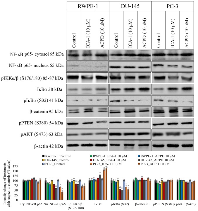
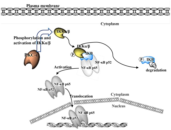
Prostate cancer (PC) is the most common type of cancer among men. Aggressive and metastatic PC results in life-threatening tumors, and represents one of the leading causes of mortality in men. Previous studies of atypical protein kinase C isoforms (aPKCs) have highlighted its role in the survival of cultured prostate cells via the nuclear factor (NF)-κB pathway. The present study showed that PKC‑ι was overexpressed in PC samples collected from cancer patients but not in non-invasive prostate tissues, indicating PKC‑ι as a possible prognostic biomarker for the progression of prostate carcinogenesis. Immunohistochemical staining further confirmed the association between PKC‑ι and the prostate malignancy. The DU‑145 and PC‑3 PC cell lines, and the non-neoplastic RWPE‑1 prostatic epithelial cell line were cultured and treated with aPKC inhibitors 2‑acetyl‑1,3-cyclopentanedione (ACPD) and 5-amino‑1-(1R,2S,3S,4R)-2,3-dihydroxy-4-methylcyclopentyl)‑1H-imidazole-4-carboxamide (ICA‑1). Western blot data demonstrated that ICA‑1 was an effective and specific inhibitor of PKC‑ι and that ACPD inhibited PKC‑ι and PKC‑ζ. Furthermore, the two inhibitors significantly decreased malignant cell proliferation and induced apoptosis. The inhibitors showed no significant cytotoxicity towards the RWPE‑1 cells, but exhibited cytostatic effects on the DU‑145 and PC‑3 cells prior to inducing apoptosis. The inhibition of aPKCs significantly reduced the translocation of NF-κB to the nucleus. Furthermore, this inhibition promoted apoptosis, reduced signaling for cell survival, and reduced the proliferation of PC cells, whereas the normal prostate epithelial cells were relatively unaffected. Overall, the results suggested that PKC‑ι and PKC‑ζ are essential for the progression of PC, and that ACPD and ICA‑1 can be effectively used as potential inhibitors in targeted therapy.


Figures

Figure 1
Figure 1
Expression of PKC-α and PKC-ι in prostate tissues. Tissue extracts (100 µg) of (A) BPH and (B) PC were immunoblotted for PKC-α and PKC‑ι. All patient tissues were maintained in liquid nitrogen and processed the same day of collection. BPH tissues showed minimal to no expression of PKC‑ι. PC tissues showed overexpression of PKC-ι compared to BPH tissues (*P=0.00048). Western blot analysis for β-actin demonstrated equal loading of each sample. Three trials were performed in triplicate. BPH, benign prostate hyperplasia; PC, prostate cancer; PKC, protein kinase C; N, normal; M, malignant.
Figure 2
Figure 2
Immunohistochemical staining of PKC-α and PKC-ι in BPH, HGPIN and PC. Tissue was stained with PKC-α antibody as a control for PKC-ι staining. Results show PKC-ι staining in (A) BPH, (B) HGPIN, and (C) PC tissue. PKC-α staining is shown in (D) BPH glands, (E) glands with HGPIN, and (F) PC glands. Tissues examined comprised BPH (n=9), HGPIN (n=8) and PC tissues (n=10). Magnification for all micrographs was x400. Three experiments were performed in triplicate. BPH, benign prostate hyperplasia; HGPIN, high grade prostate intraepithelial neoplasma; PC, prostate adenocarcinoma; PKC, protein kinase C.
Figure 3
Figure 3
Effects on PKC-ι regulation and apoptosis. Effects on PKC-ι and apoptosis were examined following inhibition of PKC-ι with either ICA-1 (10 µM) or ACPD (10 µM) for 72 h with respect to their controls. Protein expression of PKC-ι, PKC-ζ, caspase 3, Bcl-2, PARP, cytochrome c, and survivin in the RWPE-1 cell line and the DU-145 and PC-3 malignant cell lines. A total of 35 µg protein was loaded into each well and results were normalized by β-actin. Three trials were performed in triplicate. Densitometry bar graphs show the percentage change of the treated sample with respect to their controls (mean ± standard devia- tion). *P≤0.05, vs. control. PKC, protein kinase C; Bcl-2, B-cell lymphoma 2; PARP, poly (ADP-ribose); ACPD, polymerase; 2-acetyl-1,3-cyclopentanedione; ICA-1, 5-amino-1-(1R,2S,3S,4R)-2,3-dihydroxy-4-methylcyclopentyl)-1H-imidazole-4-carboxamide.
Figure 4
Figure 4
Effects of ICA-1 or ACPD on cell populations in a 72-h period. Populations of cells under normal conditions (red) and under treatment with either 10 µM ICA-1 (blue) or 10 µM ACPD (yellow) in (A) RWPE-1, (B) DU-145, and (C) PC-3 cell lines. Effects of ICA-1 and ACPD on in vitro cytotoxicity over 8 h were measured by absorbance in nm under normal conditions (red) and under the effects of either 10 µM ICA-1 (blue) or 10 µM ACPD (yellow), in (D) RWPE-1, (E) DU-145, and (F) PC-3 cell lines. There were 24 replicates per sample performed over three trials. ACPD, polymerase; 2-acetyl-1,3-cyclopentanedione; ICA-1, 5-amino-1-(1R,2S,3S,4R)-2,3-dihydroxy-4-methylcyclopentyl)-1H-imidazole-4-carboxamide.
Figure 5
Figure 5
Flow cytometric analysis of PE Annexin V staining for prostate cancer cells. Graphs show fluorescent emission of PC-3 cells in the (A) control, (B), ICA-1- and (C) ACPD-treated groups, and DU-145 cells in the (D) control, (E) ICA-1- and (F) ACPD-treated groups. Cells were incubated with PE Annexin V (x-axis) against 7AAD (y-axis). Cells were treated with ICA-1 and ACPD with respective IC50 concentrations for 24 h and 50,000 events were analyzed and recorded to obtain FL3-A (PE-Annexin V), vs. FL-2A (7-AAD). Three experiments were performed with 24 replicates and representative plots are shown. ACPD, polymerase; 2-acetyl-1,3-cyclopentanedione; ICA-1, 5-amino-1-(1R,2S,3S,4R)-2,3-dihydroxy-4-methylcyclopentyl)-1H-imidazole- 4-carboxamide; 7AAD, 7-amino-actinomycin; Ctrl, control.
Figure 6
Figure 6
Effects of 10 µM ICA-1 and 10 µM ACPD on NF-κB translocation in RWPE-1, DU-145, and PC-3 cells. Protein expression of NF-κB p65 is shown in nuclear and cytoplasmic cell fractions following 72 h of treatment. Cells were exposed to 10 nM of TNF-α 30 min prior to harvest to induce NF-κB transloca- tion. Other targets in the proposed pathway and targets known to be affected by PKC-ι activation were assessed, including pIKKα/β, IKKα/β, pIκBα, IκBα, β-catenin, pPTEN, and pAKT. A total of 50 µg protein was loaded into each well and the results were normalized by β-actin. Three trials were performed in triplicate. Densitometry bar graphs are shown as the percentage change of the treated sample with respect to their controls (mean ± standard deviation). *P≤0.05, vs. control. NF-κB, nuclear factor-κB; IκBα, inhibitor of NF-κBα; IKK, IκBα kinase; PTEN, phosphatase and tensin homolog; p, phosphorylated; ACPD, polymerase; 2-acetyl-1,3-cyclopentanedione; ICA-1, 5-amino-1-(1R,2S,3S,4R)-2,3-dihydroxy-4-methylcyclopentyl)-1H-imidazole-4-carboxamide.
Figure 7
Figure 7
Details the proposed pathway where PKC-ι acts on IKK α/β to release NF-κB, causing IKK to be degraded and allowing NF-κB to translocate to the nucleus. PKC, protein kinase C: NF-κB, nuclear factor-κB; IκBα, inhibitor of NF-κBα; IKK, IκBα kinase.

References

    1. American Cancer Society Key statistics for prostate cancer. Prostate Cancer Facts. https://www.cancer.org/cancer/prostate-cancer/about/key-statistics.html
    1. Kume H, Kawai T, Nagata M, Azuma T, Miyazaki H, Suzuki M, Fujimura T, Nakagawa T, Fukuhara H, Homma Y. Intermittent docetaxel chemotherapy is feasible for castration-resistant prostate cancer. Mol Clin Oncol. 2015;3:303–307. doi: 10.3892/mco.2014.469. - DOI - PMC - PubMed
    1. Kharaziha P, Chioureas D, Rutishauser D, Baltatzis G, Lennartsson L, Fonseca P, Azimi A, Hultenby K, Zubarev R, Ullén A, et al. Molecular profiling of prostate cancer derived exosomes may reveal a predictive signature for response to docetaxel. Oncotarget. 2015;6:21740–21754. doi: 10.18632/oncotarget.3226. - DOI - PMC - PubMed
    1. Newton AC. Protein kinase C: Poised to signal. Am J Physiol Endocrinol Metab. 2010;298:E395–E402. doi: 10.1152/ajpendo.00477.2009. - DOI - PMC - PubMed
    1. Nishizuka Y. Intracellular signaling by hydrolysis of phospholipids and activation of protein kinase C. Science. 1992;258:607–614. doi: 10.1126/science.1411571. - DOI - PubMed

MeSH terms