. 2018 Sep 18;13(9):e0204159.
doi: 10.1371/journal.pone.0204159. eCollection 2018.

ATRX loss induces multiple hallmarks of the alternative lengthening of telomeres (ALT) phenotype in human glioma cell lines in a cell line-specific manner

Affiliations

ATRX loss induces multiple hallmarks of the alternative lengthening of telomeres (ALT) phenotype in human glioma cell lines in a cell line-specific manner

Jacqueline A Brosnan-Cashman et al. PLoS One. .

Abstract

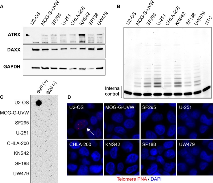
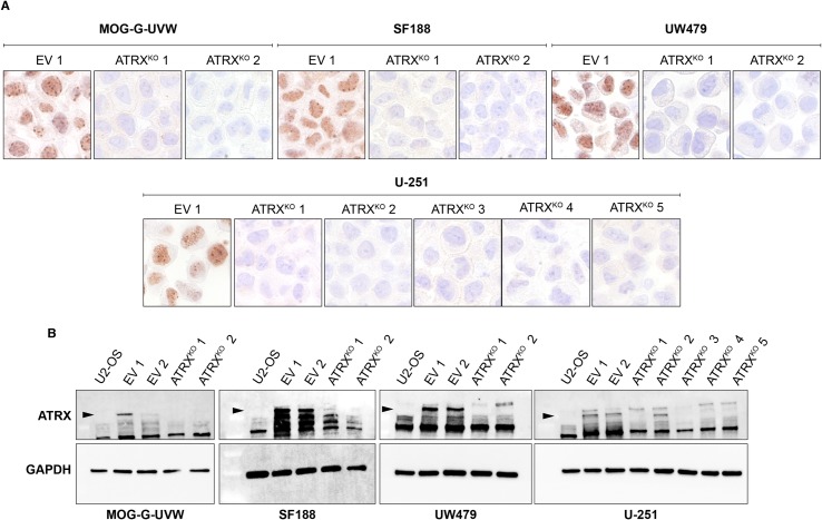
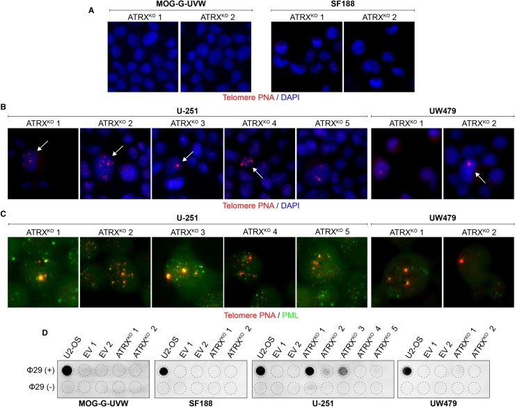
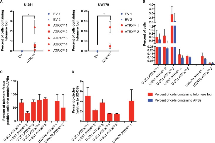
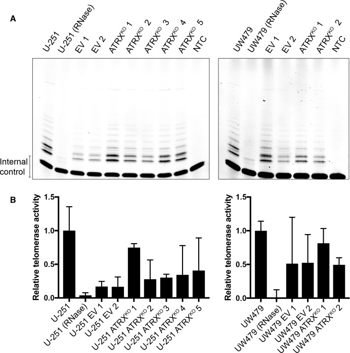
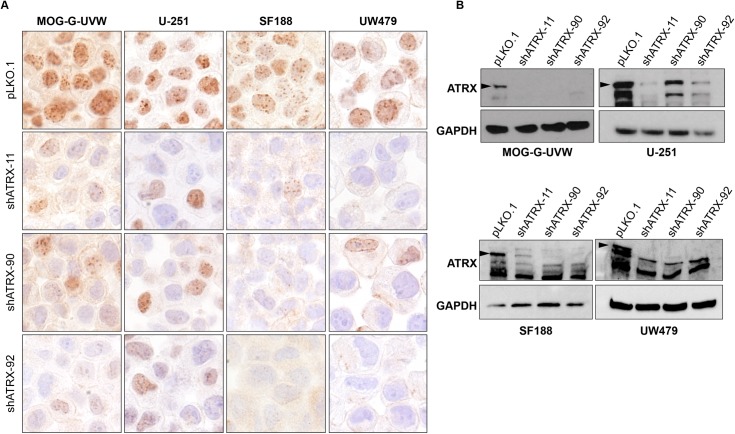
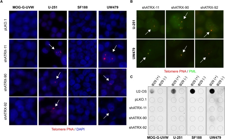
Cancers must maintain their telomeres at lengths sufficient for cell survival. In several cancer subtypes, a recombination-like mechanism termed alternative lengthening of telomeres (ALT), is frequently used for telomere length maintenance. Cancers utilizing ALT often have lost functional ATRX, a chromatin remodeling protein, through mutation or deletion, thereby strongly implicating ATRX as an ALT suppressor. Herein, we have generated functional ATRX knockouts in four telomerase-positive, ALT-negative human glioma cell lines: MOG-G-UVW, SF188, U-251 and UW479. After loss of ATRX, two of the four cell lines (U-251 and UW479) show multiple characteristics of ALT-positive cells, including ultrabright telomeric DNA foci, ALT-associated PML bodies, and c-circles. However, telomerase activity and overall telomere length heterogeneity are unaffected after ATRX loss, regardless of cellular context. The two cell lines that showed ALT hallmarks after complete ATRX loss also did so upon ATRX depletion via shRNA-mediated knockdown. These results suggest that other genomic or epigenetic events, in addition to ATRX loss, are necessary for the induction of ALT in human cancer.


Conflict of interest statement

The authors have declared that no competing interests exist.

Figures

Fig 1
Fig 1. Glioma cell lines chosen for ATRX modulation.
(A) Immunoblotting for known ALT suppressors ATRX and DAXX in seven glioma cell lines, as well as U2-OS, a known ALT-positive cell line with deletion of ATRX [24]. Arrowhead indicates band representing wild-type ATRX. (B) TRAP assay shows telomerase activity in all seven glioma cell lines, but not U2-OS. NTC indicates no template control. All seven glioma cell lines lack characteristics of ALT, including (C) c-circles, as measured by phi29-mediated rolling circle amplification and dot blot, and (D) ALT-associated telomere DNA foci (arrow), as assessed by telomere-specific FISH.
Fig 2
Fig 2. Successful elimination of ATRX expression in MOG-G-UVW, U-251, SF188, and UW479.
ATRXKO clones were validated by (A) immunohistochemistry and (B) immunoblotting against ATRX, compared to empty vector (EV) clones. Arrowheads indicate the band representing wild-type ATRX. For immunohistochemistry, one representative EV clone is shown. EV clones maintain ATRX protein expression and thus serve as positive controls for ATRX expression. For immunoblotting, U2-OS is included as a negative control for ATRX expression.
Fig 3
Fig 3. Cell line-specific induction of ALT characteristics after ATRX knockout.
(A) Representative telomere FISH images indicate the absence of ultrabright telomere DNA foci in MOG-G-UVW and SF188 ATRXKO clones. (B) Representative telomere FISH images indicate the presence of ultrabright telomeric DNA foci (arrows) in ATRXKO U-251 and UW479 clones. (C) Colocalization of ultrabright telomere DNA foci with PML was observed in ATRXKO U-251 and UW479 clones. (D) C-circles were detected in ATRXKO U-251 and UW479 clones, but not ATRXKO MOG-G-UVW and SF188 clones. A smaller input of U2-OS DNA (30 ng, compared to 150 ng) was included as a positive control.
Fig 4
Fig 4. Quantification of ALT characteristics in U-251 and UW479 ATRXKO clones.
Telomere FISH plus DAPI nuclear staining was performed on EV and ATRXKO clones, and 36–100 images (magnification = 400X) per experiment were obtained via scanning microscopy. Total cell number was determined by nuclear segmentation in TissueQuest software, and a minimum of 1000 cells were included in each analysis. (A) Cells containing ultrabright telomeric foci were identified by image analysis using pixel intensity and particle size thresholds. Analysis was limited to segmented nuclei. For both U-251 and UW479, ATRXKO clones have significantly more cells containing ultrabright telomeric DNA foci than EV clones (p = 0.0004 and 0.01, respectively). Significance was calculated using Wilcoxon rank-sum analysis. Data are from three independent measurements, and error bars represent standard error of the mean. (B) Telomere FISH plus PML immunofluorescence and DAPI nuclear staining was performed on EV and ATRXKO clones, and 36–100 images (magnification = 400X) per experiment were obtained via scanning microscopy. Total cell number was determined by nuclear segmentation in TissueQuest software, and a minimum of 1000 cells were analyzed for each experiment. Cells containing ultrabright telomeric foci were identified by image analysis using pixel intensity and particle size, and colocalization events were identified using the Image J Colocalization plugin[46]. Analysis was limited to segmented nuclei. The percent of all cells containing ultrabright foci and APBs were calculated (B), as was the percent of total focus-positive cells containing an APB (C). Data are from two independent experiments, and error bars represent standard error of the mean. (D) C-circle levels were quantified in ATRXKO cells by densitometry and compared to U2-OS. Data were generated from three independent measurements. Error bars represent standard error of the mean.
Fig 5
Fig 5. Retention of telomerase activity in ATRXKO clones that display ALT-associated hallmarks.
(A) TRAP analysis shows retention of telomerase activity in all EV and ATRXKO clones from U-251 and UW479. NTC indicates no template control. Parental U-251 and UW479 lysates were RNase treated as specificity controls. (B) Quantification of TRAP signals reveals clonal variability in telomerase activity within EV and ATRXKO clones from U-251 and UW479. Mean telomerase activity levels are shown from two independent experiments, and error bars represent standard error of the mean.
Fig 6
Fig 6. Reduction in ATRX expression using shRNAs against ATRX.
ATRX knockdown using 3 different anti-ATRX shRNA lentiviral constructs in MOG-G-UVW, U-251, SF188, and UW479 was confirmed using (A) immunohistochemistry and (B) immunoblotting against ATRX. Arrowheads indicate the band representing full-length, wild-type ATRX. Empty lentiviral vector (pLKO.1) serves as a non-knockdown control for ATRX expression.
Fig 7
Fig 7. ATRX knockdown induces ALT characteristics in U-251 and UW479.
(A) Representative telomere FISH images display ultrabright telomere DNA foci after ATRX knockdown in U-251 and UW479 (arrows), but not MOG-G-UVW and SF188. (B) A subset of telomeric foci that arise after ATRX knockdown colocalize with PML (arrows). (C) One out of three ATRX knockdown cells derived from UW479 (shATRX-90) shows c-circle positivity, while no other line with ATRX reduction consistently shows this feature. A lower input of U2-OS DNA (30 ng, compared to 150 ng) was included as a positive control.

References

    1. Shay JW, Wright WE. Role of telomeres and telomerase in cancer. Semin Cancer Biol. 2011;21(6):349–53. 10.1016/j.semcancer.2011.10.001 ; PubMed Central PMCID: PMCPMC3370415. - DOI - PMC - PubMed
    1. Hayflick L. The limited in vitro lifetime of human diploid cell strains. Exp Cell Res. 1965;37:614–36. . - PubMed
    1. Allsopp RC, Chang E, Kashefi-Aazam M, Rogaev EI, Piatyszek MA, Shay JW, et al. Telomere shortening is associated with cell division in vitro and in vivo. Exp Cell Res. 1995;220(1):194–200. 10.1006/excr.1995.1306 . - DOI - PubMed
    1. Allsopp RC, Harley CB. Evidence for a critical telomere length in senescent human fibroblasts. Exp Cell Res. 1995;219(1):130–6. 10.1006/excr.1995.1213 . - DOI - PubMed
    1. Cesare AJ, Reddel RR. Alternative lengthening of telomeres: models, mechanisms and implications. Nat Rev Genet. 2010;11(5):319–30. 10.1038/nrg2763 . - DOI - PubMed

Publication types