

Increased Myeloid Dendritic Cells and TNF-α Expression Predicts Poor Response to Hydroxychloroquine in Cutaneous Lupus Erythematosus
- PMID: 30227141
- PMCID: PMC7428867
- DOI: 10.1016/j.jid.2018.07.041
Increased Myeloid Dendritic Cells and TNF-α Expression Predicts Poor Response to Hydroxychloroquine in Cutaneous Lupus Erythematosus
Abstract
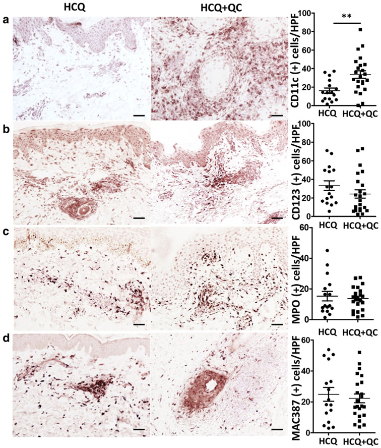
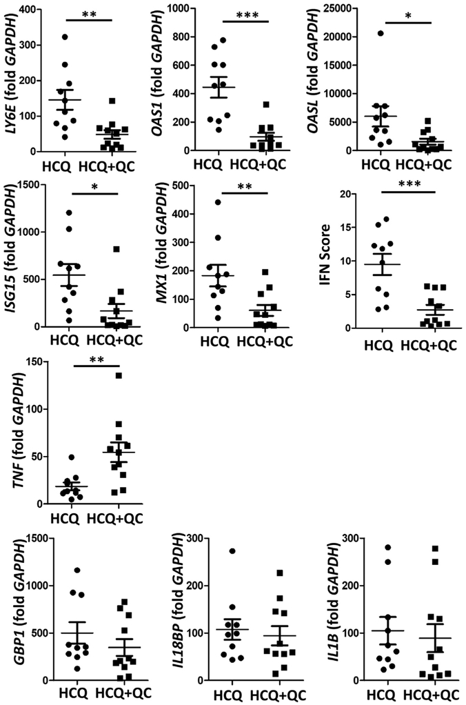
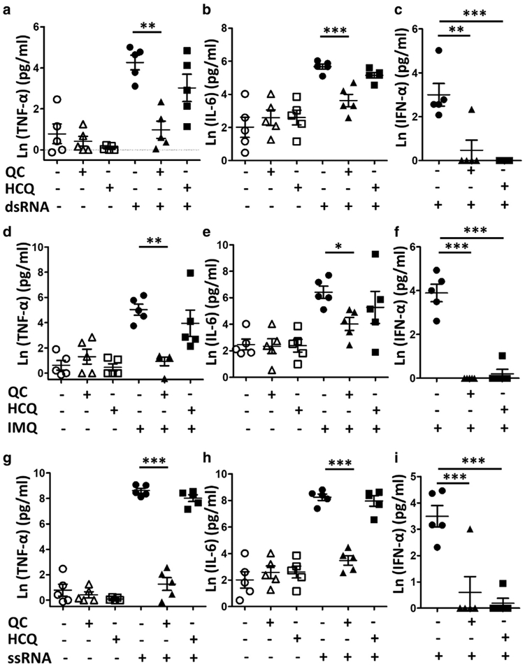
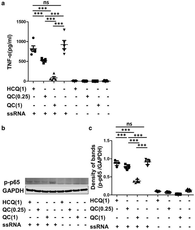
Although antimalarials are the primary treatment for cutaneous lupus erythematosus, not all patients are equally responsive. We investigated whether different inflammatory cell population and cytokine profiles in lesional cutaneous lupus erythematosus skin could affect antimalarial responsiveness, and whether hydroxychloroquine (HCQ) and quinacrine (QC) differentially suppress inflammatory cytokines. Cutaneous lupus erythematosus patients were grouped according to their response to antimalarials (HCQ vs. HCQ+QC). On immunohistochemistry, only the myeloid dendritic cell population was significantly increased in the HCQ+QC group compared to HCQ group. While the IFN scores calculated for the selected type I IFN-regulated genes (LYE6, OAS1, OASL, ISG15, and MX1) were significantly higher in the HCQ group than the HCQ+QC group, the TNF-α level was higher in the HCQ+QC group. QC was more effective than HCQ at inhibiting the toll receptor-mediated production of TNF-α and IL-6 in the peripheral blood mononuclear cells isolated from cutaneous lupus erythematosus patients, whereas QC and HCQ inhibited IFN-α equally. QC also suppressed phospho-NF-κB p65 more profoundly than HCQ. In conclusion, increased myeloid dendritic cell population with higher TNF-α expression might contribute to HCQ refractoriness and a better response to QC. Differential suppressive effects of HCQ and QC could also affect antimalarial responses in cutaneous lupus erythematosus patients.
Copyright © 2018 The Authors. Published by Elsevier Inc. All rights reserved.
Conflict of interest statement
CONFLICT OF INTEREST
The authors state no conflict of interest.
Figures
Similar articles
-
Increased CD69+CCR7+ circulating activated T cells and STAT3 expression in cutaneous lupus erythematosus patients recalcitrant to antimalarials.Lupus. 2022 Apr;31(4):472-481. doi: 10.1177/09612033221084093. Epub 2022 Mar 8. Lupus. 2022. PMID: 35258358 Free PMC article.
-
Quinacrine Suppresses Tumor Necrosis Factor-α and IFN-α in Dermatomyositis and Cutaneous Lupus Erythematosus.J Investig Dermatol Symp Proc. 2017 Oct;18(2):S57-S63. doi: 10.1016/j.jisp.2016.11.001. J Investig Dermatol Symp Proc. 2017. PMID: 28941496 Free PMC article.
-
Multidimensional Immune Profiling of Cutaneous Lupus Erythematosus In Vivo Stratified by Patient Response to Antimalarials.Arthritis Rheumatol. 2022 Oct;74(10):1687-1698. doi: 10.1002/art.42235. Epub 2022 Sep 1. Arthritis Rheumatol. 2022. PMID: 35583812
-
Recent findings about antimalarials in cutaneous lupus erythematosus: What dermatologists should know.J Dermatol. 2024 Jul;51(7):895-903. doi: 10.1111/1346-8138.17177. Epub 2024 Mar 14. J Dermatol. 2024. PMID: 38482997 Review.
-
Hydroxychloroquine and smoking in patients with cutaneous lupus erythematosus.Clin Exp Dermatol. 2012 Jun;37(4):327-34. doi: 10.1111/j.1365-2230.2011.04266.x. Clin Exp Dermatol. 2012. PMID: 22582908 Review.
Cited by
-
Increased MxA protein expression and dendritic cells in spongiotic dermatitis differentiates dermatomyositis from eczema in a single-center case-control study.J Cutan Pathol. 2021 Mar;48(3):364-373. doi: 10.1111/cup.13880. Epub 2020 Oct 23. J Cutan Pathol. 2021. PMID: 32954523 Free PMC article.
-
A double-blind, randomized, placebo-controlled, phase II trial of baricitinib for systemic lupus erythematosus: how to optimize lupus trials to examine effects on cutaneous lupus erythematosus.Br J Dermatol. 2019 May;180(5):964-965. doi: 10.1111/bjd.17344. Br J Dermatol. 2019. PMID: 31025748 Free PMC article. No abstract available.
-
Modular gene analysis reveals distinct molecular signatures for subsets of patients with cutaneous lupus erythematosus.Br J Dermatol. 2021 Sep;185(3):563-572. doi: 10.1111/bjd.19800. Epub 2021 Mar 3. Br J Dermatol. 2021. PMID: 33400293 Free PMC article.
-
The Genetic Landscape of Cutaneous Lupus Erythematosus.Front Med (Lausanne). 2022 Jun 2;9:916011. doi: 10.3389/fmed.2022.916011. eCollection 2022. Front Med (Lausanne). 2022. PMID: 35721085 Free PMC article. Review.
-
Understanding the Role of Type I Interferons in Cutaneous Lupus and Dermatomyositis: Toward Better Therapeutics.Arthritis Rheumatol. 2025 Jan;77(1):1-11. doi: 10.1002/art.42983. Epub 2024 Oct 29. Arthritis Rheumatol. 2025. PMID: 39262215 Free PMC article. Review.
References
-
- Baud V, Karin M. Signal transduction by tumor necrosis factor and its relatives. Trends Cell Biol 2001;11:372–7. - PubMed
-
- Chasset F, Frances C, Barete S, Amoura Z, Arnaud L. Influence of smoking on the efficacy of antimalarials in cutaneous lupus: a meta-analysis of the literature. J Am Acad Dermatol 2015;72:634–9. - PubMed
Publication types
MeSH terms
Substances
Grants and funding
LinkOut - more resources
Full Text Sources
Other Literature Sources
Miscellaneous
