. 2018 Sep 17;10(9):334.
doi: 10.3390/cancers10090334.

Factors Secreted by Cancer-Associated Fibroblasts that Sustain Cancer Stem Properties in Head and Neck Squamous Carcinoma Cells as Potential Therapeutic Targets

Affiliations

Factors Secreted by Cancer-Associated Fibroblasts that Sustain Cancer Stem Properties in Head and Neck Squamous Carcinoma Cells as Potential Therapeutic Targets

Saúl Álvarez-Teijeiro et al. Cancers (Basel). .

Abstract

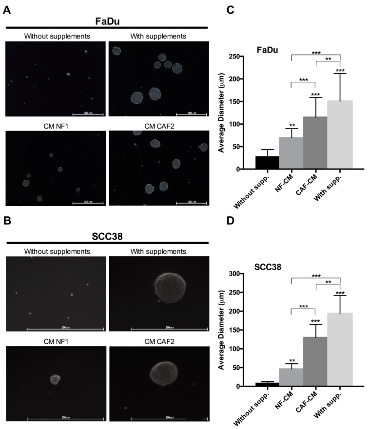
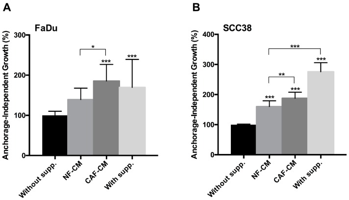
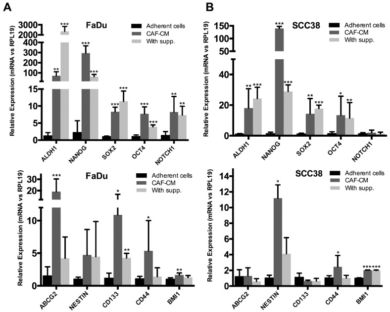
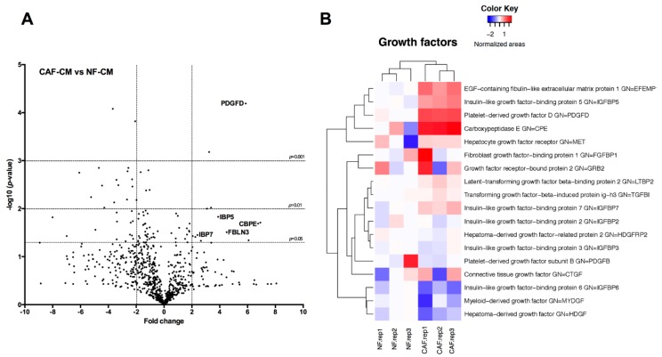
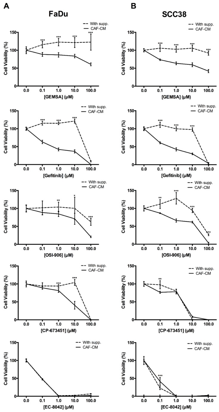
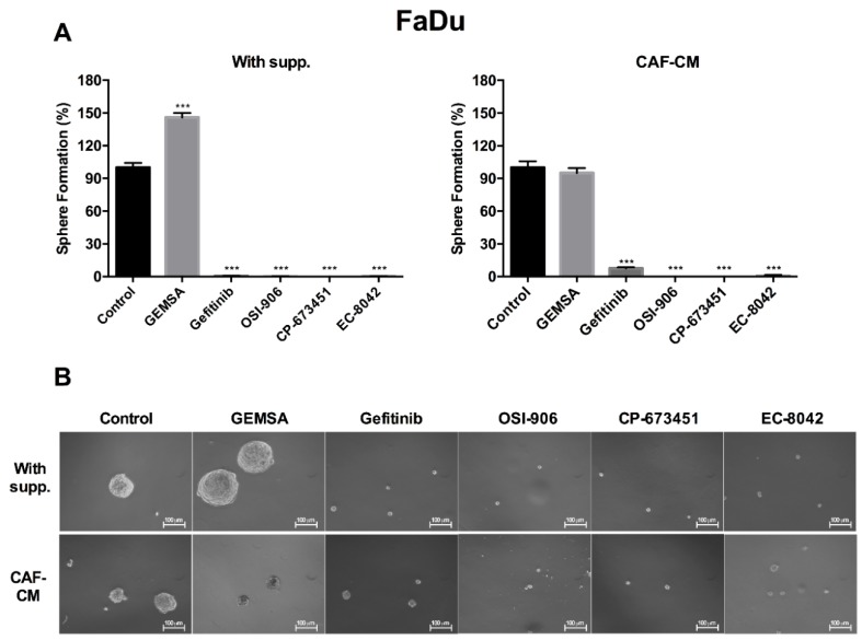
This study investigates for the first time the crosstalk between stromal fibroblasts and cancer stem cell (CSC) biology in head and neck squamous cell carcinomas (HNSCC), with the ultimate goal of identifying effective therapeutic targets. The effects of conditioned media from cancer-associated fibroblasts (CAFs) and normal fibroblasts (NFs) on the CSC phenotype were assessed by combining functional and expression analyses in HNSCC-derived cell lines. Further characterization of CAFs and NFs secretomes by mass spectrometry was followed by pharmacologic target inhibition. We demonstrate that factors secreted by CAFs but not NFs, in the absence of serum/supplements, robustly increased anchorage-independent growth, tumorsphere formation, and CSC-marker expression. Modulators of epidermal growth factor receptor (EGFR), insulin-like growth factor receptor (IGFR), and platelet-derived growth factor receptor (PDGFR) activity were identified as paracrine cytokines/factors differentially secreted between CAFs and NFs, in a mass spectrometry analysis. Furthermore, pharmacologic inhibition of EGFR, IGFR, and PDGFR significantly reduced CAF-induced tumorsphere formation and anchorage-independent growth suggesting a role of these receptor tyrosine kinases in sustaining the CSC phenotype. These findings provide novel insights into tumor stroma⁻CSC communication, and potential therapeutic targets to effectively block the CAF-enhanced CSC niche signaling circuit.

Keywords: cancer stem cells; cancer-associated fibroblasts; head and neck squamous cell carcinoma; secretome; therapeutic target; tumor microenvironment.


Conflict of interest statement

F.M. reports ownership of stock in EntreChem SL. All other authors declare they have no competing interests.

Figures

Figure 1
Figure 1
Effect of fibroblast-CM (conditioned media) on the tumorsphere formation capacity of head and neck squamous cell carcinomas (HNSCC) cells. Representative images of orospheres formed by (A) FaDu and (B) SCC38 cells in non-supplemented medium, supplemented medium, and CM from normal fibroblasts (NFs) or cancer-associated fibroblasts (CAFs). Bar chart showing the average diameter of spheroids formed by (C) FaDu and (D) SCC38 cells in the previous conditions. All data were expressed as the mean ± SD of at least three independent experiments performed. Scale bar: 500 μm. *** p < 0.001 and ** p < 0.01 by Holm-Sidak’s multiple comparisons test.
Figure 2
Figure 2
Effect of fibroblast-CM on anchorage-independent growth of HNSCC cells. (A) FaDu and (B) SCC38 cells were seeded in plates coated with Poly(2-hydroxyethyl methacrylate) (polyHEMA) and grown in non-supplemented medium, CM from NFs, CM from CAFs, or supplemented medium. Cell proliferation was estimated by tetrazolium-based MTS assay after 4 days. Data were normalized to the absorbance at day 0 and relative to control (non-supplemented) cells. All data were expressed as the mean ± SD of at least three independent experiments performed in quadruplicate. *** p < 0.001, ** p < 0.01 and * p < 0.05 by Holm-Sidak’s multiple comparisons test.
Figure 3
Figure 3
Effect of fibroblast-CM on the expression of stem-related genes in HNSCC cells. Bar chart showing the expression analysis of CSC-related genes by qRT-PCR analysis in FaDu (A) and SCC38 (B) orospheres formed in CAF-CM and supplemented medium. Adherent monolayer cultures of FaDu or SCC38 cells were used as control. Data were normalized to RPL19 levels and relative to control cells. All data were expressed as the mean ± SD of at least three independent experiments performed in triplicate. * p < 0.05, ** p < 0.01 and *** p < 0.001 by Student’s t-test.
Figure 4
Figure 4
Mass Spec analysis of extracellular proteins differentially secreted by CAFs versus NFs. (A) Volcano plot showing the global secretome changes, illustrating fold change (log base 2) and p-value (−log base 10), between CAFs and NFs. Horizontal bars represent the significance p = 0.05, p = 0.01 and p = 0.001 (proteins under horizontal bar of p = 0.05 did not reach significance). Vertical bars represent the proteins with a fold change higher than 2 or −2; (B) Heatmap represents the changes in the growth factors related-proteins found in the secretome. Three independent experiments are shown; red indicates fold changes >0 and blue indicates fold changes <0.
Figure 5
Figure 5
Effect of 2-guanidinoethylmercaptosuccinic acid (GEMSA), Gefitinib, OSI-906, CP-673451, and EC-8042 on CAF-CM-mediated anchorage-independent growth. (A) FaDu and (B) SCC38 cells were seeded in polyHEMA-coated plates and grown in CAF-CM or supplemented medium. After 24 h, cells were treated with increasing concentrations of the indicated drugs (GEMSA, Gefitinib, OSI-906, CP-673451, and EC-8042). Cell proliferation was estimated by tetrazolium-based MTS assay after 4 days. Data were normalized to the absorbance at day 0 and relative to control (vehicle-treated) cells. All data were expressed as the mean ± SD of at least three independent experiments performed in quadruplicate. * p < 0.05, ** p < 0.01 and *** p < 0.001 by Student’s t-test.
Figure 6
Figure 6
Effect of GEMSA, Gefitinib, OSI-906, CP-673451, and EC-8042 on FaDu orosphere formation. (A) Bar chart showing orospheres formation ability of FaDu grown in supplemented medium (left) or CAF-CM (right) and treated with GEMSA (10 μM), Gefitinib (1 μM), OSI-906 (10 μM), CP-673451 (5 μM), and EC-8042 (0.01 μM); (B) Representative images of FaDu orospheres for each condition shown in the bar chart. Sphere formation was estimated by tetrazolium-based MTS assay after 10–12 days. All data were expressed as the mean ± SD of at least three independent experiments performed in quadruplicate. *** p < 0.001 by Student’s t-test. Scale bar: 100 μm.

References

    1. Koontongkaew S. The tumor microenvironment contribution to development, growth, invasion and metastasis of head and neck squamous cell carcinomas. J. Cancer. 2013;4:66–83. doi: 10.7150/jca.5112. - DOI - PMC - PubMed
    1. Calvo F., Ege N., Grande-Garcia A., Hooper S., Jenkins R.P., Chaudhry S.I., Harrington K., Williamson P., Moeendarbary E., Charras G., et al. Mechanotransduction and YAP-dependent matrix remodelling is required for the generation and maintenance of cancer-associated fibroblasts. Nat. Cell Biol. 2013;15:637–646. doi: 10.1038/ncb2756. - DOI - PMC - PubMed
    1. Koczorowska M.M., Tholen S., Bucher F., Lutz L., Kizhakkedathu J.N., De Wever O., Wellner U.F., Biniossek M.L., Stahl A., Lassmann S., et al. Fibroblast activation protein-alpha, a stromal cell surface protease, shapes key features of cancer associated fibroblasts through proteome and degradome alterations. Mol. Oncol. 2016;10:40–58. doi: 10.1016/j.molonc.2015.08.001. - DOI - PMC - PubMed
    1. De Wever O., Mareel M. Role of tissue stroma in cancer cell invasion. J. Pathol. 2003;200:429–447. doi: 10.1002/path.1398. - DOI - PubMed
    1. Bello I.O., Vered M., Dayan D., Dobriyan A., Yahalom R., Alanen K., Nieminen P., Kantola S., Laara E., Salo T. Cancer-associated fibroblasts, a parameter of the tumor microenvironment, overcomes carcinoma-associated parameters in the prognosis of patients with mobile tongue cancer. Oral Oncol. 2011;47:33–38. doi: 10.1016/j.oraloncology.2010.10.013. - DOI - PubMed