

Glycosylation in the Thyroid Gland: Vital Aspects of Glycoprotein Function in Thyrocyte Physiology and Thyroid Disorders
- PMID: 30227620
- PMCID: PMC6163523
- DOI: 10.3390/ijms19092792
Glycosylation in the Thyroid Gland: Vital Aspects of Glycoprotein Function in Thyrocyte Physiology and Thyroid Disorders
Abstract
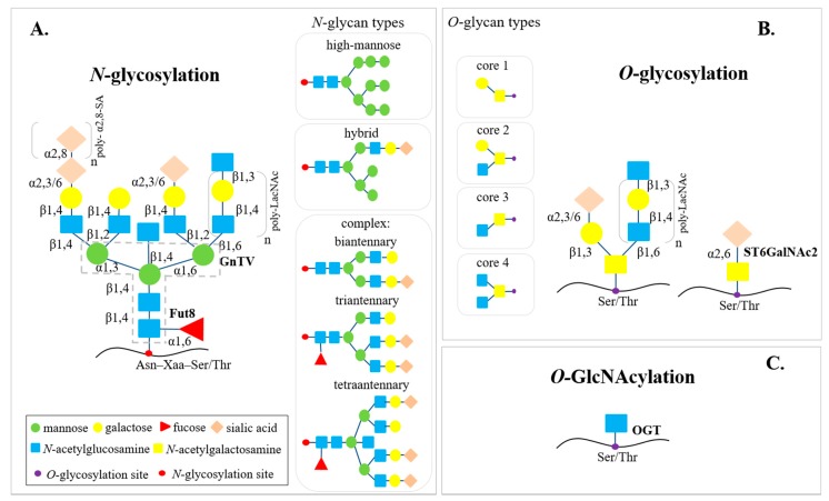
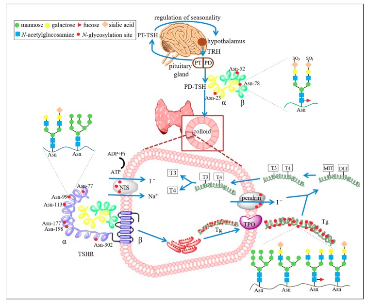
The key proteins responsible for hormone synthesis in the thyroid are glycosylated. Oligosaccharides strongly affect the function of glycosylated proteins. Both thyroid-stimulating hormone (TSH) secreted by the pituitary gland and TSH receptors on the surface of thyrocytes contain N-glycans, which are crucial to their proper activity. Thyroglobulin (Tg), the protein backbone for synthesis of thyroid hormones, is a heavily N-glycosylated protein, containing 20 putative N-glycosylated sites. N-oligosaccharides play a role in Tg transport into the follicular lumen, where thyroid hormones are produced, and into thyrocytes, where hyposialylated Tg is degraded. N-glycans of the cell membrane transporters sodium/iodide symporter and pendrin are necessary for iodide transport. Some changes in glycosylation result in abnormal activity of the thyroid and alteration of the metabolic clearance rate of hormones. Alteration of glycan structures is a pathological process related to the progression of chronic diseases such as thyroid cancers and autoimmunity. Thyroid carcinogenesis is accompanied by changes in sialylation and fucosylation, β1,6-branching of glycans, the content and structure of poly-LacNAc chains, as well as O-GlcNAcylation, while in thyroid autoimmunity the main processes affected are sialylation and fucosylation. The glycobiology of the thyroid gland is an intensively studied field of research, providing new data helpful in understanding the role of the sugar component in thyroid protein biology and disorders.
Keywords: NIS; TSHR; glycosylation; pendrin; thyroglobulin; thyroid; thyroid autoimmunity; thyroid cancers; thyroid-stimulating hormone.
Conflict of interest statement
The authors declare no conflict of interest.
Figures
Similar articles
-
The Human Thyroid-Derived CI-huThyrEC Cell Line Expresses the Thyrotropin (TSH) Receptor and Thyroglobulin but Lacks Other Essential Characteristics of Thyroid Follicular Cells.Biomolecules. 2025 Mar 5;15(3):375. doi: 10.3390/biom15030375. Biomolecules. 2025. PMID: 40149910 Free PMC article.
-
Thyrotropin, but Not Thyroid-Stimulating Antibodies, Induces Biphasic Regulation of Gene Expression in Human Thyrocytes.Thyroid. 2020 Feb;30(2):270-276. doi: 10.1089/thy.2019.0418. Epub 2020 Jan 28. Thyroid. 2020. PMID: 31805824 Free PMC article.
-
Iodinated TG in Thyroid Follicles Regulate TSH/TSHR Signaling for NIS Expression.Biol Trace Elem Res. 2017 Dec;180(2):206-213. doi: 10.1007/s12011-017-1017-z. Epub 2017 Apr 10. Biol Trace Elem Res. 2017. PMID: 28396984
-
Minireview: The sodium-iodide symporter NIS and pendrin in iodide homeostasis of the thyroid.Endocrinology. 2009 Mar;150(3):1084-90. doi: 10.1210/en.2008-1437. Epub 2009 Feb 5. Endocrinology. 2009. PMID: 19196800 Free PMC article. Review.
-
Iodide handling by the thyroid epithelial cell.Exp Clin Endocrinol Diabetes. 2001;109(1):13-7. doi: 10.1055/s-2001-11014. Exp Clin Endocrinol Diabetes. 2001. PMID: 11573132 Review.
Cited by
-
Serum Anti-Thyroglobulin Autoantibodies Are Specific in Predicting the Presence of Papillary-like Nuclear Features and Lymphocytic Infiltrate in the Thyroid Gland.Diagnostics (Basel). 2023 Jun 13;13(12):2042. doi: 10.3390/diagnostics13122042. Diagnostics (Basel). 2023. PMID: 37370937 Free PMC article.
-
An N-glycome tissue atlas of 15 human normal and cancer tissue types determined by MALDI-imaging mass spectrometry.Sci Rep. 2024 Jan 4;14(1):489. doi: 10.1038/s41598-023-50957-w. Sci Rep. 2024. PMID: 38177192 Free PMC article.
-
Oncometabolites as biomarkers in thyroid cancer: a systematic review.Cancer Manag Res. 2019 Feb 25;11:1829-1841. doi: 10.2147/CMAR.S188661. eCollection 2019. Cancer Manag Res. 2019. PMID: 30881111 Free PMC article. Review.
-
Thyroid Carcinoma Glycoproteins Express Altered N-Glycans with 3-O-Sulfated Galactose Residues.Biomolecules. 2024 Nov 21;14(12):1482. doi: 10.3390/biom14121482. Biomolecules. 2024. PMID: 39766189 Free PMC article.
-
Hyperthyroidism Associated with Gestational Trophoblastic Neoplasia: Systematic Literature Review and Pathways Analysis.Cancers (Basel). 2025 Apr 22;17(9):1398. doi: 10.3390/cancers17091398. Cancers (Basel). 2025. PMID: 40361325 Free PMC article. Review.
References
-
- Le S.N., Porebski B.T., McCoey J., Fodor J., Riley B., Godlewska M., Góra M., Czarnocka B., Banga J.P., Hoke D.E., et al. Modelling of Thyroid Peroxidase Reveals Insights into Its Enzyme Function and Autoantigenicity. PLoS ONE. 2015;10:e0142615. doi: 10.1371/journal.pone.0142615. - DOI - PMC - PubMed
Publication types
MeSH terms
Substances
Grants and funding
LinkOut - more resources
Full Text Sources
Other Literature Sources
Medical
Miscellaneous
