. 2018 Sep 18;18(1):901.
doi: 10.1186/s12885-018-4820-9.

Extracellular matrix collagen I promotes the tumor progression of residual hepatocellular carcinoma after heat treatment

Affiliations

Extracellular matrix collagen I promotes the tumor progression of residual hepatocellular carcinoma after heat treatment

Rui Zhang et al. BMC Cancer. .

Abstract

Background: Accelerated malignant behaviors induced by insufficient thermal ablation have been increasingly reported, however, the exact mechanisms are still unclear. Here, we investigated the importance of the extracellular matrix (ECM) in modulating the progression of residual hepatocellular carcinoma (HCC) after heat treatment.

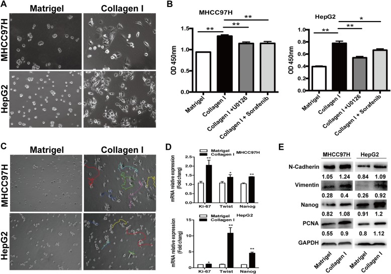
Methods: Heat-exposed residual HCC cells were cultured in different ECM gels. We used basement membrane gel (Matrigel) to simulate the normal microenvironment and collagen I to model the pathological stromal ECM. The alterations of morphology and parameters of proliferation, epithelial-mesenchymal transition (EMT) and stemness were analyzed in vitro and in vivo.

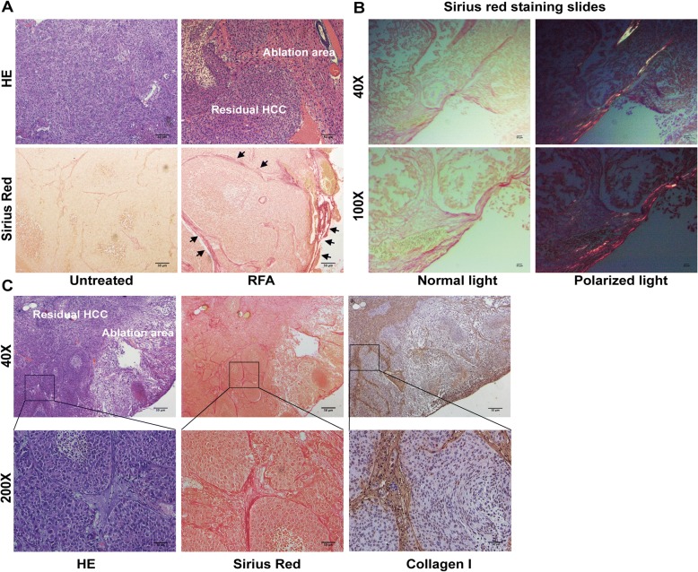
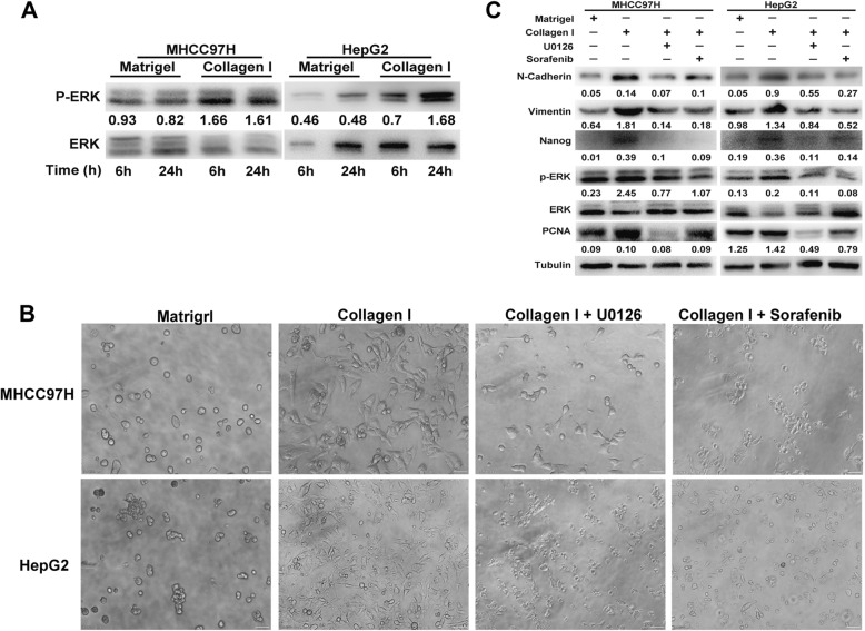
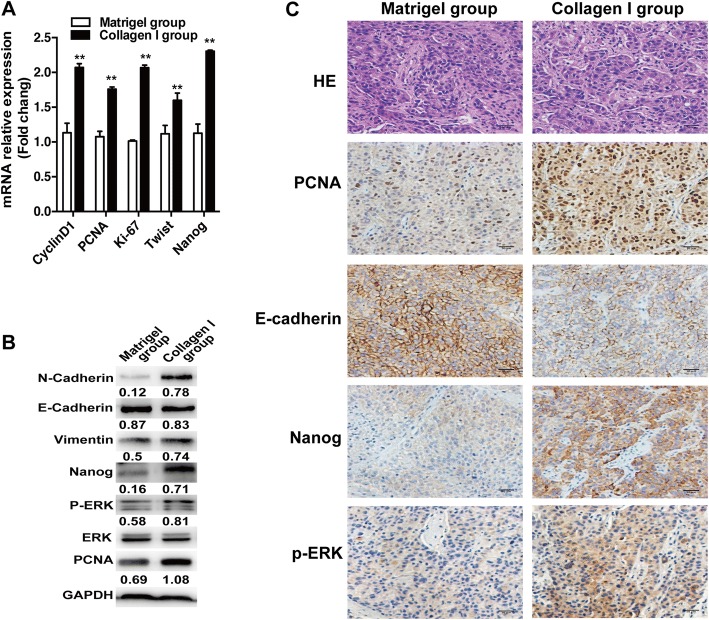
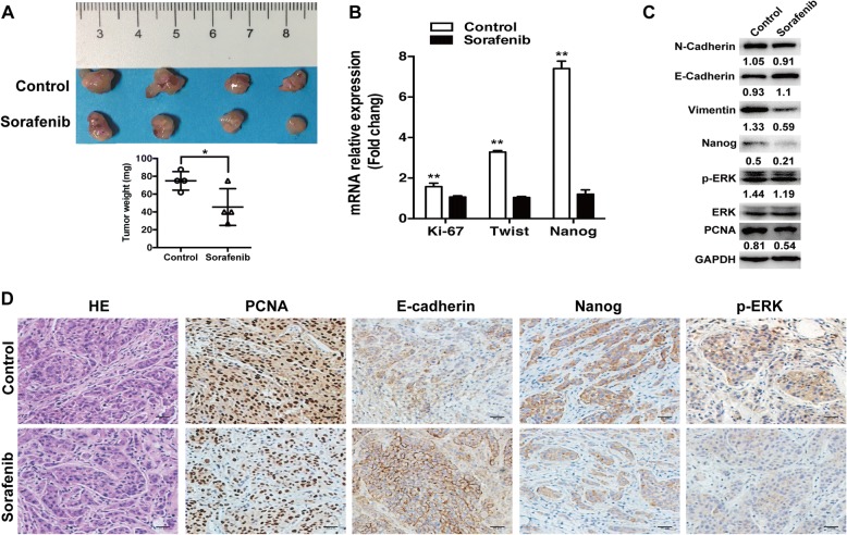
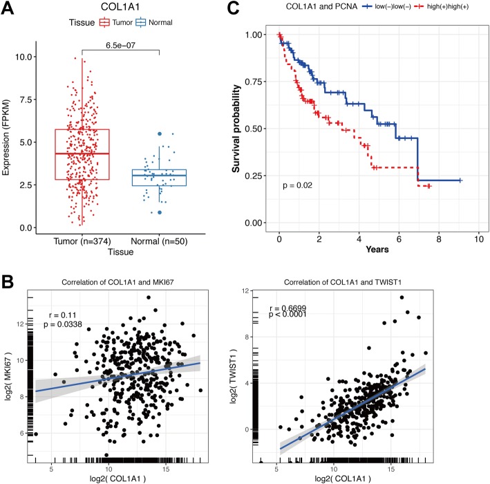
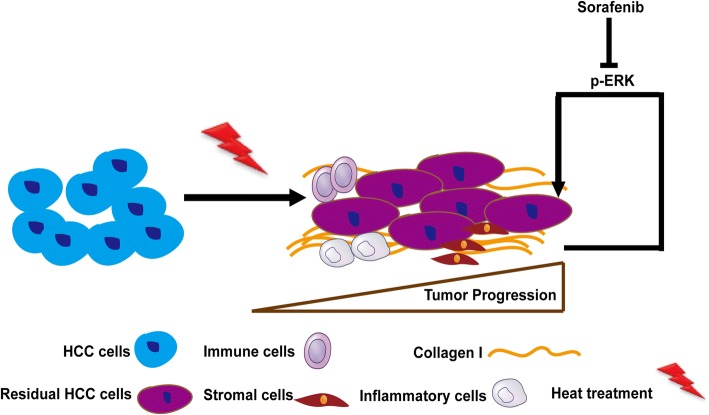
Results: Increased collagen I deposition was observed at the periablational zone after incomplete RFA of HCC in a xenograft model. The markers of cell proliferation, EMT, motility and progenitor-like traits of heat-exposed residual HCC cells were significantly induced by collagen I as compared to Matrigel (p values all < 0.05). Importantly, collagen I induced the activation of ERK phosphorylation in heat-exposed residual HCC cells. ERK1/2 inhibitor reversed the collagen I-promoted ERK phosphorylation, cell proliferative, protrusive and spindle-like appearance of heat-treated residual HCC cells in vitro. Moreover, collagen I promoted the in vivo tumor progression of heat-exposed residual HCC cells, and sorafenib markedly reversed the collagen I-mediated protumor effects.

Conclusions: Our findings demonstrate that collagen I could enhance the aggressive progression of residual HCC cells after suboptimal heat treatment and sorafenib may be a treatment approach to thwart this process.

Keywords: Collagen I; ERK; Heat treatment; Hepatocellular carcinoma.


Conflict of interest statement

Ethics approval

All animal experiments were carried out in compliance with the guidelines by the Shanghai Medical Experimental Animal Care Commission. The experimental protocols were approved by the Ethical Committee on Animal Experiments of Fudan University, Shanghai China (Permit Number: 201807002Z).

Consent for publication

Not applicable.

Competing interests

The authors declare that they have no competing interests.

Publisher’s Note

Springer Nature remains neutral with regard to jurisdictional claims in published maps and institutional affiliations.

Figures

Fig. 1
Fig. 1
Increased collagen I deposition at the periablational zone after incomplete RFA of HCC in an orthotopic animal model. a The increased collagen stained by Sirius Red was located at the periablational zone (black arrow) and surrounded the residual HCC foci. b Collagen I deposit (red-yellow color area) was observed using polarized light microphotographs of Sirius red staining-stained slides. c The increased collagen I at the periablational zone and around the residual HCC foci was verified by immunohistochemical staining. Scale bar, 50 μm
Fig. 2
Fig. 2
Collagen I stimulated the proliferation, motility, and the expression of EMT and progenitor-like markers in heat-treated residual HCC cells. a Compared with the cells cultured on Matrigel, heat-treated residual HCC cells on collagen I displayed a proliferative, protrusive and spindle-like appearance. b Collagen I promoted proliferation of heat-treated residual HCC cells as determined by the WST-1 proliferation assay. The OD (optical density) was measured at 450 nm wavelength. c Compared with Matrigel, collagen I enhanced the motility of heated-exposed residual HCC cells as demonstrated by tracking analysis. d As shown by qRT-PCR, the mRNA expression of Ki-67, twist, and Nanog was increased in heat-exposed residual HCC cells cultured on collagen I versus Matrigel. e The increased expression of PCNA, vimentin, N-cadherin and Nanog protein in heat-exposed residual HCC cell cultured on collagen I was detected by Western blot. Expression levels of target proteins were normalized to the corresponding levels of GAPDH. **, P < 0.01; *, P < 0.05
Fig. 3
Fig. 3
Collagen I induced the activation of ERK in heat-exposed residual HCC cells. a The up-regulated level of ERK1/2 phosphorylation were induced in heat-exposed residual HCC cells cultured on collagen I versus Matrigel. The p-ERK content was normalized for ERK. b ERK1/2 inhibitor U0126 (25 μM) or sorafenib (5 μM) could reverse the collagen I-promoted proliferative, protrusive and spindle-like appearance of heat-treated residual HCC cells. c ERK1/2 inhibitor (U0126, 25 μM) or sorafenib (5 μM) reversed collagen I-mediated upregulation of ERK1/2 in heat-exposed residual HCC cells. Collagen I-induced upregulation of proliferation (PCNA), EMT (vimentin and N-cadherin), cancer stem cell marker Nanog was markedly reduced in heat-exposed residual HCC cells pretreated with ERK1/2 inhibitor (U0126, 25 μM) or sorafenib (5 μM). The p-ERK content was normalized for ERK. Expression levels of PCNA, vimentin, N-cadherin and Nanog were normalized to Tubulin
Fig. 4
Fig. 4
Collagen I promoted the in vivo progression of heat-treated residual HCC cells. a The mRNA expression of PCNA, cyclin D1, Ki-67, twist and Nanog was increased in the tumors from heat-exposed residual MHCC97H cells inoculated with collagen I. b The protein expression of PCNA, vimentin, N-cadherin, E-cadherin, Nanog and ERK phosphorylation were detected by Western blot. The p-ERK was normalized for ERK. Expression levels of E-cadherin, PCNA, vimentin, N-cadherin and Nanog were normalized to GAPDH. c The expression of PCNA, E-Cadherin, Nanog and phosphorylated ERK were evaluated using immunohistochemical staining (scale bar, 50 μm). **, P < 0.01; *, P < 0.05
Fig. 5
Fig. 5
Sorafenib suppressed the in vivo collagen I-induced tumor progression of heat-treated residual HCC cells. a Mice with the tumors derived from heat-exposed residual MHCC97H cells with collagen I were subjected to treatment. Compared with the control group, sorafenib significantly inhibited tumor growth. b The mRNA expression of Ki-67, twist and Nanog were down-regulated in the sorafenib group. c The changes of PCNA, Nanog, vimentin, E-cadherin, N-cadherin, and ERK activation were detected by Western blot. The p-ERK levels were normalized for ERK. Expression levels of E-cadherin, PCNA, vimentin, N-cadherin and Nanog were normalized to GAPDH. d The immunohistochemical staining of PCNA, E-Cadherin, Nanog and phosphorylated ERK in the tumors (scale bar, 50 μm). **, P < 0.01; *, P < 0.05
Fig. 6
Fig. 6
Collagen I expression in TCGA HCC patients. a COL1A1 (Collagen I α1 chain) was significantly overexpressed in HCC samples than adjacent non-tumoral tissues. b The positive correlations were observed between COL1A1 expression and expression of proliferation marker Ki-67 (r = 0.11, P = 0.0338) or EMT marker Twist (r = 0.6699, P<0.0001). c High COL1A1 expression combined with expression of PCNA (proliferation marker) predicted unfavorable survival outcomes in TCGA HCC patients
Fig. 7
Fig. 7
Schematic diagram shows that collagen I initiates ERK signaling to accelerate the aggressive progression of residual HCC cells after sublethal RFA, which could be reversed by sorafenib. More factors implicated in post-inflammation reaction after RFA promote tumor progression of residual HCC, which has been reported by the other authors [14, 15, 49, 50]

Similar articles

Cited by

References

    1. Forner A, Llovet JM, Bruix J. Hepatocellular carcinoma. Lancet. 2012;379:1245–1255. doi: 10.1016/S0140-6736(11)61347-0. - DOI - PubMed
    1. Lau WY, Lai EC. The current role of radiofrequency ablation in the management of hepatocellular carcinoma: a systematic review. Ann Surg. 2009;249:20–25. doi: 10.1097/SLA.0b013e31818eec29. - DOI - PubMed
    1. Facciorusso A, Serviddio G, Muscatiello N. Local ablative treatments for hepatocellular carcinoma: an updated review. World J Gastrointest Pharmacol Ther. 2016;7:477–489. doi: 10.4292/wjgpt.v7.i4.477. - DOI - PMC - PubMed
    1. Lau WY, Lai ECH. The current role of radiofrequency ablation in the management of hepatocellular carcinoma. Ann Surg. 2009;249:20–25. doi: 10.1097/SLA.0b013e31818eec29. - DOI - PubMed
    1. Obara K, Matsumoto N, Okamoto M, Kobayashi M, Ikeda H, Takahashi H, et al. Insufficient radiofrequency ablation therapy may induce further malignant transformation of hepatocellular carcinoma. Hepatol Int. 2008;2:116–123. doi: 10.1007/s12072-007-9040-3. - DOI - PMC - PubMed

MeSH terms