

Genomic characterization of three novel Basilisk-like phages infecting Bacillus anthracis
- PMID: 30227847
- PMCID: PMC6145125
- DOI: 10.1186/s12864-018-5056-4
Genomic characterization of three novel Basilisk-like phages infecting Bacillus anthracis
Erratum in
-
Correction to: Genomic characterization of three novel Basilisk-like phages infecting Bacillus anthracis.BMC Genomics. 2018 Sep 27;19(1):713. doi: 10.1186/s12864-018-5105-z. BMC Genomics. 2018. PMID: 30261838 Free PMC article.
Abstract
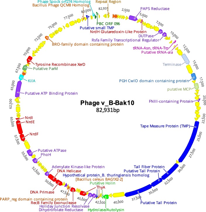
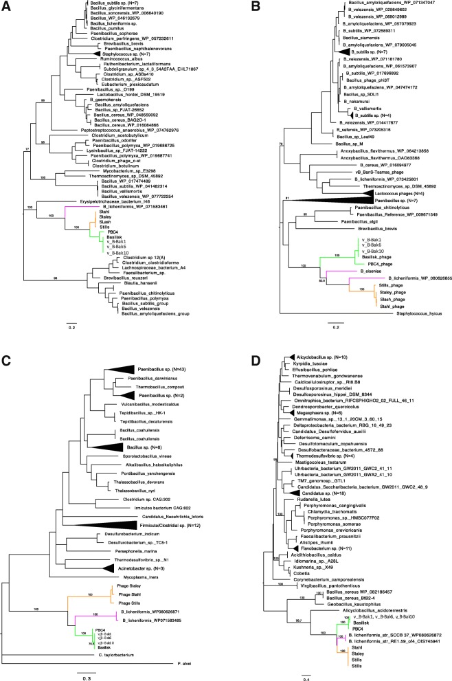
Background: In the present study, we sequenced the complete genomes of three novel bacteriophages v_B-Bak1, v_B-Bak6, v_B-Bak10 previously isolated from historical anthrax burial sites in the South Caucasus country of Georgia. We report here major trends in the molecular evolution of these phages, which we designate as "Basilisk-Like-Phages" (BLPs), and illustrate patterns in their evolution, genomic plasticity and core genome architecture.
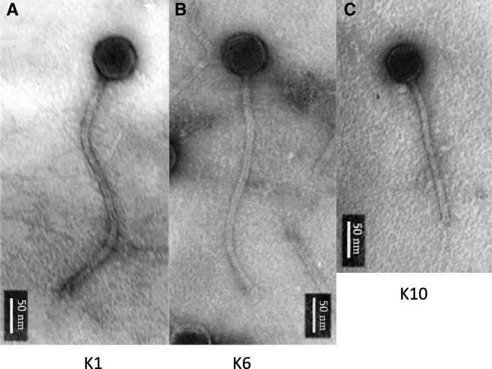
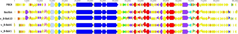
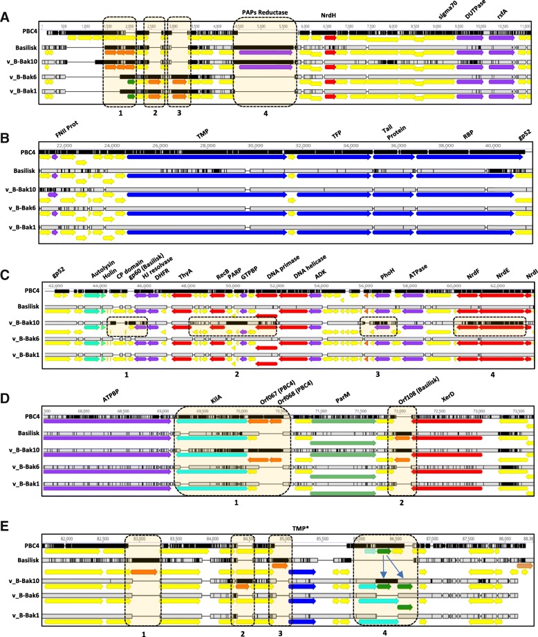
Results: Comparative whole genome sequence analysis revealed a close evolutionary relationship between our phages and two unclassified Bacillus cereus group phages, phage Basilisk, a broad host range phage (Grose JH et al., J Vir. 2014;88(20):11846-11860) and phage PBC4, a highly host-restricted phage and close relative of Basilisk (Na H. et al. FEMS Microbiol. letters. 2016;363(12)). Genome comparisons of phages v_B-Bak1, v_B-Bak6, and v_B-Bak10 revealed significant similarity in sequence, gene content, and synteny with both Basilisk and PBC4. Transmission electron microscopy (TEM) confirmed the three phages belong to the Siphoviridae family. In contrast to the broad host range of phage Basilisk and the single-strain specificity of PBC4, our three phages displayed host specificity for Bacillus anthracis. Bacillus species including Bacillus cereus, Bacillus subtilis, Bacillus anthracoides, and Bacillus megaterium were refractory to infection.
Conclusions: Data reported here provide further insight into the shared genomic architecture, host range specificity, and molecular evolution of these rare B. cereus group phages. To date, the three phages represent the only known close relatives of the Basilisk and PBC4 phages and their shared genetic attributes and unique host specificity for B. anthracis provides additional insight into candidate host range determinants.
Keywords: Bacillus anthracis; Phage evolution; Phage genome.
Conflict of interest statement
Ethics approval and consent to participate
Not applicable.
Consent for publication
Not applicable.
Competing interests
The authors declare that they have no competing interests.
Publisher’s Note
Springer Nature remains neutral with regard to jurisdictional claims in published maps and institutional affiliations.
Figures
References
-
- Okinaka RT, Keim P. The phylogeny of Bacillus cereus sensu lato. Microbiol Spectr. 2016;4:TBS–0012–2012. - PubMed
-
- Guinebretiere MH, Velge P, Couvert O, Carlin F, The DMLN. Ability of Bacillus cereus group strains to cause food poisoning varies according to phylogenetic affiliation (groups I to VII) rather than species affiliation. J Clin Microbiol. 2010;48:3388–3391. doi: 10.1128/JCM.00921-10. - DOI - PMC - PubMed
MeSH terms
Substances
LinkOut - more resources
Full Text Sources
Other Literature Sources
