. 2018 Mar 23:1:87-96.
doi: 10.1016/j.isci.2018.02.004. Epub 2018 Mar 23.

Extracellular Phosphorylation of TIMP-2 by Secreted c-Src Tyrosine Kinase Controls MMP-2 Activity

Affiliations

Extracellular Phosphorylation of TIMP-2 by Secreted c-Src Tyrosine Kinase Controls MMP-2 Activity

Javier Sánchez-Pozo et al. iScience. .

Abstract

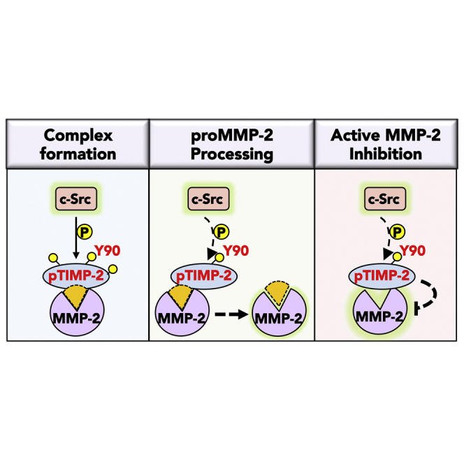
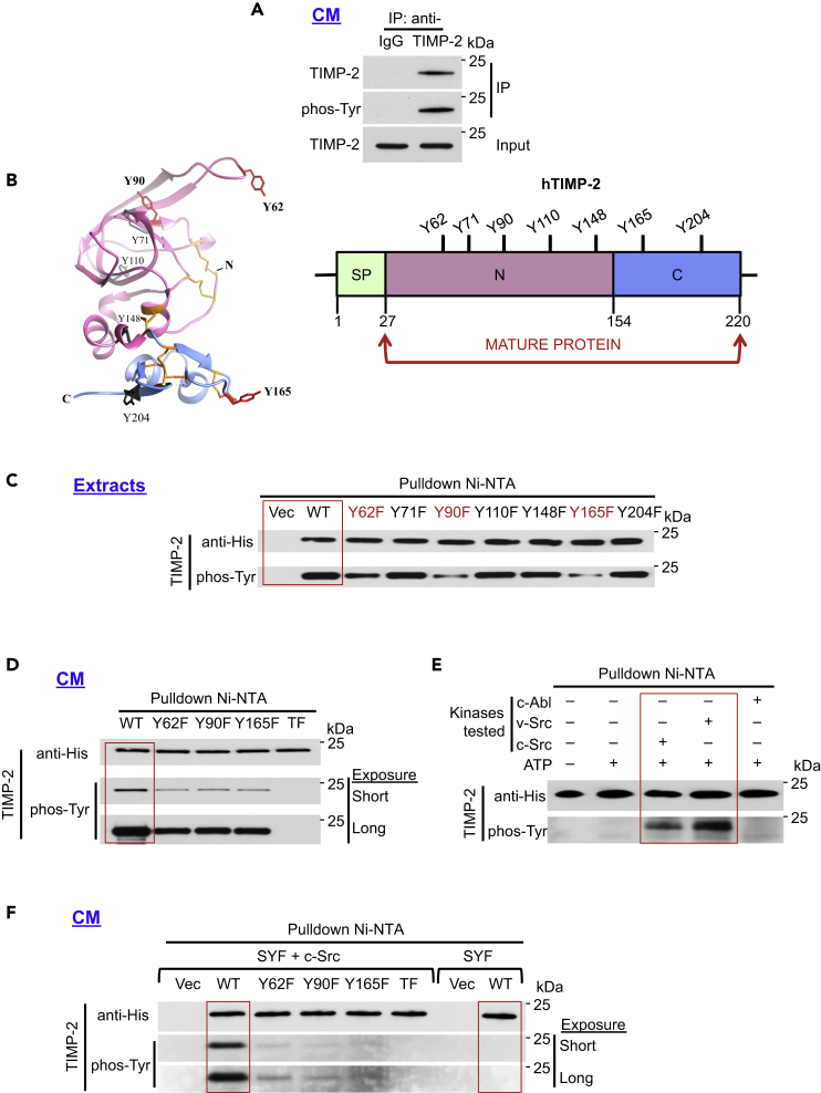
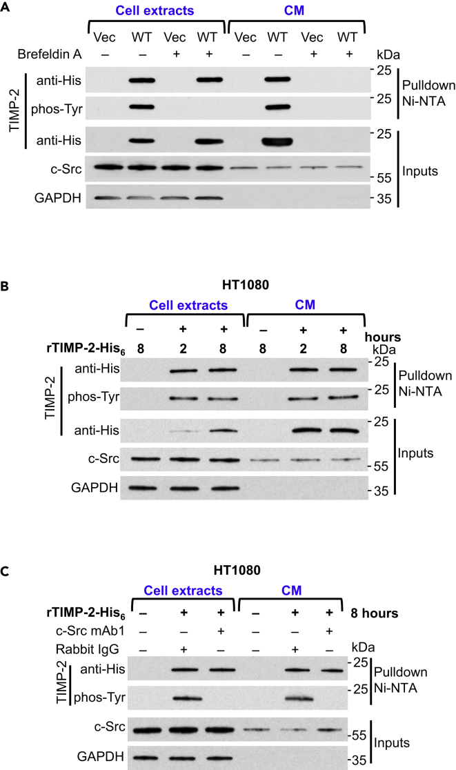
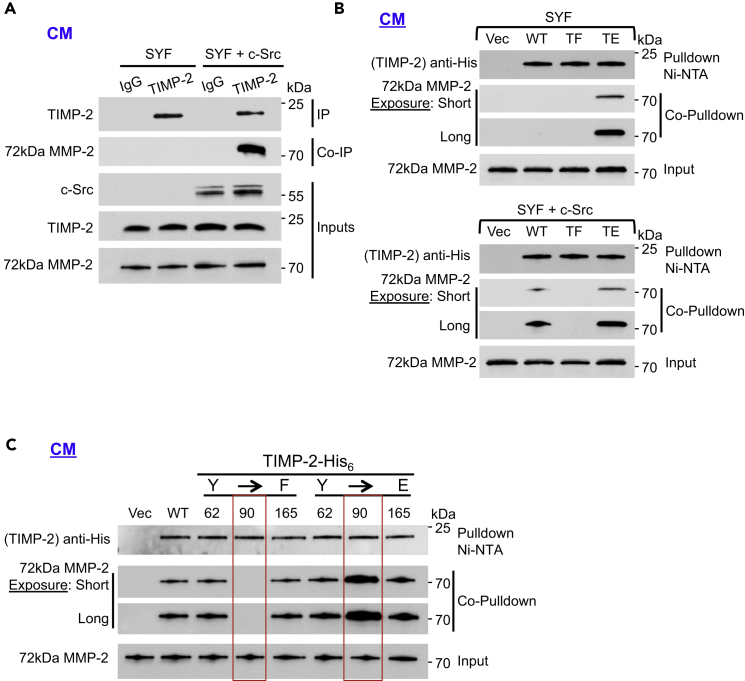
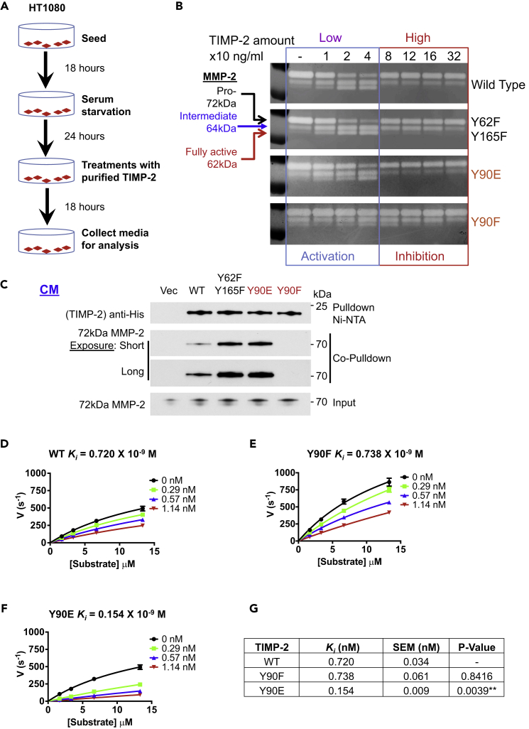
The tissue inhibitor of metalloproteinases 2 (TIMP-2) is a specific endogenous inhibitor of matrix metalloproteinase 2 (MMP-2), which is a key enzyme that degrades the extracellular matrix and promotes tumor cell invasion. Although the TIMP-2:MMP-2 complex controls proteolysis, the signaling mechanism by which the two proteins associate in the extracellular space remains unidentified. Here we report that TIMP-2 is phosphorylated outside the cell by secreted c-Src tyrosine kinase. As a consequence, phosphorylation at Y90 significantly enhances TIMP-2 potency as an MMP-2 inhibitor and weakens the catalytic action of the active enzyme. TIMP-2 phosphorylation also appears to be essential for its interaction with the latent enzyme proMMP-2 in vivo. Absence of the kinase or non-phosphorylatable Y90 abolishes TIMP-2 binding to the latent enzyme, ultimately hampering proMMP-2 activation. Together, TIMP-2 phosphorylation by secreted c-Src represents a critical extracellular regulatory mechanism that controls the proteolytic function of MMP-2.

Keywords: Biochemistry; Enzymology; Molecular Biology.


Figures

None
Graphical abstract
Figure 1
Figure 1
c-Src Phosphorylates Human TIMP-2 In Vitro and In Vivo (A) Endogenous secreted TIMP-2 was immunoprecipitated from 10X concentrated HT1080 cell-conditioned media (CM) using anti-TIMP-2 or IgG (control) and analyzed by immunoblotting for phosphorylation using an anti-pan-phosphotyrosine antibody (phos-Tyr, 4G10). (B) 3D (PDB: 1BR9) and linear domain structures of human TIMP-2 (hTIMP-2). All seven TIMP-2 tyrosine residues (Y) (black) are shown. Numbering is based on the full-length protein sequence (aa 1–220). (C and D) (C) TIMP-2 His6-tagged wild type (WT), vector control (Vec), and individual mutant plasmids were transiently expressed in HEK293H cells and pulled down from cell extracts (D) or CM and immunoblotted with indicated antibodies to assess phosphorylation. (E) Recombinant (rTIMP-2-His6) was used as the substrate in an in vitro kinase assay in the presence of full-length c-Src, v-Src, or c-Abl tyrosine kinases. Following pulldown, immunoblotting was performed to assess tyrosine phosphorylation using phos-Tyr, 4G10 antibody. (F) TIMP-2 constructs were transiently expressed in SYF and SYF + c-Src cells, pulled down from 10X concentrated CM and immunoblotted to determine TIMP-2 tyrosine phosphorylation. See also Figure S1.
Figure 2
Figure 2
c-Src Phosphorylates TIMP-2 Extracellularly (A) HEK293H cells were transiently transfected with vector (Vec) control or wild-type (WT) TIMP-2-His6. Cells were serum starved in the presence (+) and absence (−) of brefeldin A for 12 hr, and cell extracts and CM were collected for immunoblotting and pulldown Ni-NTA to determine c-Src secretion and tyrosine phosphorylation of TIMP-2. (B) HT1080 cells were serum starved for 24 hr and then treated with rTIMP-2-His6 for 2 and 8 hr. Cell extracts and CM were collected for immunoblotting and pulldown Ni-NTA analyses to determine phosphorylation of TIMP-2. (C) Anti-c-Src monoclonal antibody (mAb1) or Rabbit IgG control were added in the CM for 1 hr before addition of recombinant TIMP-2-His6 for 8 hr. Cell extracts and CM were collected for analyses. GAPDH was analyzed for equal loading in all blots. See also Figure S2.
Figure 3
Figure 3
Phosphorylation of Y90 Is Essential for TIMP-2:proMMP-2 Interaction (A) SYF and SYF + c-Src cells were serum starved for 18 hr. Immunoprecipitation (IP) of endogenous TIMP-2 from the cell CM was followed by co-immunoprecipitation (co-IP) of 72 kDa proMMP-2 to determine interaction. (B) SYF (top) and SYF + c-Src (bottom) were transiently transfected with Vec control, WT TIMP-2-His6, and mutants (TF and TE). Pulldown was performed from the CM followed by immunoblotting and co-pulldown to assess protein interaction. (C) HEK293H cells were transiently transfected with the indicated plasmids. Vec control, WT, and non-phosphorylatable (F) and phosphomimetic (E) TIMP-2 mutants were pulled down from the CM. Interaction of TIMP-2 proteins with secreted 72 kDa proMMP-2 was determined by co-pulldown and immunoblotting. See also Figure S3.
Figure 4
Figure 4
TIMP-2 Tyr90 Regulates MMP-2 Function (A) Flowchart followed in proMMP-2 activation studies. (B) HEK293H-purified WT TIMP-2 and indicated mutant proteins were added at increasing concentrations in the CM of HT1080 cells. Media were collected and analyzed by gelatin zymography. MMP-2 forms shown include the 72 kDa proMMP-2, 64 kDa intermediate form, and 62 kDa fully active form. Reduction of the 72 kDa form and increase of the 64 kDa and 62 kDa forms indicate proMMP-2 activation. (C) TIMP-2-His6 proteins were transiently transfected in HT1080 cells followed by pulldown and co-pulldown for proMMP-2 to assess protein interaction. (D–F) (D) HEK293H-purified WT TIMP-2, (E) Y90F, (F) and Y90E were tested for their ability to inhibit active MMP-2. Enzyme and inhibitors were pre-incubated at 25°C for 15 min before substrate addition. Experiments were performed at least twice. Error bars represent mean ± SD of three technical replicates. (G) WT TIMP-2, Y90F, and Y90E Ki were determined to assess TIMP-2 inhibitory potency and represent the mean average of at least two independent experiments. A Student's t-test was performed between the WT and each of the two mutants. p Values were estimated to assess statistical significance (**p < 0.01). See also Figure S4.

References

    1. Batra J., Soares A.S., Mehner C., Radisky E.S. Matrix metalloproteinase-10/TIMP-2 structure and analyses define conserved core interactions and diverse exosite interactions in MMP/TIMP complexes. PLoS One. 2013;8:e75836. - PMC - PubMed
    1. Beebe K., Mollapour M., Scroggins B., Prodromou C., Xu W., Tokita M., Taldone T., Pullen L., Zierer B.K., Lee M.J. Posttranslational modification and conformational state of heat shock protein 90 differentially affect binding of chemically diverse small molecule inhibitors. Oncotarget. 2013;4:1065–1074. - PMC - PubMed
    1. Benham A.M. Protein secretion and the endoplasmic reticulum. Cold Spring Harb. Perspect. Biol. 2012;4:a012872. - PMC - PubMed
    1. Bernardo M.M., Fridman R. TIMP-2 (tissue inhibitor of metalloproteinase-2) regulates MMP-2 (matrix metalloproteinase-2) activity in the extracellular environment after pro-MMP-2 activation by MT1 (membrane type 1)-MMP. Biochem. J. 2003;374:739–745. - PMC - PubMed
    1. Betts M.J., Russell R.B. Amino acid properties and consequences of substitutions. In: Barnes M.R., Gray I.C., editors. Bioinformatics for Geneticists. John Wiley & Sons, Ltd; 2003. pp. 291–316.