Review
. 2019 Mar;26(3):424-430.
doi: 10.1016/j.acra.2018.08.006. Epub 2018 Sep 15.

Structural and Functional Pulmonary Magnetic Resonance Imaging in Pediatrics-From the Neonate to the Young Adult

Affiliations
Review

Structural and Functional Pulmonary Magnetic Resonance Imaging in Pediatrics-From the Neonate to the Young Adult

Laura L Walkup et al. Acad Radiol. 2019 Mar.

Abstract

The clinical imaging modalities available to investigate pediatric pulmonary conditions such as bronchopulmonary dysplasia, cystic fibrosis, and asthma are limited primarily to chest x-ray radiograph and computed tomography. As the challenges that historically limited the application of magnetic resonance imaging (MRI) to the lung have been overcome, its clinical potential has greatly expanded. In this review article, recent advances in pulmonary MRI including ultrashort echo time and hyperpolarized-gas MRI techniques are discussed with an emphasis on pediatric research and translational applications.

Keywords: MRI; Pediatrics; Pulmonary.


Figures

Fig. 1.
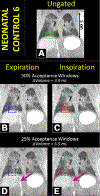
Fig. 1.
Coronal views of 3D UTE MR images in a neonatal control patient demonstrating ungated reconstructions (A) and retrospectively respiratory-gated reconstructions at 50% and 25% acceptance windows for end-expiration (B and D) and end-inspiration (C and E). The boxes indicate a region of lung-diaphragm transition, and the arrows indicate the inferior vena cava, the movement of which is more finely resolved in the 25%-windowed images. A tidal volume measurement—not easily assessed in infants by any clinical means—is measured via the segmentation of these images. Reprinted from Ref (19) “Retrospective respiratory self-gating and removal of bulk motion in pulmonary UTE MRI of neonates and adults,” Mara S. Higano, Andrew D. Hahn, Jean A. Tkach, Xuefeng Cao, Laura L. Walkup, Robert P. Thomen, Stephanie L. Merhar, Paul S. Kingma, Sean B. Fain, and Jason C. Woods, Magnetic Resonance in Medicine, 2017, Volume 77, pages 1284–1295, with permission from John Wiley and Sons. MR, magnetic resonance;UTE, ultrashort echo time. (Color version of figure is available online.)
Fig. 2.
Fig. 2.
Slice-matched comparison of axial x-ray CT (left column) and UTE MR (right column) images of Infants with cystic fibrosis and a control (ages 33–47 months old), demonstrating how both CT and UTE MRI can visualize CF pathology: bronchiectasis (1), ground-glass opacity (2), bronchial-wall thickening (3), mucus plugging (4), consolidation (5), and air trapping (6). Anesthesia-Induced atelectasis is circled in the blue. Reprinted from Ref (29) with permission of the American Thoracic Society. Copyright 2017 American Thoracic Society. David J. Roach, Yannick Crémillieux, Robert J, Fleck, Alan S. Brody, Suraj D. Serai, Rhonda D. Szczesniak, Stephanie Kerla-kian, John P. Clancy, and Jason C. Woods, 2016, “Ultrashort Echo-Time Magnetic Resonance Imaging Is a Sensitive Method for the Evaluation of Early Cystic Fibrosis Lung Disease”, Annals of the American Thoracic Society Volume 13, Issue 11, pages 1923–1931. Annals of the American Thoracic Society is an official journal of the American Thoracic Society. CF, cystic fibrosis; CT, computed tomography; MR, magnetic resonance; UTE, ultrashort echo time. (Color version of figure is available online.)
Fig. 3.
Fig. 3.
Maps of the root-mean-square diffusion length (χrms), a measurement of alveolar-airspace size) of hyperpolarized 3He in four pediatric subjects (9–10 years old). In this study, children with a history of wheezing illness with human rhinovirus (HRV) infection in early childhood (Subjects C and D) had smaller airways as compared to those who did not (Subjects A and B). Reprinted from Ref (59), 2013 with permission from Elsevier. (Color version of figure is available online.)
Fig. 4.
Fig. 4.
Hyperpolarized 3He ventilation MR images in three nonsedated, nonrestrained young children demonstrating early ventilation deficits (arrows) associated with premature birth, CF, and asthma. Figure is a composite of images reprinted from Ref (61), Clinical Imaging, 2017, Volume 45, pages 105–110, “Hyperpolarized helium-3 magnetic resonance lung imaging of nonsedated infants and young children: a proof-of-concept study”, Talissa A. Altes, Craig H. Meyer, Jaime F. Mata, Deborah K. Froh, Alix Paget-Brown, W. Gerald Teague, Sean B. Fain, Eduard E, de Lange, Kai Ruppert, Martyn C. Botfield, Mac A. Johnson, John P. Mugler (https://doi.org/10.1016/j.clinimag.2017.04.004) with permission from Elsevier under the Creative Commons license (HYPERLINK” https://creativecommons.org/licenses/by-nc-nd/4.0/). CF, cystic fibrosis; MR, magnetic resonance.

Similar articles

Cited by

References

    1. Brenner D, Elliston C, Hall E, et al. Estimated risks of radiation-induced fatal cancer from pediatric CT. AJR Am J Roentgenol 2001; 176:289–296. - PubMed
    1. Miglioretti DL, Johnson E, Williams A, et al. The use of computed tomography in pediatrics and the associated radiation exposure and estimated cancer risk. JAMA Pediatr 2013; 167:700–707. - PMC - PubMed
    1. Pearce MS, Salotti JA, Little MP, et al. Radiation exposure from CT scans in childhood and subsequent risk of leukaemia and brain tumours: a retrospective cohort study. Lancet 2012; 380:499–505. - PMC - PubMed
    1. Hatabu H, Alsop DC, Listerud J, et al. T2* and proton density measurement of normal human lung parenchyma using submillisecond echo time gradient echo magnetic resonance imaging. Eur J Radiol 1999; 29: 245–252. - PubMed
    1. Ohno Y, Koyama H, Yoshikawa T, et al. T2* measurements of 3-T MRI with ultrashort TEs: capabilities of pulmonary function assessment and clinical stage classification in smokers. AJR Am J Roentgenol 2011; 197: W279–W285. - PubMed