. 2018 Oct;8(10):1316-1331.
doi: 10.1158/2159-8290.CD-17-0987. Epub 2018 Sep 18.

Intertumoral Heterogeneity in SCLC Is Influenced by the Cell Type of Origin

Affiliations

Intertumoral Heterogeneity in SCLC Is Influenced by the Cell Type of Origin

Dian Yang et al. Cancer Discov. 2018 Oct.

Abstract

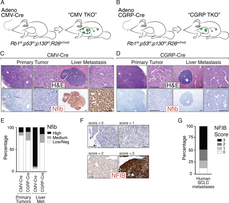
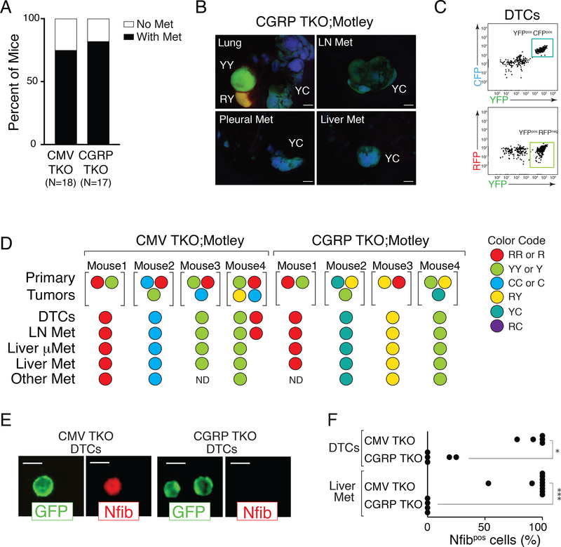
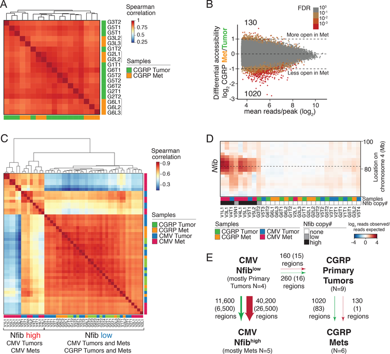
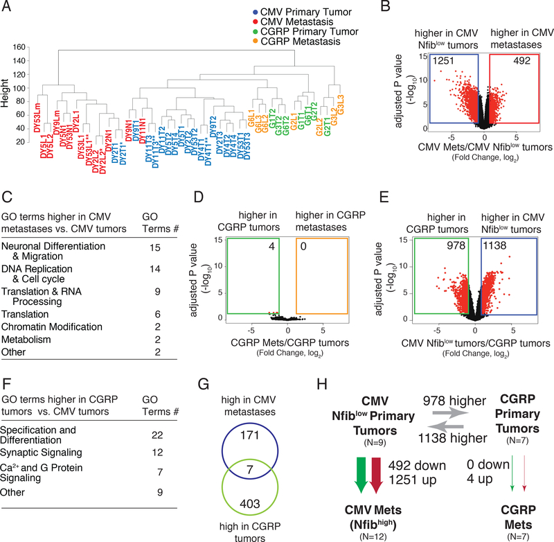
The extent to which early events shape tumor evolution is largely uncharacterized, even though a better understanding of these early events may help identify key vulnerabilities in advanced tumors. Here, using genetically defined mouse models of small cell lung cancer (SCLC), we uncovered distinct metastatic programs attributable to the cell type of origin. In one model, tumors gain metastatic ability through amplification of the transcription factor NFIB and a widespread increase in chromatin accessibility, whereas in the other model, tumors become metastatic in the absence of NFIB-driven chromatin alterations. Gene-expression and chromatin accessibility analyses identify distinct mechanisms as well as markers predictive of metastatic progression in both groups. Underlying the difference between the two programs was the cell type of origin of the tumors, with NFIB-independent metastases arising from mature neuroendocrine cells. Our findings underscore the importance of the identity of cell type of origin in influencing tumor evolution and metastatic mechanisms.Significance: We show that SCLC can arise from different cell types of origin, which profoundly influences the eventual genetic and epigenetic changes that enable metastatic progression. Understanding intertumoral heterogeneity in SCLC, and across cancer types, may illuminate mechanisms of tumor progression and uncover how the cell type of origin affects tumor evolution. Cancer Discov; 8(10); 1316-31. ©2018 AACR. See related commentary by Pozo et al., p. 1216 This article is highlighted in the In This Issue feature, p. 1195.


Conflict of interest statement

Disclosure of Potential Conflicts of Interests: W.J.G. is the scientific co-founder of Epinomics. A.K. is on the scientific advisory board of Epinomics. J.S. receives research funding from Abbvie. H.C.R. received consulting and lecture fees from Abbvie, Astra-Zeneca, Vertex and Merck and research funding from Gilead Pharmaceuticals.

Figures

Figure 1:
Figure 1:. SCLC initiated from pulmonary neuroendocrine cells metastasizes without upregulating Nfib.
A-B. Mouse models of SCLC. Rb1flox/flox;p53 flox/flox;p130 flox/flox;R26mTmG (TKO;mTmG) mice were transduced with either Adeno-CMV-Cre (“CMV TKO”) or Adeno-CGRP-Cre (“CGRP TKO”) to initiate SCLC. C-D. Representative H&E images of SCLC tumors from CMV TKO and CGRP TKO mice. Immunostaining for Nfib on primary tumors and metastases is shown. Scale bars = 100μm. E. Quantification of Nfib expression in primary tumors and liver metastases from CMV TKO and CGRP TKO mice. Most of the metastases in CMV TKO mice are Nfibpositive, while most of the in CGRP TKO mice are Nfiblow/negative. F. Representative immunostaining for NFIB on human SCLC brain metastases. Expression score for NFIB is indicated. Scale bars = 100μm. G. Quantification of NFIB expression in human SCLC metastases (including lymph node and brain metastases, N=43).
Figure 2:
Figure 2:. Tumor progression is required in both CMV TKO and CGRP TKO mice.
A. Occurrence of metastasis in CMV TKO and CGRP TKO mice. Not every mouse developed metastatic disease at the time of analysis even though each mouse in this analysis had several large primary tumors (7–11 months after initiation). B. Representative images of tumors and metastases from TKO;Motley mice with CGRP-Cre-initiated SCLC. Primary tumors existed in multiple colors, while all metastases from the same mouse had the same color, suggesting that these metastases are clonally related to one primary tumor. Scale bar = 2mm. Color code, R = RFP, Y = YFP, C = CFP. C. Representative FACS analysis on disseminated tumor cells (DTCs) from a CGRP TKO;Motley mouse. All The DTCs of this mouse have the same color (CFPpositive and YFPpositive). D. Summary of results from 4 CMV TKO;Motley mice and 4 CGRP TKO;Motley mice. The colors of the largest primary tumors, disseminated tumors cells (DTCs), and metastases are shown. mMet, micrometastases. ND, not detected. E-F. Immunofluorescence staining for Nfib on FACS-isolated GFPpositive DTCs from the pleural cavity and cancer cells from liver metastases from CMV TKO and CGRP TKO mice. (E) Representative images of DTCs from CMV TKO and CGRP TKO mice. Membrane GFP staining is encoded by the recombined R26mTmG allele. Scale bars = 10 μm. (F) Quantification of Nfib expression in DTCs and cancer cells from liver metastases from CMV TKO and CGRP TKO mice. CMV TKO data is from Denny, Yang et al, 2016. Each dot represents one mouse. * P = 0.0097, *** P=0.001, Mann-Whitney test.
Figure 3:
Figure 3:. Limited chromatin accessibility changes and lack of Nfib amplification during metastatic progression of CGRP TKO tumors.
A. Correlation of chromatin accessibility of 9 primary tumors and 7 liver metastases from CGRP TKO mice. Replicate samples from the same tumors have the same name. Metastases from the same mice (indicated with brackets) correlate most closely with one another. B. Differential accessibility between CGRP primary tumors and metastases. Log2 fold change in reads per peak between groups plotted against the mean number of reads per peak is shown. Number of regions that have significant accessibility changes (FDR < 0.1, |log2fold change| > 0) is indicated. Color of each point represents the false discovery rate (FDR) that the absolute value of the log2 fold change was greater than 0. C. Correlation of chromatin accessibility between primary tumors and metastases from CMV TKO and CGRP TKO mice. D. DNA copy number analysis at the Nfib locus on Chromosome 4 in CMV TKO and CGRP TKO primary tumors and metastases. Heatmap shows copy number variation (CNV) at the Nfib locus. none = CNV < 1, low= CNV >1 and CNV <1.5, high = CNV >1.5. E. Summary of the chromatin accessibility changes during metastatic progression of CMV TKO and CGRP TKO tumors. Numbers of peaks with changed accessibility are listed (|lfc|>0 at FDR<0.1, no peak changed < 2 fold), numbers in parentheses give the number of peaks that change in accessibility (|lfc|>0.5 at FDR<0.1, no peak changed < 2 fold).
Figure 4:
Figure 4:. Molecularly distinct SCLC subtypes.
A. Unsupervised hierarchical clustering of all CMV TKO and CGRP TKO tumors and metastases. * technical replicates, ** batch replicates. B. Differential gene expression in CMV TKO Nfiblow primary tumors and CMV TKO metastases. (|Fold change| > 2, adj. p < 0.05) C. A summary of top 50 GO terms (FDR q value < 0.05) enriched in CMV TKO metastasis in comparison to CMV Nfiblow primary tumor. D. Differential gene expression in CGRP TKO primary tumors and CGRP TKO metastases. (|Fold change| > 2, adj. p < 0.05) E. Differential gene expression in CMV TKO Nfiblow tumor and CGRP TKO tumor (|Fold change| > 2, adj. p < 0.05) F. A summary of top 50 GO terms (FDR q value < 0.05) enriched in CGRP TKO tumor in comparison to CMV Nfiblow primary tumor. G. Genes that are significantly upregulated (|Log2(Fold change)| > 1.5) in CMV TKO metastasis and CGRP TKO tumors comparing to CMV TKO Nfiblow tumors are mostly mutually exclusive. H. Summary of RNA expression differences between the 4 different tumor states. Numbers of genes with changed expression are listed (|Fold change| > 2, adj.p < 0.05).
Figure 5:
Figure 5:. Multilineage differentiation in CMV TKO Nfiblow primary tumors.
A. Gene expression of multilineage markers in tumors and metastases from CMV TKO and CGRP TKO mice. CMV TKO primary tumors express markers of Club cells, alveolar type II cells (AT II), and alveolar type I (AT I) cells. B. Immunohistochemical staining for Uchl1, Nfib, and CC10. Representative images show the presence of CC10pos cells specifically in Nfiblow primary tumors in CMV TKO mice. Scale bars = 100μm. C-D. Quantification of CC10 and Nfib expression. CC10 and Nfib are mostly mutually exclusive in CMV TKO lung tumors (C), while CC10 and Nfib are only rarely expressed in CGRP TKO lung tumors (D). E. Immunohistochemical staining for tumor clonal lineage markers (RFP, YFP/CFP) as well as CC10 and Selenbp1 on tumors from CMV TKO;Motley mice. The uniform staining of tumor lineage markers in individual tumors is consistent with their clonal origin. CC10positive and Selenbp1positive cells are cancer cells that are clonally related to the CC10negative and Selenbp1negative cells. Scale bars top = 100 μm, bottom = 20 μm.
Figure 6:
Figure 6:. SCLC in CMV TKO mice arises from an alternative cell of origin.
A. Representative immunofluorescent images of whole-mount staining and confocal imaging of tumors in CMV TKO and CGRP TKO mice. Tumors are circled. Number and percent of lesions at each location is indicated. T = tumor, B = bronchi, TB = terminal bronchiole, A = alveolar space, SA = streptavidin (which labels lung epithelial cells). Scale bars = 200 μm. B. Representative H&E images of the lungs of mice 3.5 months after transduction by same titer of adenoviral vectors with distinct promoters driving Cre expression. Scale bars = 1 mm. C. Quantification of SCLC tumor number 3.5 months and 10 months after transduction with same total titer of different types and combinations of adenoviral vectors with distinct promoters driving Cre expression. Tumor number is quantified from H&E lung sections. The number of mice in each cohort is indicated in parentheses. D. Model of SCLC development and progression from two distinct cell types of origin.

Comment in

References

    1. Lambert AW, Pattabiraman DR, Weinberg RA. Emerging Biological Principles of Metastasis. Cell. 2017;168:670–91. - PMC - PubMed
    1. Nguyen DX, Massague J. Genetic determinants of cancer metastasis. Nat Rev Genet. 2007;8:341–52. - PubMed
    1. Bunn PA, Minna JD Jr., Augustyn A, Gazdar AF, Ouadah Y, Krasnow MA, et al. Small Cell Lung Cancer: Can Recent Advances in Biology and Molecular Biology Be Translated into Improved Outcomes? J Thorac Oncol. 2016;11:453–74. - PMC - PubMed
    1. Semenova EA, Nagel R, Berns A. Origins, genetic landscape, and emerging therapies of small cell lung cancer. Genes Dev. 2015;29:1447–62. - PMC - PubMed
    1. Wu N, Jia D, Ibrahim AH, Bachurski CJ, Gronostajski RM, MacPherson D. NFIB overexpression cooperates with Rb/p53 deletion to promote small cell lung cancer. Oncotarget. 2016;7:57514–24. - PMC - PubMed

Publication types