. 2018 Nov 16;293(46):17663-17675.
doi: 10.1074/jbc.RA118.002691. Epub 2018 Sep 18.

The C-terminal GGAP motif of Hsp70 mediates substrate recognition and stress response in yeast

Affiliations

The C-terminal GGAP motif of Hsp70 mediates substrate recognition and stress response in yeast

Weibin Gong et al. J Biol Chem. .

Abstract

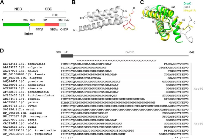
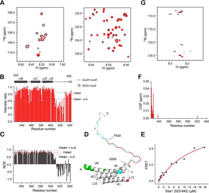
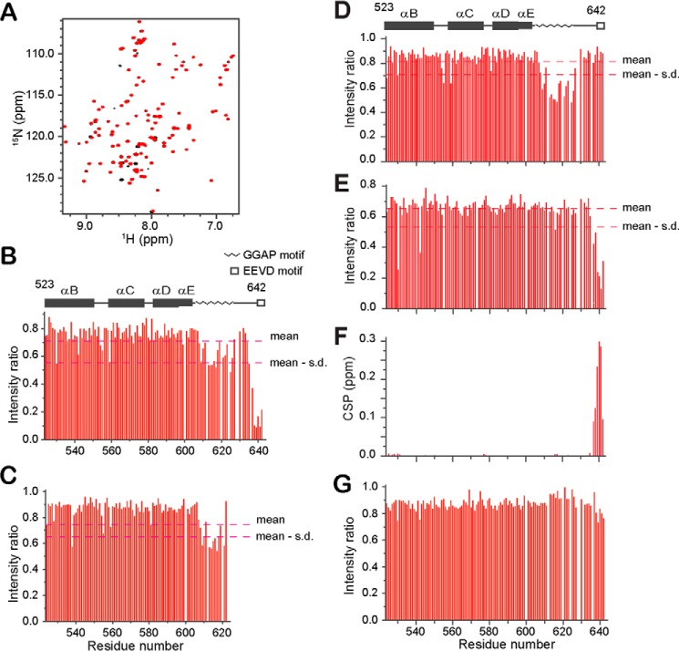
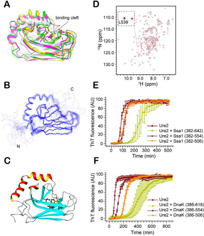
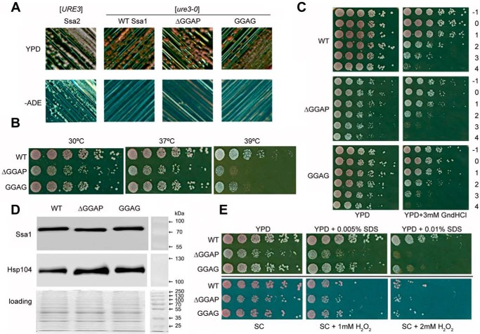
The allosteric coupling of the highly conserved nucleotide- and substrate-binding domains of Hsp70 has been studied intensively. In contrast, the role of the disordered, highly variable C-terminal region of Hsp70 remains unclear. In many eukaryotic Hsp70s, the extreme C-terminal EEVD motif binds to the tetratricopeptide-repeat domains of Hsp70 co-chaperones. Here, we discovered that the TVEEVD sequence of Saccharomyces cerevisiae cytoplasmic Hsp70 (Ssa1) functions as a SUMO-interacting motif. A second C-terminal motif of ∼15 amino acids between the α-helical lid and the extreme C terminus, previously identified in bacterial and eukaryotic organellar Hsp70s, is known to enhance chaperone function by transiently interacting with folding clients. Using structural analysis, interaction studies, fibril formation assays, and in vivo functional assays, we investigated the individual contributions of the α-helical bundle and the C-terminal disordered region of Ssa1 in the inhibition of fibril formation of the prion protein Ure2. Our results revealed that although the α-helical bundle of the Ssa1 substrate-binding domain (SBDα) does not directly bind to Ure2, the SBDα enhances the ability of Hsp70 to inhibit fibril formation. We found that a 20-residue C-terminal motif in Ssa1, containing GGAP and GGAP-like tetrapeptide repeats, can directly bind to Ure2, the Hsp40 co-chaperone Ydj1, and α-synuclein, but not to the SUMO-like protein SMT3 or BSA. Deletion or substitution of the Ssa1 GGAP motif impaired yeast cell tolerance to temperature and cell-wall damage stress. This study highlights that the C-terminal GGAP motif of Hsp70 is important for substrate recognition and mediation of the heat shock response.

Keywords: SUMO-interacting motif (SIM); Saccharomyces cerevisiae; amyloid; chaperone; heat shock protein (HSP); nuclear magnetic resonance (NMR); prion; proline-rich motif; protein structure; stress response.


Conflict of interest statement

The authors declare that they have no conflicts of interest with the contents of this article

Figures

Figure 1.
Figure 1.
Structure of Ssa1 CTD. A, schematic of Ssa1 domain structure. B, ribbon representation of the Ssa1(523–622) structure. Red, intensity ratio < mean − S.D. caused by addition of Ure2 with ratio 1:1; yellow, proline or severely overlapped residues. C, superimposition of the SBDα of yeast Ssa1 (gray), E. coli DnaK (green), and human HspA1A (yellow). D, the GGAP-like motif and EEVD motif in eukaryotic cytosolic members of the Hsp70 family. The C-terminal GGAP-like repetitive sequence is indicated by a jagged line. The numbers indicate residues of yeast Ssa1.
Figure 2.
Figure 2.
Titration of Ssa1(523–642) and GGAP motif with Ure2. A, 1H-15N HSQC spectra of Ssa1(523–642) titrated with Ure2 at a 1:2 ratio. B, bar diagram of intensity ratio versus residue number. C, steady-state 1H-15N NOE value per residue of Ssa1(523–642). D, mapping of intensity ratio and CSP onto the Ssa1(523–642) structure. Red, intensity ratio < mean − S.D. in B; cyan, proline and unassigned residues; green, CSP > 0.003 ppm in F. E, KD fitting (14.7 ± 3.6 μm) of binding of Ssa1(523–642) to Ure2 measured by FRET. F, bar diagram of CSP versus residue number. G, 1H-15N HSQC spectra of the 20-residue GGAP motif in Ssa1 C-IDR titrated with Ure2 at a 1:2 ratio.
Figure 3.
Figure 3.
Titration of Ssa1 SBD(523–642) with Ydj1 or BSA. A, 1H-15N HSQC spectra of Ssa1(523–642) titrated with Ydj1 at a ratio of 1:3. B, bar diagram of intensity ratio versus residue number of Ssa1(523–642) in A. C, bar diagram of intensity ratio of Ssa1(523–622) caused by addition of Ydj1 with ratio 1:7. D and E, bar diagram of intensity ratio of Ssa1(523–642) caused by addition of α-synuclein (D) and SMT3 (E) both with ratio 1:4. F, bar diagram of CSP versus residue number of Ssa1(523–642) caused by addition of SMT3 as in E. G, bar diagram of intensity ratio of Ssa1(523–642) caused by addition of BSA at a ratio of 1:3.
Figure 4.
Figure 4.
Effect of SBD truncation on the structure and fibril inhibition ability of Ssa1. A, structural superimposition of truncation mutants of SBDβ of different Hsp70 homologues, including S. cerevisiae Ssa1(382–554) (magenta), E. coli DnaK (white, PDB code 1BPR), G. kaustophilus DnaK (green, PDB code 2V7Y), rat Hsc70 (light blue, PDB code 1CKR), and bovine Hsc70 (yellow, PDB code 1YUW). B, the backbone ensemble of 20 structures of Ssa1(382–554). C, ribbon representation of the Ssa1(382–554) structure. Side chain of Leu539 (yellow) and surrounding hydrophobic side chains are shown by ball-and-stick model. The structural figures (A–C) were generated using MOLMOL (67) and PyMOL (68). D, overlay of the 2D 1H-15N HSQC spectra of Ssa1(382–554) and Ssa1(382–582). E, fibril formation of 30 μm Ure2 in the absence or presence of 50 μm Ssa1(382–642), Ssa1(382–554), and Ssa1(382–506), as indicated. F, fibril formation of 30 μm Ure2 in the absence or presence of 50 μm DnaK(384–638), DnaK(384–554), and DnaK(384–506), as indicated. E and F, the buffer conditions were 50 mm Tris-HCl, pH 8.4, 200 mm NaCl. The error bars indicate the standard error from at least three parallel experiments.
Figure 5.
Figure 5.
Effects of Ssa1 ΔGGAP and GGAG mutations in S. cerevisiae [URE3] strain under different conditions. A, assessment of [URE3] propagation. Single colonies containing Ssa2, WT Ssa1, ΔGGAP, or GGAG mutant Ssa1 as the sole source of Ssa were streaked on YPD and −ADE plates, which were then incubated at 30 °C for 2 days. Red colonies on YPD and nonviability to grow on −ADE plates indicate a [ure3-0] state; white colonies on YPD and viability on −ADE plates indicate a [URE3] state. B, growth assay at elevated temperatures. Fresh cultures were spotted onto YPD after a one-fifth serial dilution. The plates were incubated at 30, 37, or 39 °C for 2 days. C, acquired thermotolerance assay of ΔGGAP and GGAG mutations. Yeast cells were cultured at 30 °C to reach an A600 of 0.4 before pretreating at 39 °C for 1 h. Then 39 °C pretreated cells were heat-shocked at 47 °C for a time course. −1, cultures without 39 °C incubation; 0–4, cultures were first incubated 39 °C for 1 h and then moved to 47 °C incubator for 0, 10, 20, 30, or 40 min. D, expression levels of Ssa1 and Hsp104. The experiment was repeated three times, consistently showing that the mutant Ssa1 proteins are expressed at a similar level to WT and that slightly more Hsp104 is expressed in cells with GGAP deletion or substitution. E, assessment of ΔGGAP and GGAG mutations under cell-wall damage reagent SDS and oxidative damage reagent H2O2.

References

    1. Kim Y. E., Hipp M. S., Bracher A., Hayer-Hartl M., and Hartl F. U. (2013) Molecular chaperone functions in protein folding and proteostasis. Annu. Rev. Biochem. 82, 323–355 10.1146/annurev-biochem-060208-092442 - DOI - PubMed
    1. Clerico E. M., Tilitsky J. M., Meng W., and Gierasch L. M. (2015) How hsp70 molecular machines interact with their substrates to mediate diverse physiological functions. J. Mol. Biol. 427, 1575–1588 10.1016/j.jmb.2015.02.004 - DOI - PMC - PubMed
    1. Bertelsen E. B., Chang L., Gestwicki J. E., and Zuiderweg E. R. (2009) Solution conformation of wild-type E. coli Hsp70 (DnaK) chaperone complexed with ADP and substrate. Proc. Natl. Acad. Sci. U.S.A. 106, 8471–8476 10.1073/pnas.0903503106 - DOI - PMC - PubMed
    1. Zuiderweg E. R., Bertelsen E. B., Rousaki A., Mayer M. P., Gestwicki J. E., and Ahmad A. (2013) Allostery in the Hsp70 chaperone proteins. Top. Curr. Chem. 328, 99–153 - PMC - PubMed
    1. Alderson T. R., Kim J. H., and Markley J. L. (2016) Dynamical structures of Hsp70 and Hsp70–Hsp40 complexes. Structure 24, 1014–1030 10.1016/j.str.2016.05.011 - DOI - PMC - PubMed

Publication types

MeSH terms

LinkOut - more resources