. 2018 Sep 18;9(1):3795.
doi: 10.1038/s41467-018-06237-7.

Paf1 and Ctr9 subcomplex formation is essential for Paf1 complex assembly and functional regulation

Affiliations

Paf1 and Ctr9 subcomplex formation is essential for Paf1 complex assembly and functional regulation

Ying Xie et al. Nat Commun. .

Abstract

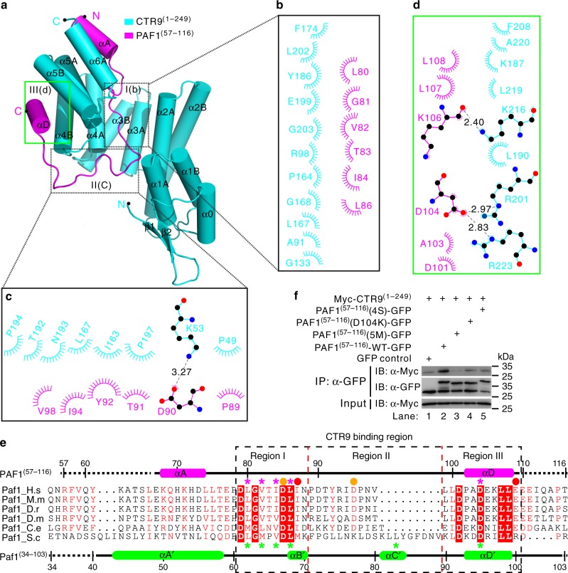
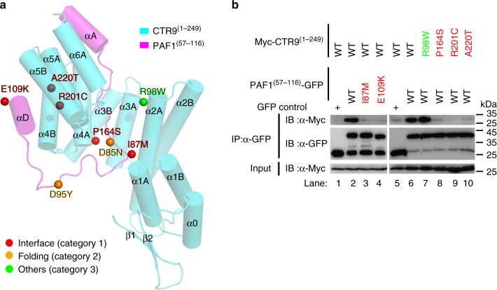
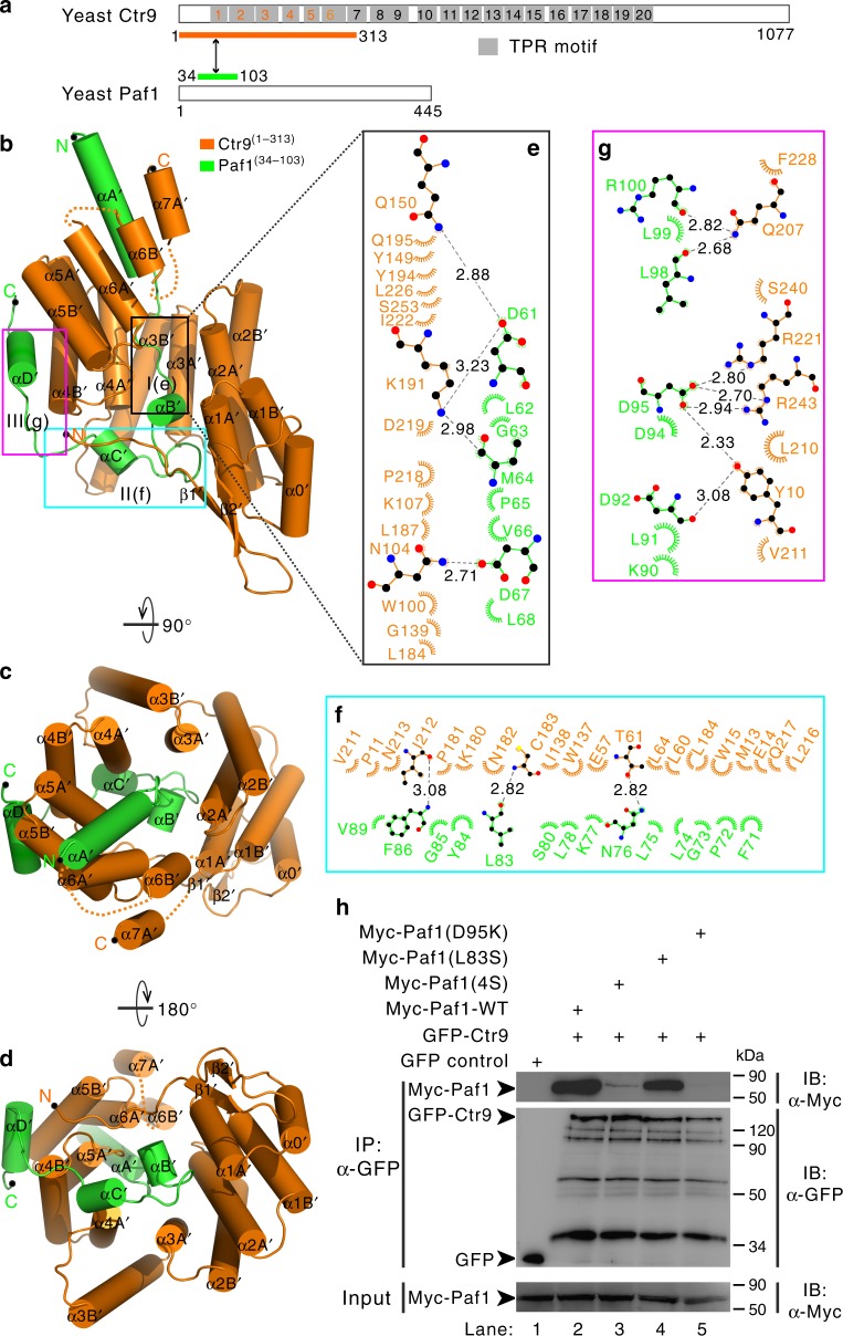
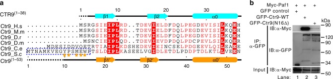
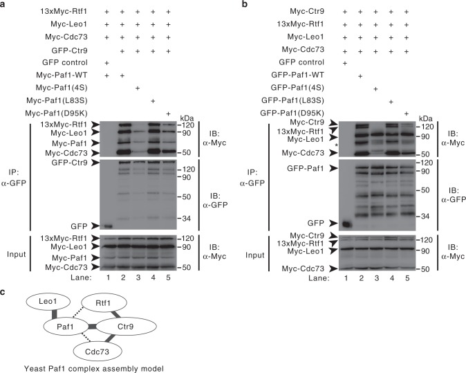
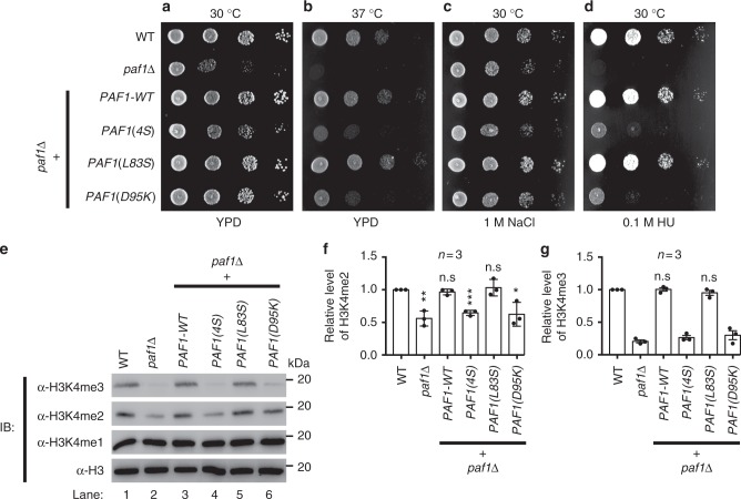
The evolutionarily conserved multifunctional polymerase-associated factor 1 (Paf1) complex (Paf1C), which is composed of at least five subunits (Paf1, Leo1, Ctr9, Cdc73, and Rtf1), plays vital roles in gene regulation and has connections to development and human diseases. Here, we report two structures of each of the human and yeast Ctr9/Paf1 subcomplexes, which assemble into heterodimers with very similar conformations, revealing an interface between the tetratricopeptide repeat module in Ctr9 and Paf1. The structure of the Ctr9/Paf1 subcomplex may provide mechanistic explanations for disease-associated mutations in human PAF1 and CTR9. Our study reveals that the formation of the Ctr9/Paf1 heterodimer is required for the assembly of yeast Paf1C, and is essential for yeast viability. In addition, disruption of the interaction between Paf1 and Ctr9 greatly affects the level of histone H3 methylation in vivo. Collectively, our results shed light on Paf1C assembly and functional regulation.


Conflict of interest statement

The authors declare no competing interests.

Figures

Fig. 1
Fig. 1
Crystal structure of the human CTR9/PAF1 heterodimer. a Schematic representation of full-length CTR9 and PAF1. A LEO1-interacting region (blue) and a histone H3-interacting region (orange) are shown in PAF1. The predicted 19 TPR motifs (gray) were defined using TPRpred (https://toolkit.tuebingen.mpg.de/#/tools/tprpred). The protein fragments of the CTR9(1–249)/PAF1(57–116) complex used for structural determination are indicated by a two-way arrow and are colored cyan and magenta, respectively. b Ribbon diagram representation of the CTR9(1–249) (cyan)/PAF1(57–116) (magenta) complex viewed from the side. The N- and C-termini of the two proteins are labeled. Cylinder representation of the CTR9(1–249)/PAF1(57–116) complex structure viewed from the top (c) or the bottom (d). e Surface representation of the CTR9(1–249)/PAF1(57–116) complex with its orientation corresponding to the other side from that shown in (b)
Fig. 2
Fig. 2
The interaction interface of the human CTR9/PAF1 heterodimer. ae The CTR9(1–249)/PAF1(57–116) interface is divided into three regions corresponding to the CTR9 TPR1–5 concave channel/N-terminal loop of PAF1 (b, e), the side of the TPR1–5/C-terminal loop of PAF1 (c, e), and the convex surface of the TPR4–5/αD helix of PAF1 (d, e). The interaction details between CTR9 and PAF1 in the three regions are shown in (b) to (d). Charge–charge or hydrogen-bonding and hydrophobic interactions are shown as gray dotted lines and spoked arcs, respectively. e Structural-based sequence alignment of the CTR9-binding fragments of PAF1 in various species. In this alignment, the secondary structures of human PAF1 and yeast Paf1 are shown at the top and bottom, respectively, according to the crystal structures of CTR9(1–249)–PAF1(57–116) and Ctr9(1–313)–Paf1(34–103), and conserved residues are shaded in red. The highly conserved residues, which are mutated in PAF1(57–116)(4S), PAF1(57–116)(D104K), or PAF1(57–116)(5M) and the Paf1(4S), Paf1(L83S) or Paf1(D95K) construct, are indicated with magenta and green asterisks, respectively. The disease-associated amino acid substitutions I87M or E109K (category 1 shown in Fig. 3a) and D85N or D95Y (category 2 shown in Fig. 3a) are indicated with red and orange spheres, respectively. Three regions of CTR9-binding in PAF1 are indicated with dotted boxes. Species abbreviations: H.s, Homo sapiens; M.m, Mus musculus; D.r, Danio rerio; D.m, Drosophila melanogaster; C.e, Caenorhabditis elegans; S.c, Saccharomyces cerevisiae. f Co-IP experiments testing the interaction between CTR9(1–249) and PAF1(57–116) wild-type (WT) or mutants. The PAF1(57–116)(4S) mutant contains four amino acid substitutions L80S, V82S, I84S, and L86S. The PAF1(57–116)(5M) mutant contains five amino acid substitutions L80S, V82S, I84S, L86S, and D104K. Myc was tagged to the N-terminal of CTR9(1–249) and GFP was tagged to the C-terminal of PAF1(57–116) WT or mutant. Extracts were prepared from HEK293T cells transfected with various combinations of plasmids, as indicated. The bottom panel shows 3% of the Myc fusion proteins for each IP. Uncropped blots are shown in Supplementary Fig. 8
Fig. 3
Fig. 3
Disease-associated mutations affect the interaction between CTR9 and PAF1. a Disease-associated mutations in the CTR9(1–129)/PAF1(57–116) complex. For clarify, only five missense-mutation sites in category 1 (interface), two sites in category 2 (folding) of PAF1, and one site (R98W) in category 3 (others) of CTR9 are highlighted with spheres and colored in red, orange, and green, respectively. The full lists of disease-associated mutations in CTR9 and PAF1 are summarized in Supplementary Tables 2 and 3, respectively. b The interaction sites between CTR9(1–249) and PAF1(57–116) containing various disease-associated mutations were evaluated using a co-IP strategy. Myc was tagged to the N-terminal of CTR9(1–249) WT or mutant and GFP was tagged to the C-terminal of PAF1(57–116) WT or mutant. Extracts were prepared from HEK293T cells transfected with various combinations of plasmids, as indicated. The bottom panel shows 3% of the Myc fusion proteins for each IP. Uncropped blots are shown in Supplementary Fig. 8
Fig. 4
Fig. 4
Overall structure of the yeast Ctr9/Paf1 heterodimer. a Schematic representation of full-length Ctr9 and Paf1. The 20 predicted TPR motifs (gray) were defined using TPRpred. The protein fragments of the Ctr9(1–313)/Paf1(34–103) complex used for structural determination are indicated by a two-way arrow and are colored orange and green, respectively. b Cylinder representation of the Ctr9(1–313) (orange)/Paf1(34–103) (green) complex viewed from the side. The N- and C-termini of the two proteins are labeled. The Ctr9(1–313)/Paf1(34–103) complex structure viewed from the top (c) or the bottom (d). eg The Ctr9(1–313)/Paf1(34–103) interface is divided into three regions corresponding to the Ctr9 TPR1–5 concave channel/N-terminal loop of Paf1 (e and Fig. 2e), the side of the TPR1–5/C-terminal loop of Paf1 (f and Fig. 2e), and the convex surface of the TPR4–5/αD' helix of Paf1 (g and Fig. 2e). The interaction details between Ctr9 and Paf1 in the three regions are shown in eg. Charge–charge or hydrogen-bonding and hydrophobic interactions are shown as gray dotted lines and spoked arcs, respectively. h Co-IP experiments testing the interactions between Ctr9 and Paf1-WT or mutants. The Paf1(4S) mutant contains four amino acid substitutions L62S, M64S, V66S, and L68S. Extracts were prepared from HEK293T cells transfected with various combinations of plasmids, as indicated. Myc and GFP were tagged to the Paf1 and Ctr9, respectively. The bottom panel shows 3% of the Myc fusion proteins for each IP. Uncropped blots are shown in Supplementary Fig. 8
Fig. 5
Fig. 5
The longer N-terminal tail of Ctr9 is essential for its binding to Paf1. a Structural-based sequence alignment of the N-terminal fragments of Ctr9 in various species. In this alignment, the secondary structures of human CTR9(1–38) and yeast Ctr9(1–53) are shown at the top and bottom, respectively, and conserved residues are shaded in red. The amino acids 1–16 of yeast Ctr9, which were deleted in the GFP-Ctr9(N16Δ) construct [used in b], are marked with a dotted blue box. The amino acids Y10, P11, M13, E14, and W15 of Ctr9 involved in its binding to Paf1 are marked by orange stars. b Co-IP experiments testing the interactions between Ctr9 WT or Ctr9(N16Δ) mutant and Paf1. Extracts were prepared from HEK293T cells transfected with various combinations of plasmids, as indicated. The bottom panel shows 3% of the Myc fusion proteins for each IP. Uncropped blots are shown in Supplementary Fig. 8
Fig. 6
Fig. 6
Ctr9/Paf1 subcomplex formation is essential for yeast Paf1C assembly. a, b Co-IP experiments of Paf1C formation by Ctr9 (a) and Paf1 (b). Extracts were prepared from HEK293T cells transfected with various combinations of plasmids, as indicated, immunoprecipitated with agarose-conjugated anti-GFP and subsequently immunoblotted with anti-Myc or anti-GFP, as indicated. The top panel shows the IP results. The middle panel represents the IP of GFP and GFP fusion proteins (GFP-Ctr9 or GFP-Paf1). The bottom panel shows 3% of the Myc fusion proteins for each IP. The asterisk indicate the degradation of Myc-Ctr9. c Model of yeast Paf1C assembly. The Ctr9/Paf1 heterodimer is the core component for Paf1C assembly. The bold line represents the interaction in the crystal structure of the Ctr9(1–313)–Paf1(34–103) heterodimer or the interaction between Paf1 and Leo1 obtained from the IP results and studies,. The fine lines represent the interaction obtained from the IP results. The dotted lines represent interactions that need to be further studied. Uncropped blots are shown in Supplementary Fig. 8
Fig. 7
Fig. 7
Ctr9/Paf1 subcomplex formation is essential for the integrity of a functional yeast Paf1C. ad Ctr9/Paf1 subcomplex-mediated Paf1C assembly is essential for yeast cell viability. Strains of the indicated genotype were grown to late log phase/stationary phase (overnight) and plated in serial dilutions on a YPD plate. The plates are shown after 40 h of incubation at 30 °C (a), at 37 °C (b), on YPD containing 1 M NaCl (c), and on YPD containing 0.1 M hydroxyurea (HU) (d). e Representative immunoblot with specific H3K4me1/2/3 antibodies to detect methylation strength (top panels), or with H3 antibody to demonstrate equal loading (bottom panel). Bar graphs of H3K4me2 (f) and H3K4me3 levels (g) from various yeast strains, as indicated. All statistic data in this figure represents the results from three independent batches of experiments. Error bars represent s.e.m. (n = 3). *P < 0.05, **P < 0.01, ***P < 0.001. Uncropped blots are shown in Supplementary Fig. 8

Similar articles

Cited by

References

    1. Wade PA, et al. A novel collection of accessory factors associated with yeast RNA polymerase II. Protein Expres Purif. 1996;8:85–90. doi: 10.1006/prep.1996.0077. - DOI - PubMed
    1. Jaehning JA. The Paf1 complex: platform or player in RNA polymerase II transcription? Biochim. Biophys. Acta. 2010;1799:379–388. doi: 10.1016/j.bbagrm.2010.01.001. - DOI - PMC - PubMed
    1. Tomson BN, Arndt KM. The many roles of the conserved eukaryotic Paf1 complex in regulating transcription, histone modifications, and disease states. (BBA)—Gene Regul. Mech. 2013;1829:116–126. - PMC - PubMed
    1. Krogan NJ, et al. RNA polymerase II elongation factors of Saccharomyces cerevisiae: a targeted proteomics approach. Mol. Cell Biol. 2002;22:6979–6992. doi: 10.1128/MCB.22.20.6979-6992.2002. - DOI - PMC - PubMed
    1. Zhu B, et al. The human PAF complex coordinates transcription with events downstream of RNA synthesis. Genes Dev. 2005;19:1668–1673. doi: 10.1101/gad.1292105. - DOI - PMC - PubMed

Publication types

MeSH terms