. 2018 Sep 19;7(9):70.
doi: 10.1038/s41389-018-0079-x.

Targeting nonsense-mediated mRNA decay in colorectal cancers with microsatellite instability

Affiliations

Targeting nonsense-mediated mRNA decay in colorectal cancers with microsatellite instability

A'dem Bokhari et al. Oncogenesis. .

Abstract

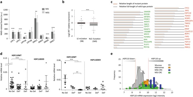
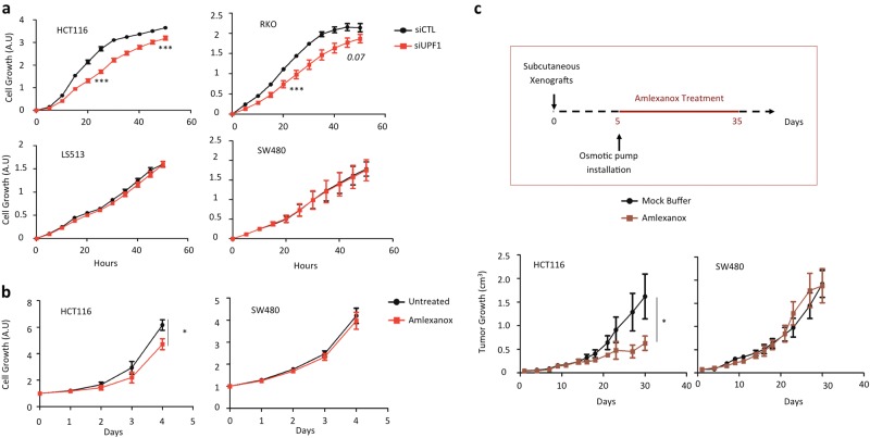
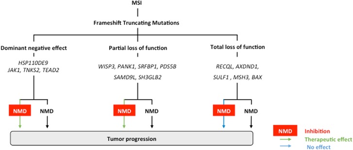
Nonsense-mediated mRNA decay (NMD) is responsible for the degradation of mRNAs with a premature termination codon (PTC). The role of this system in cancer is still quite poorly understood. In the present study, we evaluated the functional consequences of NMD activity in a subgroup of colorectal cancers (CRC) characterized by high levels of mRNAs with a PTC due to widespread instability in microsatellite sequences (MSI). In comparison to microsatellite stable (MSS) CRC, MSI CRC expressed increased levels of two critical activators of the NMD system, UPF1/2 and SMG1/6/7. Suppression of NMD activity led to the re-expression of dozens of PTC mRNAs. Amongst these, several encoded mutant proteins with putative deleterious activity against MSI tumorigenesis (e.g., HSP110DE9 chaperone mutant). Inhibition of NMD in vivo using amlexanox reduced MSI tumor growth, but not that of MSS tumors. These results suggest that inhibition of the oncogenic activity of NMD may be an effective strategy for the personalized treatment of MSI CRC.


Conflict of interest statement

The authors declare that they have no conflict of interest.

Figures

Fig. 1
Fig. 1. The overexpression of NMD factor in MSI primary CRC.
a Microarray analysis of NMD-related factors in MSI/MSS tumor tissues ([MSS primary CRC], n = 48; [MSI primary CRC], n = 40). b Microarray analysis associated with Exome sequencing data of 30 MSI tumors to compare the expression of genes with mutation in microsatellite located in the last exon (LE; n = 98) or before the last exon (NLE; n = 569). c Schematic structure of mutant proteins (15 mutated tumor suppressor genes in green or 16 mutated oncogenes in red) with NLE mutations and significant down-regulation in MSI tumors. d Left panel: relative expression levels of HSP110wt and HSP110DE9 mRNAs by quantitative RT-PCR in MSS and MSI CRC primary tumors. No Del (wild type status; MSS CRCs) n = 36; DelS (small deletion: ≤4 pb; MSI CRCs), n = 28; DelL (large deletion: >4 pb; MSI CRCs), n = 7. Right panel: relative mRNA expression levels of HSP110wt and HSP110DE9 determined by quantitative RT-PCR in MSS and MSI CRC cell lines. No T17 deletion [No Del], n = 6 CRC cell lines (ISI, SW1116, V9P, ALA, FET, SW480); small T17 Deletion [DelS], n = 4 CRC cell lines (HCT8, HCT116, TC71, LIM1215); large T17 Deletion [DelL], n = 6 CRC cell lines (TC7, Lovo, KM12, LS411, Ls174T, Co115). Data are means ± SEM. e Quantification of all HSP110 mRNAs. Densities and bar plots of HSP110 log2 intensities in normal colonic mucosa (Muc), adenomas (Ade), MSS tumors (MSS), and MSI tumors (MSI). All Data are means ± SEM. Unpaired t-test was performed to determine significance. **p < 0.01, ***p < 0.001, ****p < 0.0001
Fig. 2
Fig. 2. Inhibition of NMD system and differential RNA decay.
a Upper panel: schematic representation of the HSP110DE9-specific NMD reporter system used in this work. The NMD.reporter gene consisted of an in-frame HSP110 construction and contains the cDNA sequence from exon 1 to exon 8, intron 9, exon 10, intron 16, and exon 18. As in the case of the T17 mutation of HSP110 located near the splice acceptor site of intron 8, a nonsense mutation appears in exon 10 due to the frameshift mutation caused by skipping of exon 9, making the exogenous mRNA a target of NMD. This construction is placed in an EBV stable vector. Lower panel: relative expression of HSP110DE9-PTC mRNA from NMD reporter stably transfected in the SW480 CRC cell line treated with several NMD inhibitors (siUPF1, cycloheximide (CHX), amlexanox, and NMDI-1). b Relative expression levels of TGFBR2, MSH3, or HSP110DE9 mRNAs determined by quantitative RT-PCR in CRC cell lines. After cycloheximide [CHX] treatment (4 h, 400 μg/ml). RNA expression levels compared to untreated cells [UT] in cell lines were analyzed with the TGFBR2 probe, [No Del] (SW480 and FET), Heterozygote [Htz] (HCT8 and RKO), Homozygote [Hmz] (HCT116 and LS174T); with the MSH3 probe, [No Del] (SW480 and HCT8), Heterozygote [Htz] = (LS174T), Homozygote [Hmz] (HCT116); with HSP110DE9 probes, [No Del] (SW480 and FET); [DelS] (HCT116 and HCT8); [DelL] (RKO and LS174T). Dashed line refers to the ratio calculated between treated and untreated cells with CHX. c Relative mRNA expression levels of MSI target genes (containing coding DNA repeats) as determined by quantitative RT-PCR in HCT116 (MSI) and SW480 (MSS) CRC cell lines transfected with siUPF1 (24 h post-transfection; top panel) or treated for 24 h with 5 µM amlexanox (lower panel). IGF2R is used as an internal control (not mutated in HCT116 and SW480 CRC cell line) for experimental conditions. All data are means ± SEM. Unpaired t-test was performed to determine significance. *p < 0.05, **p < 0.01, ***p < 0.001, ****p < 0.0001
Fig. 3
Fig. 3. Impact of NMD inhibition in cell proliferation and tumor growth.
a Cell proliferation of HCT116 (MSI), RKO (MSI), SW480 (MSS), and LS513 (MSS) colon cancer cells were analyzed with Xcelligence technology (see Materials and methods). Twenty-four hours post-transfection with a control (siCTL) or siRNA against UPF1 (siUPF1), cells were seeded in E-plate 96 to allow measurement of proliferation rates during 48 h. b HCT116 (MSI) and SW480 (MSS) CRC cancer cells were treated once a day during 4 days with or without 5 μM amlexanox. OD was measured every day after drug treatment using the WST-1 assay. c Upper panel: schematic representation of the protocol for treating mice with the NMD inhibitor amlexanox. The osmotic pump contained either a mock buffer made with 50% DMSO and 50% PEG400, or amlexanox diluted in the mock buffer in order to deliver 0.15 mg of amlexanox per day to each mouse during 28 days. Lower panel: comparative analysis of tumor growth (mean tumor volumes) in mice treated with or without amlexanox. Eight mice per group. Experiments were performed with MSI (HCT116) or MSS (SW480) CRC cells (left and right panels, respectively). All data are means ± SEM. Unpaired t-test was performed to determine significance. *p < 0.05, ***p < 0.001
Fig. 4
Fig. 4. Schematic representation of the role of NMD in MSI tumors and the expected therapeutic benefit of NMD blockade in this tumor model.
Numerous MSI-driven mutant PTC-mRNAs processed by NMD are generated due to frameshift-truncating mutations in coding DNA microsatellites in MMR-deficient colon tumors. These mutations usually result in loss of function effects and they inactivate tumor suppressor genes (e.g., BAX, MSH3…). Although the corresponding mutant PTC-containing mRNAs are degraded by NMD or not it is of poor functional significance when the mutant proteins have totally lost their function and therefore do not have residual biological activity. In contrast, it is expected that inhibiting NMD could be deleterious for MSI cancer cells when mutant mRNAs encode for (i) tumor-suppressor mutant proteins that have retained some residual activity (notably, partial loss of function is expected when the truncation occurs down-stream of important functional domains in the protein), or (ii) dominant negative mutant proteins like HSP110DE9

References

    1. Larrea AA, Lujan SA, Kunkel TA. SnapShot: DNA mismatch repair. Cell. 2010;141:730e1. doi: 10.1016/j.cell.2010.05.002. - DOI - PubMed
    1. Duval A, Hamelin R. Mutations at coding repeat sequences in mismatch repair-deficient human cancers: toward a new concept of target genes for instability. Cancer Res. 2002;62:2447–2454. - PubMed
    1. Hause RJ, Pritchard CC, Shendure J, Salipante SJ. Classification and characterization of microsatellite instability across 18 cancer types. Nat. Med. 2016;22:1342–1350. doi: 10.1038/nm.4191. - DOI - PubMed
    1. Liu W, et al. Mutations in AXIN2 cause colorectal cancer with defective mismatch repair by activating beta-catenin/TCF signalling. Nat. Genet. 2000;26:146–147. doi: 10.1038/79859. - DOI - PubMed
    1. Duval A, et al. The human T-cell transcription factor-4 gene: structure, extensive characterization of alternative splicings, and mutational analysis in colorectal cancer cell lines. Cancer Res. 2000;60:3872–3879. - PubMed

LinkOut - more resources