Review
. 2018 Nov;18(11):669-680.
doi: 10.1038/s41568-018-0056-x.

Stem cell fate in cancer growth, progression and therapy resistance

Affiliations
Review

Stem cell fate in cancer growth, progression and therapy resistance

Nikki K Lytle et al. Nat Rev Cancer. 2018 Nov.

Abstract

Although we have come a long way in our understanding of the signals that drive cancer growth, and how these signals can be targeted, effective control of this disease remains a key scientific and medical challenge. The therapy resistance and relapse that are commonly seen are driven in large part by the inherent heterogeneity within cancers that allows drugs to effectively eliminate some, but not all, malignant cells. Here, we focus on the fundamental drivers of this heterogeneity by examining emerging evidence that shows that these traits are often controlled by the disruption of normal cell fate and aberrant adoption of stem cell signals. We discuss how undifferentiated cells are preferentially primed for transformation and often serve as the cell of origin for cancers. We also consider evidence showing that activation of stem cell programmes in cancers can lead to progression, therapy resistance and metastatic growth and that targeting these attributes may enable better control over a difficult disease.


Conflict of interest statement

Competing interests

The authors declare no competing interests.

Figures

Fig. 1 |
Fig. 1 |. Impact of the cell of origin on cancer development.
a | Oncogenic mutation can drive distinct cancer subtypes depending on the epigenetic and transcriptomic profile of the cell of origin. For example, in haematologic malignancies, when BCR–ABL is introduced into stem cells, it results in chronic myeloid leukaemia (CML); however, when this same mutation is introduced into progenitor cells, it results in B cell acute lymphoblastic leukaemia (B-ALL). b | Alternatively, oncogenic mutation in distinct cells of origin can lead to a convergence of cell states that results in the same cancer subtype. For example, in medulloblastoma, deletion of protein patched homologue 1 (PTC1) in either neural stem cells or granule neural precursors leads to the development of aggressive medulloblastoma. P, phosphorylation.
Fig. 2 |
Fig. 2 |. Epigenetic regulation of the stem cell state in cancer.
a | During normal development, stem cell programmes are extinguished during differentiation; in cancers, such as myeloid leukaemia, epigenetic reactivation of stem cell programmes can promote propagation and progression to an aggressive state. The activation of these programmes in a subpopulation (cancer stem cells (CSCs), shown in orange) is associated with chronic myeloid leukaemia (CML), a low-grade disease, while widespread activation of these programmes — illustrated by the expanded pool of CSCs in the figure — is associated with blast crisis CML, an aggressive, high-grade disease. b | Epigenetic regulation of stem cell programmes may also be mediated through modification of DNA. For example, mutation of the DNA methyltransferase DNA (cytosine-5)-methyltransferase 3A (DNMT3A) can promote the stem cell state through either loss of function mutations (which can lead to hypomethylation and activation of genes that promote the stem cell state; shown on the left) or gain of function mutations (which can lead to hypermethylation and silencing of genes associated with differentiation; shown on the right). Me, methylation.
Fig. 3 |
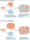
Fig. 3 |. Asymmetric division in cancer.
The disruption of asymmetric division is one way in which cancer may progress to an aggressive state. In low-grade cancers, symmetric renewal and asymmetric divisions are fairly balanced, resulting in both tumour heterogeneity and the maintenance of cancer stem cells (CSCs). However, in high-grade cancers, this balance may be shifted towards increased symmetric renewal, resulting in the expansion of CSCs, which may result in a more aggressive disease state. While imbalances in asymmetric division leading to the progression of cancer have been clearly demonstrated in haematologic malignancies, there is evidence to suggest that disruption of asymmetric division can promote an aggressive state in some solid tumours as well.
Fig. 4 |
Fig. 4 |. Metastasis and cancer stem cells.
The classic epithelial–mesenchymal (EMT) model of metastasis (top) posits that the dissemination of cancer cells requires loss of epithelial cell traits commensurate with gain of mesenchymal cell traits (dark blue), which enables the cells to detach from the primary tumour and invade surrounding tissue, intravasate and survive in circulation, and, finally, extravasate and localize to a distant metastatic site. Several genes (shown in the centre box) have been shown to drive EMT, and their expression serves as a marker of the process. Interestingly, cancer stem cells (CSCs) (bottom) are also enriched in disseminated tumour cells and express the EMT gene signature. Further, the capacity for tumour propagation, which is required for establishment of a tumour at a distant site, is a salient feature of CSCs. The parallels between EMT cells and CSCs raise the possibility that they represent overlapping concepts.
Fig. 5 |
Fig. 5 |. Therapy resistance in cancer stem cells.
a | Cytotoxic agents such as radiation and chemotherapy are commonly used to treat cancer, efficiently targeting bulk cancer cells (blue cells) but not cancer stem cells (CSCs) (orange cells). The residual disease can be enriched in CSC populations that can drive a more aggressive disease, triggering recurrence. b | Stem cell properties are commonly hijacked in cancer. One such property is increased drug efflux. Chemotherapeutic agents target bulk cancer cells with normal levels of drug efflux, resulting in cell death (top). In CSCs, higher expression of ATP-binding cassette (ABC) transporters can increase drug efflux capacity, increasing cell survival (bottom). c | Enhanced DNA repair can also be hijacked in cancer. In glioblastoma, radiation generates unrepaired double strand breaks in CD133 bulk cancer cells, leading to cell death (top). In CD133+ CSCs (bottom), the DNA damage checkpoint is activated, allowing for repair that leads to increased cell survival. d | CSCs utilize the tumour microenvironment for increased survival. In brain tumours, the endothelial cells of the perivascular niche promote the survival of CSCs. Endothelial cell signalling supports the stem cell properties of the cancer, which allows CSC expansion. CSCs can promote angiogenesis by secreting factors such as vascular endothelial growth factor (VEGF) and stromal cell-derived factor 1 (SDF1). e | Hypoxic environments can support CSCs. Although hypoxia (represented by the descending oxygen gradient shown in blue) induces some cell death within the tumour, it also promotes CSC expansion and triggers expression of genes that promote therapy resistance.
None

References

    1. Hunger SP, Winick NJ, Sather HN & Carroll WL Therapy of low-risk subsets of childhood acute lymphoblastic leukemia: when do we say enough? Pediatr. Blood Cancer 45, 876–880 (2005). - PubMed
    1. Hodgson DC, Hudson MM & Constine LS Pediatric hodgkin lymphoma: maximizing efficacy and minimizing toxicity. Semin. Radiat. Oncol 17, 230–242 (2007). - PubMed
    1. Barker N et al.Crypt stem cells as the cells-of-origin of intestinal cancer. Nature 457, 608–611 (2009). - PubMed
    1. Gibson P et al.Subtypes of medulloblastoma have distinct developmental origins. Nature 468, 1095–1099 (2010). - PMC - PubMed
    1. Lapouge G et al.Identifying the cellular origin of squamous skin tumors. Proc. Natl Acad. Sci. USA 108, 7431–7436 (2011). - PMC - PubMed

Publication types