. 2018 Sep 18;8(1):13958.
doi: 10.1038/s41598-018-32080-3.

Sharing of human milk oligosaccharides degradants within bifidobacterial communities in faecal cultures supplemented with Bifidobacterium bifidum

Affiliations

Sharing of human milk oligosaccharides degradants within bifidobacterial communities in faecal cultures supplemented with Bifidobacterium bifidum

Aina Gotoh et al. Sci Rep. .

Abstract

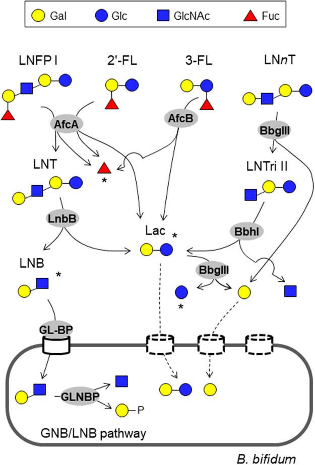
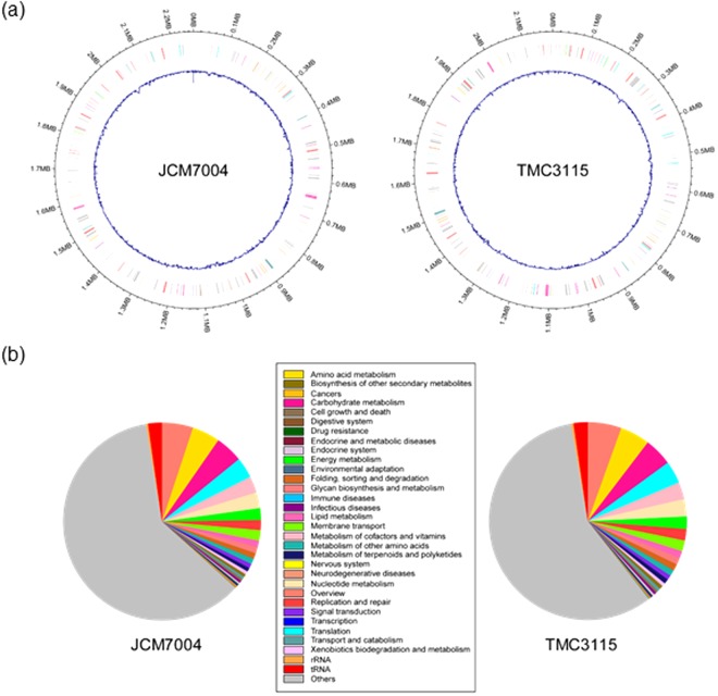
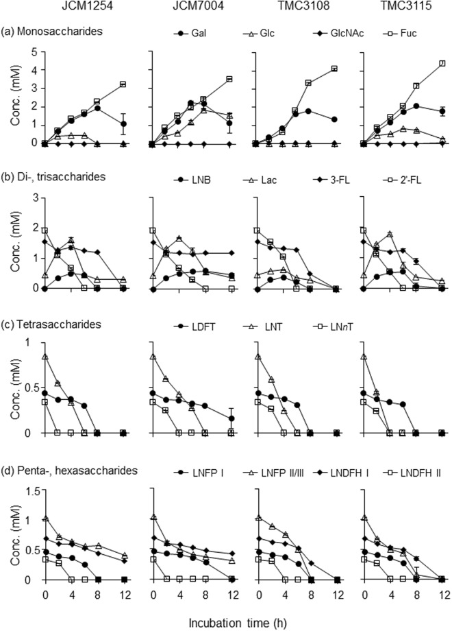
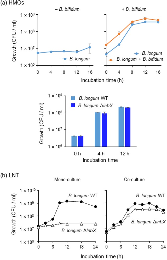
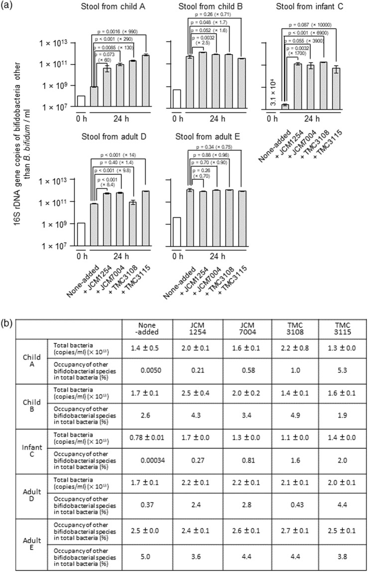
Gut microbiota of breast-fed infants are generally rich in bifidobacteria. Recent studies show that infant gut-associated bifidobacteria can assimilate human milk oligosaccharides (HMOs) specifically among the gut microbes. Nonetheless, little is known about how bifidobacterial-rich communities are shaped in the gut. Interestingly, HMOs assimilation ability is not related to the dominance of each species. Bifidobacterium longum susbp. longum and Bifidobacterium breve are commonly found as the dominant species in infant stools; however, they show limited HMOs assimilation ability in vitro. In contrast, avid in vitro HMOs consumers, Bifidobacterium bifidum and Bifidobacterium longum subsp. infantis, are less abundant in infant stools. In this study, we observed altruistic behaviour by B. bifidum when incubated in HMOs-containing faecal cultures. Four B. bifidum strains, all of which contained complete sets of HMO-degrading genes, commonly left HMOs degradants unconsumed during in vitro growth. These strains stimulated the growth of other Bifidobacterium species when added to faecal cultures supplemented with HMOs, thereby increasing the prevalence of bifidobacteria in faecal communities. Enhanced HMOs consumption by B. bifidum-supplemented cultures was also observed. We also determined the complete genome sequences of B. bifidum strains JCM7004 and TMC3115. Our results suggest B. bifidum-mediated cross-feeding of HMOs degradants within bifidobacterial communities.


Conflict of interest statement

The authors declare no competing interests.

Figures

Figure 1
Figure 1
Schematic representation of the minimum enzymatic set required to degrade different sugar linkages found in neutral HMOs by B. bifidum (See also Table 1). Sugars are depicted according to the nomenclature committee of the Consortium for Function Glycomics. LNFP I (Fucα1-2Galβ1-3GlcNAcβ1-3Galβ1-4Glc), 2′-FL (Fucα1-2Galβ1-4Glc), 3-FL (Galβ1-4(Fucα1-3)Glc), and LNnT (Galβ1-4GlcNAcβ1-3Galβ1-4Glc) are shown as representatives of the different sugar linkages. AfcA: 1,2-α-l-fucosidase; AfcB: 1,3-1,4-α-l-fucosidase; LnbB: lacto-N-biosiadse; BbgIII: β-1,4-galactosidase; BbhI: β-N-acetylglucosaminidase; GL-BP: galacto-N-biose/lacto-N-biose I-binding protein of ABC transporter; GLNBP: galacto-N-biose/lacto-N-biose I phosphorylase. Dashed lines indicate pathways that have not been identified in B. bifidum. Degradants that accumulate in the spent media during culture in the presence of HMOs are indicated by asterisks (See Fig. 3 and Table S3).
Figure 2
Figure 2
Schematic representation of the genomic structures (a) and gene functions (KEGG Orthology) (b) of B. bifidum strains JCM7004 and TMC3115. The genome structures were depicted using the R package “circlize”. Blue lines represent GC content. See also Tables S1 and S2.
Figure 3
Figure 3
In vitro HMO degradation behaviour of four B. bifidum strains. Each strain was cultured in HMOs-containing basal media in triplicate, and samples were collected at the indicated time points (see Fig. S2). The sugars in the culture supernatants were labelled with 2-AA and analysed by HPLC, as described in the Methods section. Note that LNB was not accurately quantified due to its heat lability. Concentrations of (a) monosaccharides (Fuc, Gal, Glc, and GlcNAc), (b) di- and trisaccharides (2′-FL, 3-FL, Lac, and LNB), (c) tetrasaccharides (LNT, LNnT, and LDFT), and (d) penta- and hexasaccharides (LNFP I, LNFP II/III, LNDFH I, and LNDFH II) are shown. The data are means ± SD of the labelled sugars obtained from three separate cultures.
Figure 4
Figure 4
Inter-species cross-feeding of HMO degradants among bifidobacteria (a). WT strain of B. longum 105-A carrying pBFS38 (CamR) was grown in basal medium supplemented with 1% HMOs in the absence (upper left panel) and presence (upper right panel) of B. bifidum JCM1254. Samples were taken at the indicated time points, and CFUs were determined by spreading the diluted cultures on Cam-containing (for B. longum cells) and not containing (for B. longum + B. bifidum cells) agar plates. For competition assay in the presence of B. bifidum JCM1254, WT and its isogenic ΔlnbX strains of B. longum 105-A were transformed with pBFS38 (CamR) and pBFO2 (SpR), respectively, to distinguish between them (lower panel). (b) Intra-species cross-feeding of LNT degradants. WT strain of B. longum 105-A carrying pBFS38 (CamR) and its ΔlnbX variant carrying pBFO2 (SpR) were cultured in basal medium containing 1% LNT as a carbon source, separately (mono-culture) or in combination (co-culture). Growth was monitored by calculation of CFUs. Identical results were obtained when the marker genes were exchanged between the strains. The data are means ± SD of three independents experiments (a,b).
Figure 5
Figure 5
Addition of B. bifidum to faecal suspensions incubated in the presence of HMOs enriches the Bifidobacterium population (species other than B. bifidum) in the culture. (a) Stool suspensions from two children (4-year-old female and 5-year-old male), one infant (caesarean delivered 4-month-old preweaning female), and two adults (30-year-old male and 39-year-old male) were cultured in basal medium containing 1% HMOs with and without the addition of four B. bifidum strains for 24 h. The abundance of 16S rRNA gene sequences corresponding to Bifidobacterium species other than B. bifidum were determined by qPCR at 0 h (white bars) and 24 h (grey bars) post-inoculation, as described in the Methods section. The data are means ± SD of three independents assays. Dunnett’s test was used to examine the statistical significance. (b) Prevalence of Bifidobacterium species other than B. bifidum in the culture. The total bacterial population was determined as described in the Methods section. Prevalence was calculated by dividing bifidobacterial 16S rRNA gene counts (except for B. bifidum) by total bacterial 16S rRNA gene counts. See also Figs S5–S8 and Tables S4 and S5.

Similar articles

Cited by

References

    1. Kau AL, Ahern PP, Griffin NW, Goodman AL, Gordon JI. Human nutrition, the gut microbiome and the immune system. Nature. 2011;474:327–336. doi: 10.1038/nature10213. - DOI - PMC - PubMed
    1. Yatsunenko T, et al. Human gut microbiome viewed across age and geography. Nature. 2012;486:222–227. doi: 10.1038/nature11053. - DOI - PMC - PubMed
    1. Olszak T, et al. Microbial exposure during early life has persistent effects on natural killer T cell function. MBio. 2012;336:489–493. - PMC - PubMed
    1. Cox LMM, et al. Altering the intestinal microbiota during a critical developmental window has lasting metabolic consequences. Cell. 2014;158:705–721. doi: 10.1016/j.cell.2014.05.052. - DOI - PMC - PubMed
    1. Tannock GW, et al. Comparison of the compositions of the stool microbiotas of infants fed goat milk formula, cow milk-based formula, or breast milk. Appl. Environ. Microbiol. 2013;79:3040–3048. doi: 10.1128/AEM.03910-12. - DOI - PMC - PubMed

Publication types

MeSH terms