Comparative Study
. 2018 Sep 18;8(1):14010.
doi: 10.1038/s41598-018-32361-x.

Comparison of initial oral microbiomes of young adults with and without cavitated dentin caries lesions using an in situ biofilm model

Affiliations
Comparative Study

Comparison of initial oral microbiomes of young adults with and without cavitated dentin caries lesions using an in situ biofilm model

Stefan Rupf et al. Sci Rep. .

Abstract

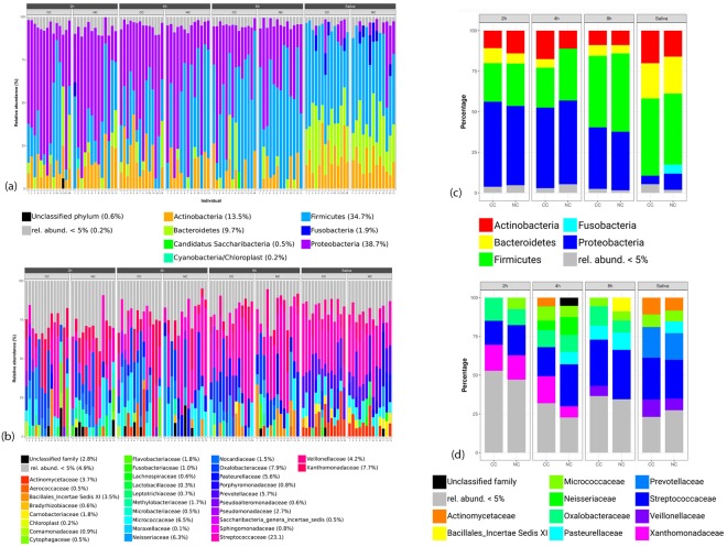
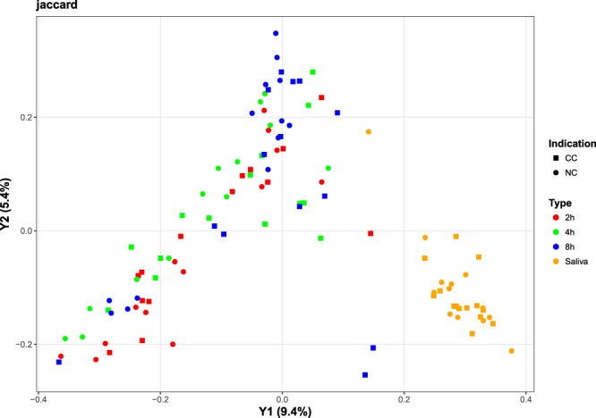
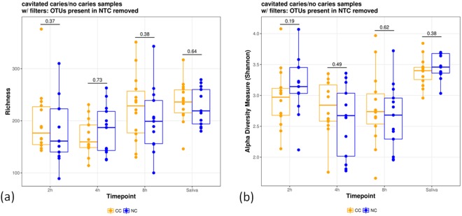
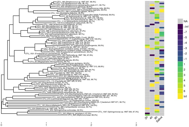
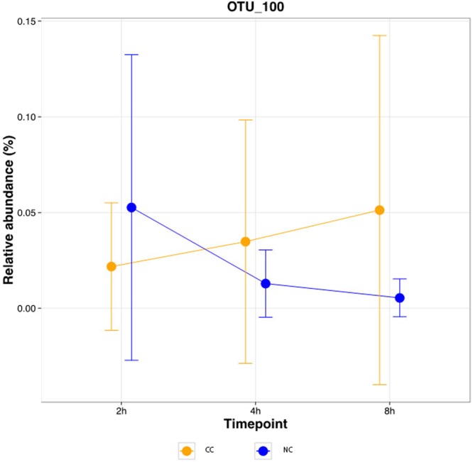
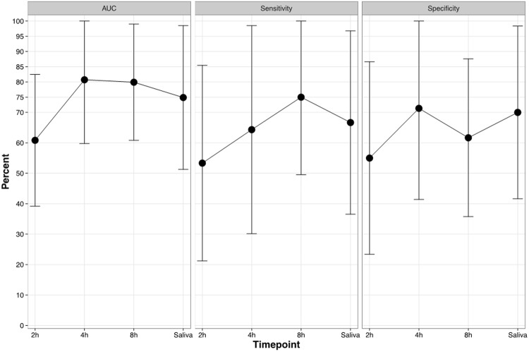
Dental caries is caused by acids released from bacterial biofilms. However, the in vivo formation of initial biofilms in relation to caries remains largely unexplored. The aim of this study was to compare the oral microbiome during the initial phase of bacterial colonization for individuals with (CC) and without (NC) cavitated dentin caries lesions. Bovine enamel slabs on acrylic splints were worn by the volunteers (CC: 14, NC: 13) for in situ biofilm formation (2 h, 4 h, 8 h, 1 ml saliva as reference). Sequencing of the V1/V2 regions of the 16S rRNA gene was performed (MiSeq). The relative abundances of individual operational taxonomic units (OTUs) were compared between samples from the CC group and the NC group. Random forests models were furthermore trained to separate the groups. While the overall heterogeneity did not differ substantially between CC and NC individuals, several individual OTUs were found to have significantly different relative abundances. For the 8 h samples, most of the significant OTUs showed higher relative abundances in the CC group, while the majority of significant OTUs in the saliva samples were more abundant in the NC group. Furthermore, using OTU signatures enabled a separation between both groups, with area-under-the-curve (AUC) values of ~0.8. In summary, the results suggest that initial oral biofilms provide the potential to differentiate between CC and NC individuals.


Conflict of interest statement

The authors declare no competing interests.

Figures

Figure 1
Figure 1
Relative abundances of individual taxa per sample at phylum (a) and family (b) level, and summarized per indication at phylum (c) and family (d) level. The abundances were computed over all OTUs before filtering out OTUs present in controls. Samples were grouped by indication (CC: cavitated caries, NC: no cavitated caries) and time point (2 h-, 4 h- and 8 h-biofilms, and saliva) in (a) and (b); in (c) and (d), the indication is shown per time point. Taxa with a relative abundance below 5% per sample (a,b) and indication (c,d) were grouped into one category (“rel. abund. <5%”). In panels (a) and (b), the relative taxon abundance across all non-control samples and OTUs is shown in brackets next to the taxon name in the legend. It was computed as the sum of OTU counts corresponding to the respective taxon divided by the total OTU count across all non-control samples.
Figure 2
Figure 2
PCoA-based ordination of the Jaccard distance matrix. Colors reflect the sample type (2 h, 4 h, 8 h, and saliva) and shapes reflect the CC and NC groups. The percentage of variation explained is included in the axis labels.
Figure 3
Figure 3
Richness (a) and alpha diversity (b) measures of different time points (2 h-, 4 h-, and 8 h-biofilms) and for saliva and indications (CC: cavitated caries, NC: no cavitated caries). OTUs present in the NTC were removed and richness and Shannon’s alpha diversity measures are shown. The individual points represent individual diversity estimates.
Figure 4
Figure 4
Phylogenetic OTU tree created by LotuS with taxonomic assignment from the eHOMD database. Only OTUs with significantly different relative abundance are depicted with a heat map showing the log2(fold change) within the respective groups used for comparison. The values were highlighted as missing (“NA”), or colored w.r.t. their rounded value. A positive log fold-change indicates a higher relative abundance in the cavitated-caries group, a negative log fold-change stands for higher abundances in the no cavitated-caries group. Match identities according to eHOMD are given following the taxonomic name.
Figure 5
Figure 5
Relative OTU abundance over time per indication for an exemplary OTU. The mean +/− standard deviation of OTU 100 (Moryella sp. according to LotuS, Stomatobaculum longum according to eHOMD); absolute difference of the correlation of the mean values = 2 are shown.
Figure 6
Figure 6
Classification performances. The cross-validated (5-fold, 10 repetitions) means +/− standard deviations (capped at 100%) of AUC, sensitivity, and specificity are shown per time points for the classification of samples as “cavitated caries”.

Similar articles

Cited by

References

    1. Global Burden of Disease Study 2013 Collaborators. Global, regional, and national incidence, prevalence, and years lived with disability for 301 acute and chronic diseases and injuries in 188 countries, 1990–2013: a systematic analysis for the Global Burden of Disease Study 2013. Lancet. 386, 743–800 (2015). - PMC - PubMed
    1. Kassebaum NJ, et al. Global burden of untreated caries: a systematic review and metaregression. J. Dent. Res. 2015;94:650–658. doi: 10.1177/0022034515573272. - DOI - PubMed
    1. Marthaler TM. Changes in dental caries 1953–2003. Caries Res. 2004;38:173–181. doi: 10.1159/000077752. - DOI - PubMed
    1. Mejàre I, Stenlund H, Zelezny-Holmlund C. Caries incidence and lesion progression from adolescence to young adulthood: a prospective 15-year cohort study in Sweden. Caries Res. 2004;38:130–141. doi: 10.1159/000075937. - DOI - PubMed
    1. Hugoson A, Koch G, Helkimo AN, Lundin SA. Caries prevalence and distribution in individuals aged 3–20 years in Jönköping, Sweden, over a 30-year period (1973–2003) Int. J. Paediatr. Dent. 2008;18:18–26. - PubMed

Publication types