

Case Reports
Digital Tomosynthesis Applications in Pediatric Orthopedic Imaging: A Case Series
Affiliations
- PMID: 30228765
- PMCID: PMC6140262
Item in Clipboard
Case Reports
Digital Tomosynthesis Applications in Pediatric Orthopedic Imaging: A Case Series
Mo Med.
2018 Jul-Aug.
Abstract
Digital tomosynthesis (DTS) is an emerging technology that provides cross-sectional, three-dimensional imaging similar to computed tomography (CT) at a fraction of the radiation dose and cost. In this article, we describe multiple cases where our pediatric orthopedic surgeons have used DTS imaging to help in clinical management of fracture healing.
Figures
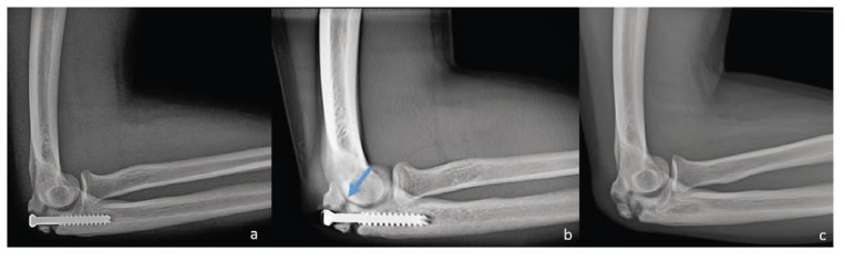
15-year-old male with history of right olecranon stress fracture was treated with screw fixation. His course was complicated by a post-operative infection. (a) Lateral radiograph of the elbow demonstrates a screw across an olecranon fracture. (b) A selected slice from a digital tomosynthesis exam demonstrates a bony bridge between the proximal fracture fragments (arrow). (c) Lateral radiograph of the elbow two months after the initial radiographs demonstrates healing bony bridging of the olecranon fracture after screw removal.
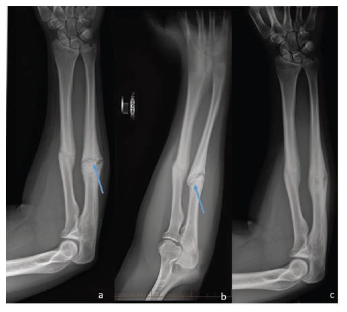
15-year-old male who presents for a 6-month follow-up of his left forearm fractures. (a) Frontal radiograph of the forearm demonstrates a healing radial diaphysis fracture (white arrow) and a persistent lucent fracture line spanning the width of an ulnar diaphysis fracture (blue arrow). (b) A selected slice from a digital tomosynthesis exam demonstrates a bony bridge between the two fragments of the ulnar fracture (blue arrow). (c) A follow up frontal radiograph of the forearm demonstrates both the radial and ulnar fractures are healing with bony bridging across both fractures with no persistent fracture lines.
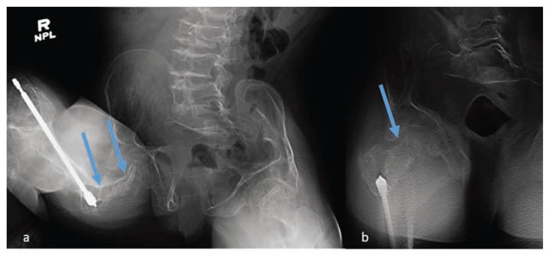
13-year-old female with a history of osteogenesis imperfecta presents with right hip pain after a wheelchair injury. (a) Frontal radiograph of the pelvis is suspicious for femoral neck fracture because of contour deformity in the femoral neck (arrows). there is also diff use bony demineralization making interpretation more challenging. (b) A selected slice from a digital tomosynthesis exam demonstrates that the femoral neck cortex in question is intact (arrow).
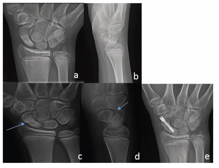
13-year-old male presents with right wrist pain three months after a rollover motor vehicle accident. Frontal (a) and lateral (b) radiographs of the right wrist demonstrates a nondisplaced scaphoid waist fracture with some sclerosis around the fracture line. No discrete fracture line lucency is imaged. selected slices from a frontal (c) and lateral (d) digital tomosynthesis exam of the wrist demonstrate a 3 mm gap between the two fragments of the scaphoid fracture indicating non-union. (e) A follow up frontal radiograph of the wrist demonstrates a screw across the scaphoid fracture with no avascular necrosis or remaining lucent fracture line.

References
-
- Applegate KE, Cost NG. Image Gently: a campaign to reduce children’s and adolescents’ risk for cancer during adulthood. J Adolesc Health. 2013 May;52(5 Suppl):S93–97. - PubMed
-
- Cohen MD. ALARA, image gently and CT-induced cancer. Pediatr Radiol. 2015 Apr;45(4):465–470. - PubMed
-
- Frush DP, Goske MJ. Image Gently: toward optimizing the practice of pediatric CT through resources and dialogue. Pediatr Radiol. 2015 Apr;45(4):471–475. - PubMed
Publication types
MeSH terms
LinkOut - more resources
Full Text Sources
Medical