

Advances in Pediatric Cardiovascular Imaging
Affiliations
- PMID: 30228767
- PMCID: PMC6140247
Item in Clipboard
Advances in Pediatric Cardiovascular Imaging
Mo Med.
2018 Jul-Aug.
Abstract
Cardiac imaging plays a key role in the accurate diagnosis of pediatric congenital heart disease (CHD). Echocardiography and catheter angiography are traditionally used to delineate cardiac anatomy. CT and MRI imaging offer a non-invasive way to image cardiovascular anatomy which can be used in conjunction with echocardiography for the diagnosis and treatment planning for CHD. These modalities can depict the morphology and relationship to surrounding structures better than echocardiography, especially in complex congenital defects.
Figures
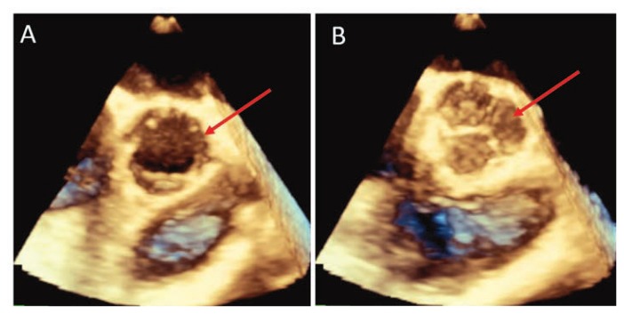
Three dimensional echocardiogram images show excellent resolution of an open (A) and closed (B) aortic valve.
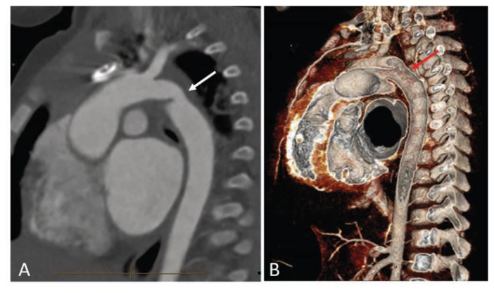
(A) Sagittal oblique CT images of a 15 month old for arch evaluation. Mild hypoplasia of the distal transverse arch and narrowing of the juxtaductal arch consistent with coarctation. Patient also had a patent ductus arteriosus. (B) 3 dimensional images show area of arch narrowing and relation to the patient’s PDA.
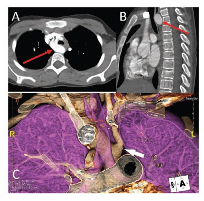
Axial (A) and sagittal (B) CT angiography demonstrates a right sided aortic arch with posterior compression on the trachea. (C) 3D reformatted image shows the relationship of the trachea and right sided aortic arch.
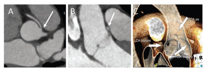
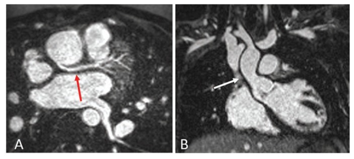
(A) Axial image from a gated cardiac CT shows an anomalous right coronary artery arising off the left aorta and coursing intra-arterial between the aorta and main pulmonary artery. (B and C) Multiplanar reconstructed images show the takeoff of the RCA is superior to the commissure between the right and left coronary cusps. The RCA origin is ovoid and has an acute angled take off
Examples of Cardiac MRI with use of Ferumoxytol as a contrast agent. (A) Shows a slightly posterior [benign variant] origin of the left coronary origin in detail on contrast MRI. (B) Excellent contrast enhancement of the ascending aorta which can be used for evaluation of vessel size and anatomy.
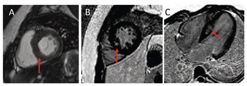
8-year-old with hypertrophic cardiomyopathy. MRI images in short axis (A) shows asymmetric thickening of the septal wall with patchy mid-septal delayed post contrast enhancement (B and C) consistent with fibrosis [red arrows].


References
-
- Hoffman J, Kaplan S. The Incidence of Congenital Heart Disease. J Am Coll Cardiol. 2002 Jun 19;39(12):1890–900. - PubMed
-
- Marelli AJ, Mackie AS, Ionescu-Ittu R, Rahme E, Pilote L. Congenital heart disease in the general population: changing prevalence and age distribution. Circulation. 2007 Jan 16;115(2):163–72. - PubMed
-
- Khairy P, Ionescu-Ittu R, Mackie AS, Abrahamowicz M, Pilote L, Marelli AJ. Changing mortality in congenital heart disease. J Am Coll Cardiol. 2010 Sep 28;56(14):1149–57. - PubMed
-
- Nelle M, Raio L, Pavlovic M, Carrel T, Surbek D, Meyer-Wittkopf M. Prenatal diagnosis and treatment planning of congenital heart defects-possibilities and limits. World J Pediatr. 2009 Feb;5(1):18–22. - PubMed
-
- Dillman JR, Hernandez RJ. Role of CT in the Evaluation of Congenital Cardiovascular Disease in Children. AJR. 2009;192:1219–1231. - PubMed
MeSH terms
LinkOut - more resources
Full Text Sources
Medical