

Review
Emerging Technology and Applications of 3D Printing in the Medical Field
Affiliations
- PMID: 30228770
- PMCID: PMC6140256
Item in Clipboard
Review
Emerging Technology and Applications of 3D Printing in the Medical Field
Mo Med.
2018 Jul-Aug.
Abstract
3D printing technology evolved in the 1980s, but has made great strides in the last decade from both a cost and accessibility standpoint. While most printers are employed for commercial uses, medical 3D printing is a growing application which serves to aid physicians in the diagnosis, therapeutic planning, and potentially the treatment of patients with complex diseases. In this article we will delineate the types of printers available to the consumer, the various materials which can be utilized, and potential applications of 3D models in the healthcare field.
Figures
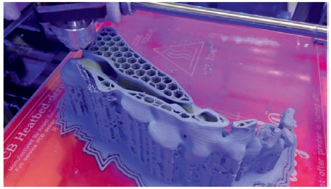
A model of a sacrum being printed on an FDM machine. The model is printed in thin layers (0.1–0.4 mm) as the printer bed slowly descends. The hexagons inside the model serve as support material to provide internal stability. Exterior support material can be removed when printing is complete.
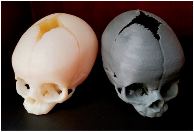
Two models of an infant skull. The model on the left was printed on a polyjet machine. The righthand model was printed on an FDM machine. Print lines are visible in the FDM print and the model is less sturdy. Additionally, the polyjet model has a higher degree of fine detail.
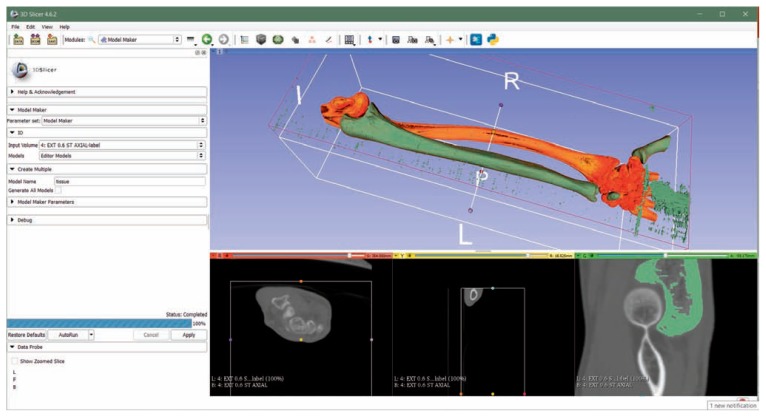
The ulna is segmented from a CT of the forearm in the open source software program 3D Slicer (www.slicer.org ).
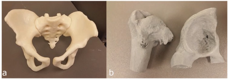
a) Full pelvic model printed on an FDM machine. Model was utilized by an orthopedic surgeon to mold a fixation plate preoperatively. b) Proximal femur and acetabulum of a patient with Perthes. Size of osseous spurs was better appreciated on the 3D model and changed the surgical approach.
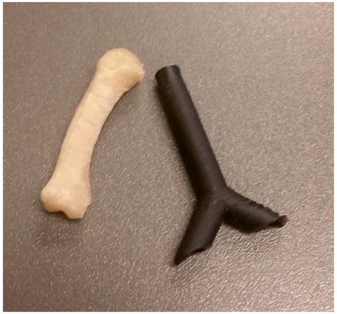
Two models printed for research purposes. The model on the left is a metacarpal used to evaluate different pinning techniques for fracture fixation. On the right is a pediatric trachea phantom used to study foreign body obstruction with tomography.
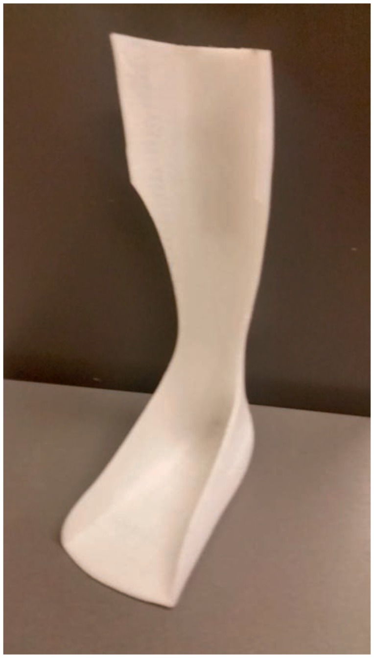
A custom ankle foot orthotic printed in nylon/thermoplastic elastomer. Orthotic was tailored to the patient’s foot and lower leg length, width, and circumference. The use of nylon/TPE provides stability and durability while retaining some degree of flexibility.

References
-
- Wenz SM, Zeichner SJ, Berg BI, Zeilhofer HF, Thieringer F. 3D Printed Surgical Simulation Models as educational tool by maxillofacial surgeons. s.l.: European Journal of Dental Education; 2018. [Epub ahead of print] - PubMed
-
- Yang T, Lin S, Tan T, Yanh J, Pan J, Hu C, Li J, Zou Y. Impact of 3D printing technology on comprehension of surgical anatomy of retroperitoneal tumor. s.l.: World Journal of Surgery; 2018. [Epub ahead of print] - PubMed
Publication types
MeSH terms
LinkOut - more resources
Full Text Sources
Medical