

Electromagnetic simulation for diagnosing damage to femoral neck vasculature: A feasibility study
- PMID: 30228775
- PMCID: PMC6140380
- DOI: 10.1016/j.jor.2018.08.036
Electromagnetic simulation for diagnosing damage to femoral neck vasculature: A feasibility study
Erratum in
-
Erratum regarding missing Declaration of Competing Interest statements in previously published articles.J Orthop. 2020 Dec 14;23:274. doi: 10.1016/j.jor.2020.12.002. eCollection 2021 Jan-Feb. J Orthop. 2020. PMID: 33746418 Free PMC article.
Abstract
Background: Femoral neck fractures are common injuries managed by orthopedic surgeons across the world. From pediatrics to geriatrics, disruption of the blood supply to the femoral neck is a well-recognized source of morbidity and mortality, oftentimes resulting in avascular necrosis of the femoral head. This devastating complication occurs in 10-45% of femoral neck fractures. Therefore, it is vital for orthopedic surgeons provide efficient treatment of this injury, in order to optimize the patient's potential outcome and prevent long-term sequelae.
Methods: In this study, the anatomy of the proximal femur, including femoral metaphysis, femoral neck, vasculature, and femoral head, were simulated in COMSOL Finite Element Analysis (FEA) software. Electric fields were generated in a fashion that exploited disruptions within the vasculature of the femoral neck. This study was aimed at developing an alternative imaging modality for narrowing or disrupting the femoral neck's vasculature. The variables used for investigation included: frequency, penetration depth, and magnitude of the electrical energy. These variables, when combined, allowed for enhanced simulated visualization of the vasculature of the femoral neck and theoretically expedited diagnosis of obvious, or occult, femoral neck injury.
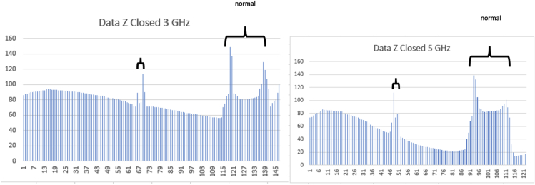
Results: Simulated blood vessels were developed in two-dimensions: the phi direction (circular), and z-direction. Two different frequencies, 3 GHz, and 5 GHz were considered, with 100-J energy pulses within blood vessels of 2.54 mm in diameter. The fat surrounding the bone to the outside surface body was simulated at 0.25 inch (0.65 cm). An additional model, with layered fat and skin above the vessels, was simulated at 2000J and successfully able to visualize the femoral neck's blood vessels. Results showed a distinguished E field across the blood boundary of nearly 170 V/M.
Conclusions: The electric field simulation data within the Phi and Z directions promises the feasibility of a subsequent practical model.
Figures


References
-
- Dedrick Dale K. Complications of Femoral Neck Fracture sin Young Adults. J Trauma. 1986 Oct;26(10):932–937. - PubMed
-
- Frandsen P.A. Garden's classification of femoral neck fractures. An assessment of inter-observer variation. J Bone Joint Surg Br. 1988 Aug;70(4):588–590. - PubMed
-
- Pauwels F. vol. 23. F. Enke. BJS; Stuttgart: 1936 April. p. 157. (Der Schenkelhalsbruch, ein mechanisches problem). 92.
-
- Tornetta 3rd Paul. Diagnosis of femoral neck fractures in patients with a femoral shaft fracture. JBJS. 2007 January;89(1):39–43. - PubMed
LinkOut - more resources
Full Text Sources
Other Literature Sources
