. 2018 Aug 28:2018:8460490.
doi: 10.1155/2018/8460490. eCollection 2018.

MeHg Causes Ultrastructural Changes in Mitochondria and Autophagy in the Spinal Cord Cells of Chicken Embryo

Affiliations

MeHg Causes Ultrastructural Changes in Mitochondria and Autophagy in the Spinal Cord Cells of Chicken Embryo

Fabiana F Ferreira et al. J Toxicol. .

Abstract

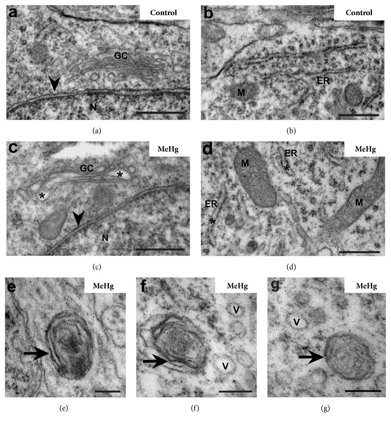
Methylmercury (MeHg) is a known neurodevelopmental toxicant, which causes changes in various structures of the central nervous system (CNS). However, ultrastructural studies of its effects on the developing CNS are still scarce. Here, we investigated the effect of MeHg on the ultrastructure of the cells in spinal cord layers. Chicken embryos at E3 were treated in ovo with 0.1 μg MeHg/50 μL saline solution and analyzed at E10. Then, we used transmission electron microscopy (TEM) to identify possible damage caused by MeHg to the structures and organelles of the spinal cord cells. After MeHg treatment, we observed, in the spinal cord mantle layer, a significant number of altered mitochondria with external membrane disruptions, crest disorganization, swelling in the mitochondrial matrix, and vacuole formation between the internal and external mitochondrial membranes. We also observed dilations in the Golgi complex and endoplasmic reticulum cisterns and the appearance of myelin-like cytoplasmic inclusions. We observed no difference in the total mitochondria number between the control and MeHg-treated groups. However, the MeHg-treated embryos showed an increased number of altered mitochondria and a decreased number of mitochondrial fusion profiles. Additionally, unusual mitochondrial shapes were found in MeHg-treated embryos as well as autophagic vacuoles similar to mitophagic profiles. In addition, we observed autophagic vacuoles with amorphous, homogeneous, and electron-dense contents, similar to the autophagy. Our results showed, for the first time, the neurotoxic effect of MeHg on the ultrastructure of the developing spinal cord. Using TEM we demonstrate that changes in the endomembrane system, mitochondrial damage, disturbance in mitochondrial dynamics, and increase in mitophagy were caused by MeHg exposure.


Figures

Figure 1
Figure 1
Effect of MeHg on the endomembrane system of the spinal cord embryos. Electron micrograph of the E10 spinal cord mantle layer showing Golgi complex (GC) and endoplasmic reticulum (ER) in control and MeHg-treated embryos. Slightly dilated cisterns () in the GC and ER and myelin-like membranous inclusions (arrows) were observed. Note the vesicles (V) close to the myelin-like inclusions. Nuclear envelope (arrowhead), nucleus (N), and mitochondria (M). Scale bars: (a–d) 0.5 μm; (e–g) 200 nm.
Figure 2
Figure 2
Effects of MeHg on mitochondrial structure in spinal cord of E10 embryos. Electron micrograph showing mitochondria in the mantle layer of control and MeHg-treated embryos. Mitochondria (M) in control embryos showed mitochondrial crests (C), internal mitochondrial membrane (IMM, white arrow), and external mitochondrial membrane (EMM, black arrow) visualized in longitudinal (a, c) and transverse planes (a, d). Mitochondria of the MeHg-treated embryos showed ruptures in the EMM (black arrowhead), loss and disorganization of crests, swelling () in the mitochondrial matrix, and vacuolization (∗∗) between IMM and EMM. Unusual mitochondrial shapes, cup-like (i) and donut-like (j-k), were observed in MeHg-treated embryos. Fusion (white arrowhead) in mitochondrial donut-like shape. The graphs show the total number of mitochondria (l) and the number of altered mitochondria (m) in control and MeHg-treated groups. indicates P < 0.05. Nucleus (N), endoplasmic reticulum (ER), and Golgi complex (GC). Scale bars: (a-b) 1 μm; (c–k) 200 nm.
Figure 3
Figure 3
Effect of MeHg on mitochondrial fusion and fission profiles in spinal cord cells of E10 embryos. Electron micrograph of the mantle layer in control and MeHg-treated embryos. The graphs show the number of the fusion (g) and fission (h) mitochondrial profiles in both analyzed groups. Mitochondrial fusion (black arrow) and fission (arrowhead). Indicates P < 0.05. Scale bars: (a–f) 200 ηm.
Figure 4
Figure 4
Effect of MeHg on vacuole formation in spinal cord cells of E10 embryos. Electron micrograph of the mantle layer showing the mitophagic profile in the black box (a). The insert in (b) shows a detail of mitochondria (M) surrounded by the autophagic vacuole membrane (white arrowhead). In detail (b) internal mitochondrial membranes (asterisk) and external mitochondrial membranes (double asterisk) are visible. Endoplasmic reticulum (ER) can be seen close to the autophagic vacuole (A) maintaining contact with it by membrane extensions (black arrowhead). Scale bars: (a) 500 μm; (b) 200 ηm.
Figure 5
Figure 5
Autophagic vacuoles in spinal cord of MeHg-treated embryos. Cells in mantle layer show autophagic vacuoles in different stages of autolysis (a–h). Plasma membranes delimit the cells (black arrow) and inner membranes delimit compartments similar to autophagic vacuoles (white arrowhead). Mitochondria (M) with IMM () and EMM (∗∗) still preserved, inside early autophagic vacuole (a-b, d). Later autophagic vacuoles with amorphous (am), homogeneous, and more electron-dense content (a, c, g, h). (a) Spinal cord cells in lower magnification. (b) Magnification of the cell inside of the white box in (a). (c) Magnification of the cell inside the black box in (a). (d) Magnification of the autophagic vacuoles of the black dotted box in (b). (e) Spinal cord mantle layer cells. In the white dotted box, there is a cell in autophagy. (f) Magnification of the cell in the death process inside the white dotted box in (e). (g) Cell with amorphous and more electron dense cytoplasm in white dashed box. (h) Magnification of the highlighted cell in (g). Axon (ax) of neuronal cell in transverse section, chromatin (cr), nucleus (N), and nucleolus (nl). Scale bars: (a, e) 2 μm; (b) 1.0 μm; (c) 0.5 μm; (d,f) 0.2 μm.

References

    1. Mansour M. M., Dyer N. C., Hoffman L. H., Schulert A. R., Brill A. B. Maternal-fetal transfer of organic and inorganic mercury via placenta and milk. Environmental Research. 1973;6(4):479–484. doi: 10.1016/0013-9351(73)90061-3. - DOI - PubMed
    1. Amin-Zaki L., Elhassani S., Majeed M. A., Clarkson T. W., Doherty R. A., Greenwood M. Intra-uterine methylmercury poisoning in Iraq. Pediatrics. 1974;54(5):587–595. - PubMed
    1. Choi B. H., Lapham L. W., Amin-Zaki L., Saleem T. Abnormal neuronal migration, deranged cerebral cortical organization, and diffuse white matter astrocytosis of human fetal brain: a major effect of methylmercury poisoning in utero. Journal of Neuropathology & Experimental Neurology. 1978;37(6):719–733. doi: 10.1097/00005072-197811000-00001. - DOI - PubMed
    1. Eto K., Oyanagi S., Itai Y., Tokunaga H., Takizawa Y., Suda I. A fetal type of Minamata disease - An autopsy case report with special reference to the nervous system. Molecular and Chemical Neuropathology. 1992;16(1-2):171–186. doi: 10.1007/BF03159968. - DOI - PubMed
    1. Myers G. J., Thurston S. W., Pearson A. T., et al. Postnatal exposure to methyl mercury from fish consumption: a review and new data from the Seychelles Child Development Study. NeuroToxicology. 2009;30(3):338–349. doi: 10.1016/j.neuro.2009.01.005. - DOI - PMC - PubMed

LinkOut - more resources