. 2018 Jul 26;7(9):e1475874.
doi: 10.1080/2162402X.2018.1475874. eCollection 2018.

High expression of PD-1 and PD-L1 in ocular adnexal sebaceous carcinoma

Affiliations

High expression of PD-1 and PD-L1 in ocular adnexal sebaceous carcinoma

Thomas J Kandl et al. Oncoimmunology. .

Abstract

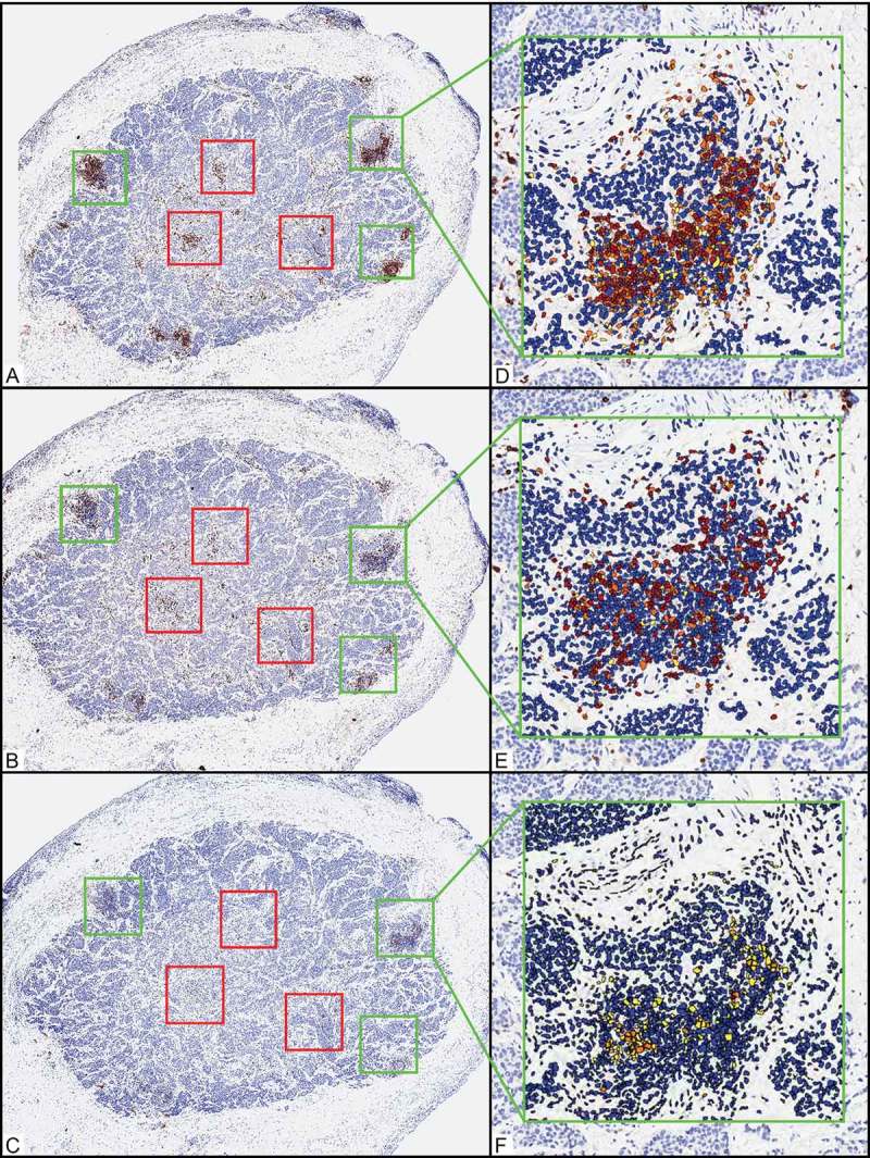
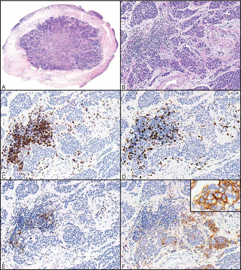
Ocular adnexal sebaceous carcinoma (OASC) is an aggressive malignancy that frequently recurs locally and metastasizes. Surgical extirpation may produce significant aesthetic morbidity, and effective systemic therapies for locally advanced or metastatic disease are largely ineffective. Immune checkpoint inhibitors have shown efficacy in the management of several solid tumors where tumor cell PD-L1 expression correlates with improved response. To determine whether OASC might be amenable to immune checkpoint blockade, we performed comprehensive immune profiling for CD3, CD8, PD-1, FOXP3, and PD-L1 in 24 patients with primary OASC. The composition, distribution and density of the tumor associated immune infiltrate were quantified by automated image analysis and correlated with measures of clinical outcome. Tumor cells in 12 OASCs (50%) expressed PD-L1. Higher densities of CD3+ (p = 0.01), CD8+ (p = 0.006), and PD-1+ (p = 0.024) tumor-associated T cells were associated with higher T category (≥T3a per the 7th edition of the American Joint Committee on Cancer staging manual). Higher tumor cell expression of PD-L1 correlated with higher density of PD-1+ tumor-associated T cells (p = 0.021). Since a CD3+ CD8+ PD-1 + T-cell infiltrate represents a "suppressed T-cell phenotype" apparently permissive toward OASC progression, our findings provide a mechanistic rationale for the effective application of immune checkpoint blockade in OASC to abrogate PD-1/PD-L1 interaction and effectively unleash the immune infiltrate to treat higher-stage tumors.

Keywords: PD-1; PD-L1; Sebaceous; biomarkers; carcinoma; immunosurveillance; inflammation and cancer; ocular.


Figures

Figure 1.
Figure 1.
Representative example of quantification of the relative composition, density, and distribution of immune infiltrates in primary ocular adnexal sebaceous carcinoma. (A-C) Scanning magnification shows an ocular adnexal sebaceous carcinoma after immunohistochemical studies for CD3 (A, 40x), CD8 (B, 40x), and PD-1 (C, 40x). Green and red boxes delineate areas of the tumor in which the highest areas of immune cell density were assessed. Green boxes designate tumor periphery (3 x 0.25-mm2 boxes), and red boxes designate central areas (3 x 0.25-mm2 boxes). (D-F) Automated image analysis quantified the CD3-positive (CD3+) (D, 200x), CD8+ (E, 200x), and PD-1+ (F, 200x) cells. Immunohistochemically positive (IHC+) cells were quantified according to intensity (1+ yellow, 2+ orange, and 3+ brown), and cells with IHC-negative nuclei (including stromal and tumor cells) in a given area were counted and designated as a blue nucleus. IHC+ cells (including the sum of 1+, 2+, and 3+ cells) were tabulated and quantified as (1) number of IHC+ cells per 0.25 mm2 or (2) percentage of the total nucleated cells in a given 0.25-mm2 area.
Figure 2.
Figure 2.
Relationships between CD3+, CD8+, and PD-1 + T cells and PD-L1-expressing tumor cells in a primary ocular adnexal sebaceous carcinoma. (A) Scanning magnification of an ocular adnexal sebaceous carcinoma (H&E, 40x). (B) Higher magnification shows infiltrating carcinoma in the same area where immunohistochemical positivity was quantified (H&E, 200x). (C-E) Antibodies for CD3 highlight a cluster of CD3-positive (CD3+) T cells associated with the tumor periphery (C, 200x), and a subset of these are CD8 + T cells (D, 200x) that also expressing PD-1 (E, 200x). (F) PD-L1+ tumor cells are seen in close proximity to these (F, 200x; inset 400x).

References

    1. Mulay K, Aggarwal E, White VA.. Periocular sebaceous gland carcinoma: A comprehensive review. Saudi J Ophthalmol. 2013;27:159–165. doi:10.1016/j.sjopt.2013.05.002. - DOI - PMC - PubMed
    1. Pfeiffer ML, Savar A, Esmaeli B. Sentinel lymph node biopsy for eyelid and conjunctival tumors: what have we learned in the past decade? Ophthal Plast Reconstr Surg. 2013;29:57–62. doi:10.1097/IOP.0b013e31827472c5. - DOI - PubMed
    1. Jakobiec FA, Mendoza PR. Eyelid sebaceous carcinoma: clinicopathologic and multiparametric immunohistochemical analysis that includes adipophilin. Am J Ophthalmol. 2014;157:186–208.e2. doi:10.1016/j.ajo.2013.08.015. - DOI - PubMed
    1. Shields JA, Saktanasate J, Lally SE, Carrasco JR, Shields CL. Sebaceous carcinoma of the ocular region: the 2014 professor winifred mao lecture. Asia Pac J Ophthalmol (Philadelphia, Pa). 2015;4:221–227. doi:10.1097/APO.0000000000000105. - DOI - PubMed
    1. Nelson BR, Hamlet KR, Gillard M, Railan D, Johnson TM. Sebaceous carcinoma. J Am Acad Dermatol. 1995;33:1–15; quiz 6–8. doi:10.1016/0190-9622(95)90001-2. - DOI - PubMed

LinkOut - more resources