. 2018 Jul 23;7(9):e1477459.
doi: 10.1080/2162402X.2018.1477459. eCollection 2018.

Combination of radiation and interleukin 12 eradicates large orthotopic hepatocellular carcinoma through immunomodulation of tumor microenvironment

Affiliations

Combination of radiation and interleukin 12 eradicates large orthotopic hepatocellular carcinoma through immunomodulation of tumor microenvironment

Chia-Jen Wu et al. Oncoimmunology. .

Abstract

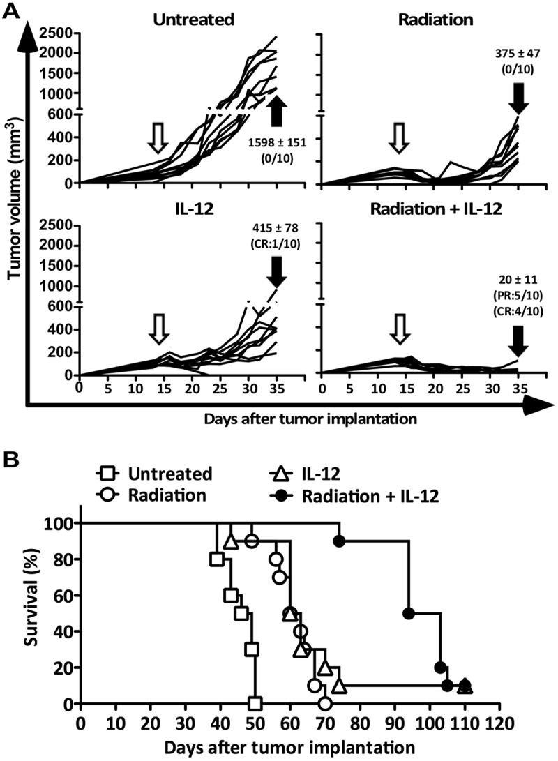
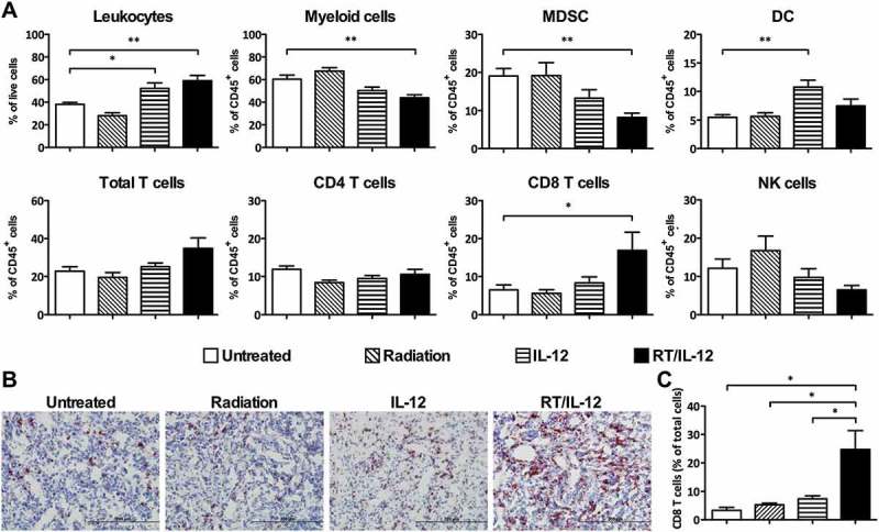
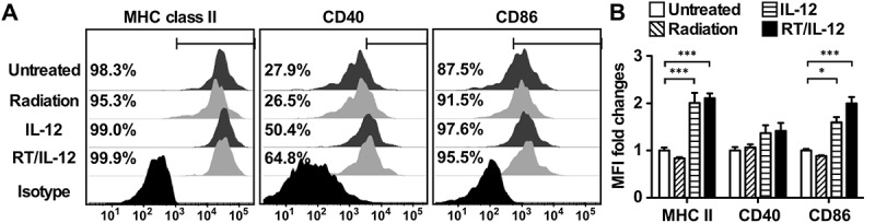
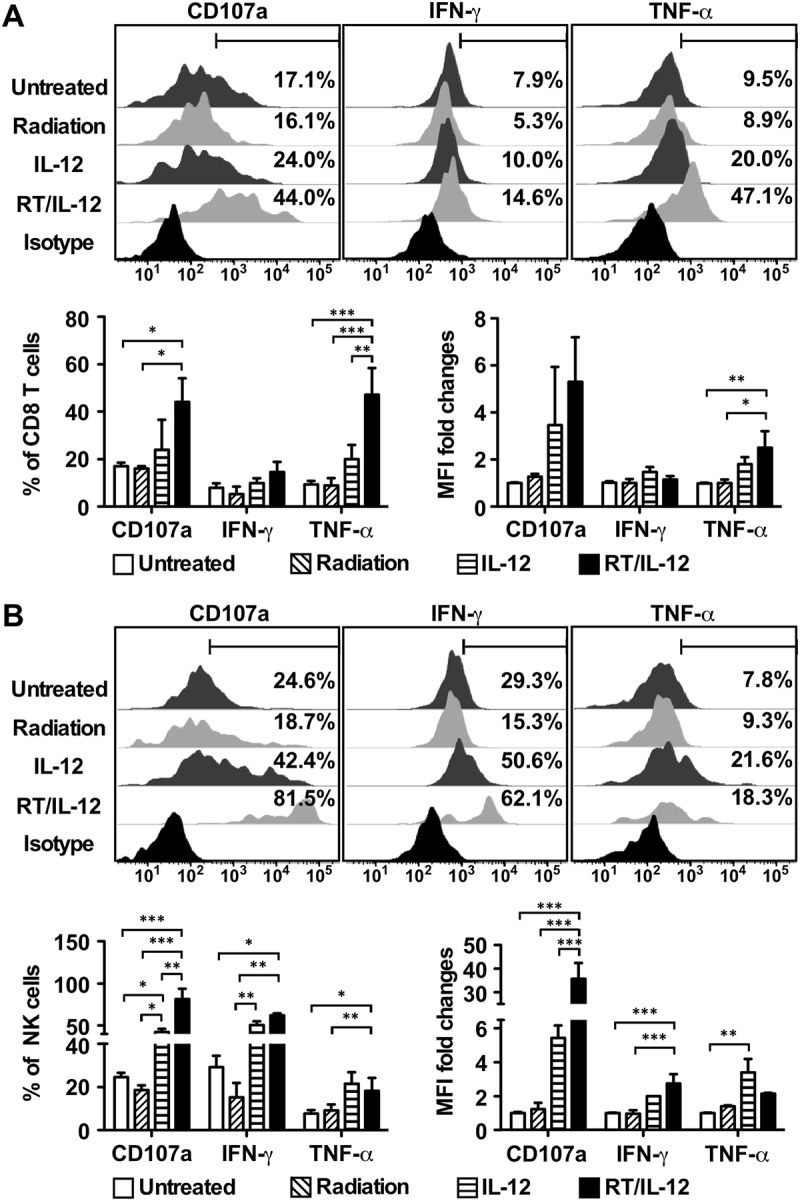
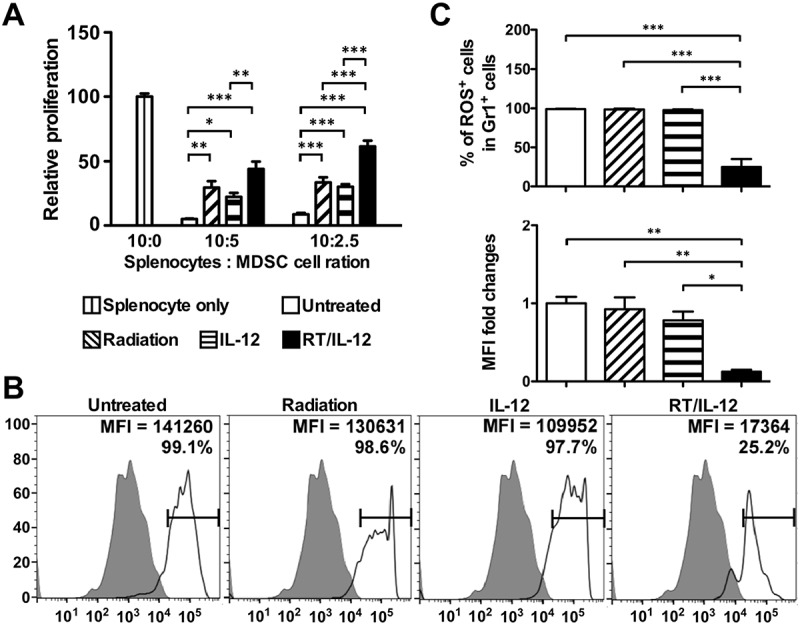
Immunotherapies have shown promising results in certain cancer patients. For hepatocellular carcinoma (HCC), the multiplicity of an immunotolerant microenvironment within both the tumor, and the liver per se may limit the efficacy of cancer immunotherapies. Since radiation induces immunogenic cell death and inflammatory reactions within the tumor microenvironment, we hypothesized that a combination therapy of radiation and lasting local immunostimulating agents, achieved by intratumoral injection of an adenoviral vector encoding interleukin 12, may reverse the immunotolerant microenvironment within a well-established orthotopic HCC toward a state favorable for inducing antitumor immunities. Our data showed that radiation and IL-12 combination therapy (RT/IL-12) led to dramatic tumor regression in animals bearing large subcutaneous or orthotopic HCC, induced systemic effect against distant tumor, and significantly prolonged survival. Radiation monotherapy induced tumor regression at early times but afterwards most tumors regained exponential growth, while IL-12 monotherapy only delayed tumor growth. Mechanistic studies revealed that RT/IL-12 increased expression of MHC class II and co-stimulatory molecules CD40 and CD86 on tumor-infiltrating dendritic cells, suggesting an improvement of their antigen presentation activity. RT/IL-12 also significantly reduced accumulation of tumor-infiltrating myeloid-derived suppressor cells (MDSCs) and impaired their suppressive functions by reducing production of reactive oxygen species. Accordingly, tumor-infiltrating CD8+ T cells and NK cells were significantly activated toward the antitumor phenotype, as revealed by increased expression of CD107a and TNF-α. Together, our data showed that RT/IL-12 treatment could reset the intratumoral immunotolerant state and stimulate activation of antitumor cellular immunity that is capable of eliminating large established HCC tumors.

Keywords: MDSC; Orthotopic HCC; combination therapy; immunomodulation; immunotherapy; radiotherapy; tumor microenvironment.


Figures

Figure 1.
Figure 1.
Antitumor activity of combined radiation and IL-12 on subcutaneous HCC. BALB/c mice (n = 10 in each group) were injected into the right flank with 1 × 106 BNL HCC cells. Fourteen days later, these mice were randomly grouped and treated with 10 Gy radiation, 1 × 108 pfu of Ad/IL-12 by intratumoral injection, combination of both or remained untreated. (A) Tumor volume was measured 3 times/week for three weeks and (B) mice were kept for long-term survival observation. The data are presented as mean ± SEM. CR: complete regression; PR: partial regression. Unfilled arrow: day of treatment. Filled arrow: 3 weeks post treatment for tumor volume measurement. Panels represent data from 2 independent experiments.
Figure 2.
Figure 2.
Systemic antitumor effect of radiation and IL-12 combination therapy. Groups of BALB/c mice (n = 5) were injected subcutaneously with 5 × 105 CT26 cells in the right flank and 1 × 105 CT26 cells in the left flank. Tumors on right flank were treated with radiation, Ad/IL-12, combination of both or remained untreated as described above, and the left flank tumor remained untreated. Tumor growth on the left flank was monitored at the indicated times. The data are presented as mean ± SEM.
Figure 3.
Figure 3.
Antitumor activity of combined radiation and IL-12 on large orthotopic HCC. BALB/c mice (n = 6–10 in different groups) were injected into the left liver lobe with 3 × 105 BNL HCC cells. Ten days later, these mice were randomly grouped and treated with 10 Gy radiation, 1 × 108 pfu of Ad/IL-12 by intratumoral injection, combination of both or remained untreated. Mice were sacrificed at day 31. For large orthotopic model, mice were treated on day 14 and sacrificed on day 35 for tumor volume measurement. (A) Tumor volumes were presented as before (day 10 or 14) and after (21 days after) treatment. (B) Representative photographs of the liver of each group. The data are presented as mean ± SEM.
Figure 4.
Figure 4.
The change of cell population after combination of radiation and IL-12 treatment in tumor microenvironment. BALB/c mice (n = 4–11 in different groups) were injected with 3 × 105 BNL HCC cells into the left liver lobe. Fourteen days later, these mice treated with 10 Gy radiation, 1 × 108 pfu of Ad/IL-12 by intratumoral injection, combination of both or remained non-treated. (A) Mice were sacrificed 7 days after treatment and the leukocyte subpopulations within the tumor were analyzed by flow cytometer. (B) Tumor tissues were removed 7 days after treatment for immunohistochemical (IHC) analysis. Cryostat sections were immunohistochemically stained with anti-CD8 and developed with AEC, then counterstained with hematoxylin. Original magnification, ×200. (C) The percentage of CD8+ cells in tumor tissues. The IHC stained slides were scanned and analyzed using a Panoramic 250 slide scanner. The data are presented as mean ± SEM. *, p < 0.05; **, p < 0.01. Representative data are shown from three experiments. RT/IL-12: radiation plus IL-12.
Figure 5.
Figure 5.
Combination of radiation and IL-12 increased the frequency and maturation of tumor-infiltrated DCs. BALB/c mice (n = 4–7 in different groups) were injected into the left liver lobe with BNL HCC cells and treated as described in Figure 3. (A) Mice were sacrificed 7 days post-treatment. CD11c+ DCs from TILs were gated and stained with anti-MHC class II CD40, and CD86 Abs, or appropriate isotype-control Abs, as described in Materials and Methods. The percentages of positive cells for surface proteins are shown. The results shown were from one representative mouse of each group. (B) Summarized MFI fold changes for surface proteins were presented as mean ± SEM. *, p < 0.05; **, p < 0.01; ***, p < 0.001. RT/IL-12: radiation plus IL-12.
Figure 6.
Figure 6.
Combination of radiation and IL-12 significantly increased CD107a expression on CD8 T cells and NK cells. BALB/c mice (n = 4–10 in different groups) were injected into the left liver lobe with 3 × 105 BNL HCC cells, and 14 days later received radiation, 1 × 108 pfu of Ad/IL-12 by intratumoral injection, combination of both or remained non-treated. The percentage and MFI fold changes of CD107a, IFN-γ and TNF-α on tumor-infiltrating (A) CD8 T cells and (B) NK cells were analyzed 7 days later. The summarized results were presented as mean ± SEM. *, p < 0.05; **, p < 0.01; ***, p < 0.001. RT/IL-12: radiation plus IL-12.
Figure 7.
Figure 7.
Combination of radiation and IL-12 subdued the suppressive activity and ROS production of tumor-infiltrated MDSC. BALB/c mice (n = 6–10 in different groups) were injected into the left liver lobe with BNL HCC cells and treated as described in Figure 3. Mice were sacrificed 7 days post-treatment. (A) The suppressive activity of MDSC obtained from TILs. Gr1± cells were purified with magnetic beads. Splenocytes from naïve mice were stimulated with αCD3/αCD28 Abs in the presence of purified Gr1± cells at a ratio of 10:5 or 10:2.5. Three days later, 1 μCi 3H-thymidine was added and cultured for additional 16 h. Cells were harvested and data were calculated by a TopCount instrument. (B) The levels of ROS in purified MDSC were measured by fluorescence intensity of 2ʹ,7ʹ-dichlorofluorescin diacetate labeling after PMA stimulation for 30 minutes. (C) Summarized results presented as means ± SEM. **, p < 0.01; ***, p < 0.001. RT/IL-12: radiation plus IL-12.

Similar articles

Cited by

References

    1. Jemal A, Bray F, Center MM, Ferlay J, Ward E, Forman D.. Global cancer statistics. CA Cancer J Clin 2011;61:69–90.doi: 10.3322/caac.20107 PMID:21296855 - DOI - PubMed
    1. Forner A, Gilabert M, Bruix J, Raoul JL. Treatment of intermediate-stage hepatocellular carcinoma. Nat Rev Clin Oncol 2014;11:525–535.doi: 10.1038/nrclinonc.2014.122 PMID:25091611 - DOI - PubMed
    1. Prieto J, Melero I, Sangro B. Immunological landscape and immunotherapy of hepatocellular carcinoma. Nat Rev Gastroenterol Hepatol 2015;12:681–700.doi: 10.1038/nrgastro.2015.173 PMID:26484443 - DOI - PubMed
    1. Fridman WH, Pages F, Sautes-Fridman C, Galon J. The immune contexture in human tumours: impact on clinical outcome. Nat Rev Cancer 2012;12:298–306.doi: 10.1038/nrc3245 PMID:22419253 - DOI - PubMed
    1. Shurin GV, Ma Y, Shurin MR. Immunosuppressive mechanisms of regulatory dendritic cells in cancer. Cancer Microenviron 2013;6:159–167.doi: 10.1007/s12307-013-0133-3 PMID:23749739 - DOI - PMC - PubMed

Publication types