

Fourteen new mutations of BCKDHA, BCKDHB and DBT genes associated with maple syrup urine disease (MSUD) in Malaysian population
- PMID: 30228974
- PMCID: PMC6140420
- DOI: 10.1016/j.ymgmr.2018.08.006
Fourteen new mutations of BCKDHA, BCKDHB and DBT genes associated with maple syrup urine disease (MSUD) in Malaysian population
Abstract
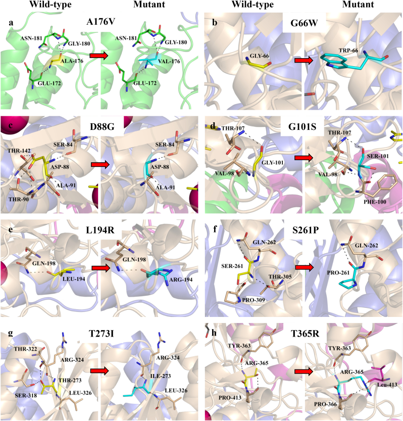
Maple syrup urine disease (MSUD) is a rare autosomal recessive metabolic disorder. This disorder is usually caused by mutations in any one of the genes; BCKDHA, BCKDHB and DBT, which represent E1α, E1β and E2 subunits of the branched-chain α-keto acid dehydrogenase (BCKDH) complex, respectively. This study presents the molecular characterization of 31 MSUD patients. Twenty one mutations including 14 new mutations were identified. The BCKDHB gene was the most commonly affected (45.2%) compared to BCKDHA gene (16.1%) and DBT gene (38.7%). In silico webservers predicted all mutations were disease-causing. In addition, structural evaluation disclosed that all new missenses in BCKDHA, BCKDHB and DBT genes affected stability and formation of E1 and E2 subunits. Majority of the patients had neonatal onset MSUD (26 of 31). Meanwhile, the new mutation; c.1196C > G (p.S399C) in DBT gene was noted to be recurrent and found in 9 patients. Conclusion: Our findings have expanded the mutational spectrum of the MSUD and revealed the genetic heterogeneity among Malaysian MSUD patients. We also discovered the p.S399C from DBT gene was noted as a recurrent mutation in Malay community and it suggested the existence of common and unique mutation in Malay population.
Keywords: Autosomal recessive; BCAAs; BCKDH; MSUD; Maple syrup urine disease; Mutation.
Figures


References
-
- Chuang D.T., Shih V.E. Maple syrup urine disease (branched-chain ketoaciduria) In: Sciver C.R., Beaudet A.L., Valle D., editors. The Metabolic and Molecular Basis of Inherited Disease. McGraw-Hill; New York, USA: 2001.
-
- Reed L.J., Daminu Z., Merryfield M.L. Regulation of mammalian pyruvate and branched-chain α-keto acid dehydrogenase complexes by phosphorylation — dephosphorylation. Curr. Top. Cell. Regul. 1985;27:41–49. - PubMed
-
- Aevarsson A., Chuang J.L., Wynn R.M., Turley S., Chuang D.T., Hol W.G. Crystal structure of human branched-chain α α-ketoacid dehydrogenase and the molecular basis of multienzyme complex deficiency in maple syrup urine disease. Structure. 2000;8:277–291. - PubMed
-
- Rodríguez-Pombo P., Navarrete R., Merinero B., Gómez-Puertas P., Ugarte M. Mutational spectrum of maple syrup urine disease in Spain. Hum. Mutat. 2006;27:715. - PubMed
Grants and funding
LinkOut - more resources
Full Text Sources
Other Literature Sources
