. 2017 Nov 28;2(4):e0034.
doi: 10.2106/JBJS.OA.17.00034. eCollection 2017 Dec 28.

The Anatomy of the Articularis Genus Muscle and Its Relation to the Extensor Apparatus of the Knee

Affiliations

The Anatomy of the Articularis Genus Muscle and Its Relation to the Extensor Apparatus of the Knee

Karl Grob et al. JB JS Open Access. .

Abstract

Background: The anatomy of the articularis genus muscle has prompted speculation that it elevates the suprapatellar bursa during extension of the knee joint. However, its architectural parameters indicate that this muscle is not capable of generating enough force to fulfill this function. The purpose of the present study was to investigate the anatomy of the articularis genus, with special emphasis on its relationship with the adjacent vastus intermedius and vastus medialis muscles.

Methods: The articularis genus muscle was investigated in 18 human cadaveric lower limbs with use of macrodissection techniques. All components of the quadriceps muscle group were traced from origin to insertion, and their affiliations were determined. Six limbs were cut transversely in the middle third of the thigh. The modes of origin and insertion of the articularis genus, its nerve supply, and its connections with the vastus intermedius and vastus medialis were studied.

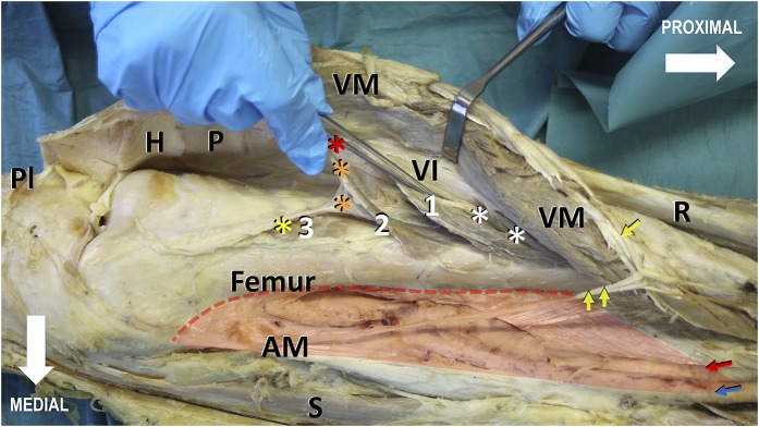
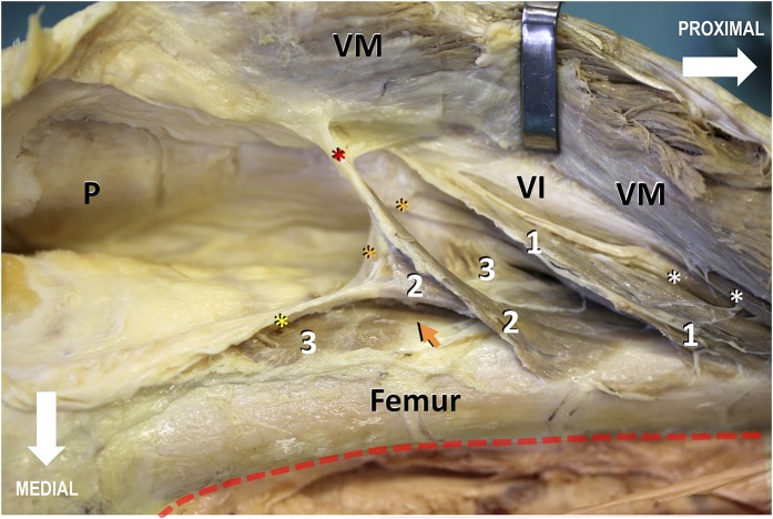
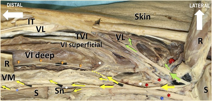
Results: The muscle bundles of the articularis genus were organized into 3 main layers: superficial, intermediate, and deep. The bundles of the superficial layer and, in 60% of the specimens, the bundles of the intermediate layer originated from both the vastus intermedius and the anterior and anterolateral surfaces of the femur. The bundles of the deep layer and, in 40% of the specimens, the bundles of the intermediate layer arose solely from the anterior surface of the femur. The distal insertion sites included different levels of the suprapatellar bursa and the joint capsule. A number of connections between the articularis genus and the vastus intermedius were found. While the vastus medialis inserted into the whole length of the vastus intermedius aponeurosis, it included muscle fibers of the articularis genus, building an intricate muscle system supplied by nerve branches of the same medial deep division of the femoral nerve.

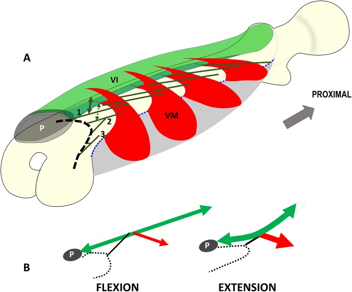
Conclusions: The articularis genus, vastus medialis, and vastus intermedius have a complex, interacting architecture, suggesting that the articularis genus most likely does not act as an independent muscle. With support of the vastus intermedius and vastus medialis, the articularis genus might be able to function as a retractor of the suprapatellar bursa. The finding of likely interplay between the articularis genus, vastus intermedius, and vastus medialis is supported by their concurrent innervation.

Clinical relevance: The association between the articularis genus, vastus medialis, and vastus intermedius may be more complex than previously believed, and this close anatomical connection could have functional implications for knee surgery. Dysfunction, scarring, or postoperative arthrofibrosis of the sophisticated interactive mechanism needs further investigation.


Figures

Fig. 1
Fig. 1
Photograph showing the medial view of the distal two-thirds of a right thigh. The vastus medialis (VM), together with its insertion into the vastus intermedius (VI), is lifted from its hammock-like origin (red transparent shading), the medial lip of the linea aspera, and the medial supracondylar line (red dotted line), thereby revealing the knee joint cavity, the articularis genus (indicated by the white numbers), the vastus intermedius aponeurosis, and the medial surface of the distal part of the femur. The articularis genus is arranged in 3 layers: superficial (indicated by the number 1), intermediate (indicated by the number 2), and deep (indicated by the number 3). Distally, the articularis genus bundles insert gradually into the suprapatellar bursa (asterisks) and the joint capsule. The bundles of the superficial layer insert into the synovial membrane adjacent to the quadriceps tendon (red asterisk), the bundles of the intermediate layer insert into the middle section of the suprapatellar bursa (orange asterisks), and the muscles of the deep layer insert into the synovial membrane (yellow asterisk) facing the femur. Distal deep muscle bundles of the vastus intermedius (white asterisks) contribute to the vastus intermedius aponeurosis and finally merge with the quadriceps tendon, inserting into the base of the patella (P). The long nerve branch to the vastus medialis (single yellow arrow), lifted with the vastus medialis muscle, courses distally along the anteromedial border of the muscle and, in contrast to the saphenous nerve (double yellow arrows), remains lateral to its superficial aponeurosis in a separate fibrotic tunnel. Red and blue arrows = superficial femoral vessels, H = Hoffa fat pad, PI = transected patellar tendon, R = rectus femoris, AM = tendon of the adductor magnus, and S = sartorius.
Fig. 2
Fig. 2
Enlarged view of Figure 1, showing the origin and insertion of the articularis genus. The bundles of the superficial layer (indicated by the number 1) and, in 60% of the specimens, the bundles of the intermediate layer (indicated by the number 2) originated from both the deep surface of the vastus intermedius (VI) aponeurosis and from the anterior and anterolateral surfaces of the femur. The bundles of the deep layer (indicated by the number 3) and, in 40% of the specimens, the bundles of the intermediate layer originated entirely from the anterior surface of the femur. Distally, the articularis genus bundles inserted gradually into the suprapatellar bursa and the joint capsule (asterisks), with the bundles of the superficial layer inserting into the synovial membrane adjacent to the quadriceps tendon (red asterisk), the bundles of the intermediate layer inserting into the middle section of the suprapatellar bursa (orange asterisks), and the bundles of the deep layer inserting into the synovial membrane facing the femur (yellow asterisk). Proximally, no distinct investing fascia separated the articularis genus from the vastus intermedius; in that area, the articularis genus muscle bundles were in continuity with the deep muscle fibers of the vastus intermedius (white asterisks). Distally, the deep and intermediate articularis genus muscle bundles were partially separated by or imbedded in a considerable amount of fatty tissue (orange arrow). For better visualization of the articularis genus muscle bundles, the fatty tissue was partially removed. VM = vastus medialis. P = Patella.
Fig. 3
Fig. 3
Figs. 3-A and 3-B Schematic drawings showing the anatomy and mode of operation of the interacting muscle complex consisting of the articularis genus, vastus intermedius (VI), and vastus medialis (VM). Fig. 3-A Schematic illustration depicting the hammock-like origin (gray) and the fleshy clip-like double-insertion of the vastus medialis units (red) into the entire vastus intermedius aponeurosis (green). The blue dotted line corresponds to the origin of the vastus medialis at the medial lip of the linea aspera. The vastus medialis clamps the vastus intermedius aponeurosis like a clip holding a sheet. The deep layer of the vastus intermedius (green lines) corresponds with the muscle bundles that continue as the articularis genus to the knee joint. The superficial layer of the vastus intermedius contributes to the layers of the quadriceps tendon and finally inserts into the base of the patella (P). In the present study, the superficial articularis genus muscle bundles (indicated by the number 1) always originated from both the anterior surface of the femur and the vastus intermedius (double arrows). The deep articularis genus muscle bundles (indicated by the number 3) arose solely from the femur. The intermediate articularis genus muscle bundles (indicated by the number 2) always arose from the anterior surface of the femur. However, in 60% of the specimens, muscle fibers also originated from the vastus intermedius aponeurosis. All muscle bundles of the articularis genus inserted into the synovial membrane of the joint capsule and the suprapatellar bursa (black dashed line). Fig. 3-B Schematic illustration depicting the mode of operation of the muscle complex consisting of the articularis genus (black lines), vastus intermedius (green double arrows), and vastus medialis (red arrows). As a derivative of the vastus intermedius, the articularis genus consists of a few muscle bundles that arise from the deep part of the vastus intermedius. Therefore, the articularis genus does not act as an independent entity. The muscle bundles of the articularis genus connect with the neighboring vastus intermedius and vastus medialis. With their support, the articularis genus retracts or elevates the suprapatellar bursa (black dotted lines) during extension of the knee, preventing entrapment of the bursa between the patella (P) and the femur.
Fig. 4
Fig. 4
Photograph showing the anterior view of the proximal part of a right thigh along with the nerve supply to the extensor apparatus of the knee joint. For better visualization of the femoral nerve branches, the sartorius (S) and rectus femoris (R) muscles were transected and elevated. Some nerve branches are identified with black paper. The red pinhead on the right side of the image indicates the middle of the neck of the femur on the intertrochanteric line. The vastus medialis (VM), the medial part of the vastus intermedius (VI), and the articularis genus are innervated by the medial division of the femoral nerve (yellow arrows). One of these terminal branches crosses the medial portion of the vastus intermedius (yellow dotted line), courses distally above the deep layer of the vastus intermedius, and finally branches to the articularis genus and the synovial pouch of the knee joint. This nerve branch (orange arrowheads) is a guide for separating the vastus medialis from the vastus intermedius proximally and the articularis genus inclusive of the deep layer of the vastus intermedius from the superficial layer of the vastus intermedius distally. The nerve branch to the vastus medialis runs along the anteromedial border of the muscle. It separates from the saphenous nerve (Sn) proximally. The green arrows indicate the lateral division of the femoral nerve to the lateral parts of the vastus intermedius, the vastus lateralis (VL), and the tensor vastus intermedius (TVI). Red asterisks = superficial, deep, and lateral circumflex femoral arteries; blue asterisk = superficial femoral vein; and IT = iliotibial tract.

References

    1. Kimura K, Takahashi Y. M. articularis genus. Observations on arrangement and consideration of function. Surg Radiol Anat. 1987;9(3):231-9. - PubMed
    1. Woodley SJ, Latimer CP, Meikle GR, Stringer MD. Articularis genus: an anatomic and MRI study in cadavers. J Bone Joint Surg Am. 2012. January 4;94(1):59-67. - PubMed
    1. Puig S, Dupuy DE, Sarmiento A, Boland GW, Grigoris P, Greene R. Articular muscle of the knee: a muscle seldom recognized on MR imaging. AJR Am J Roentgenol. 1996. May;166(5):1057-60. - PubMed
    1. Platzer W. Taschenatlas anatomie. Bewegungsapparat. 11th ed. New York: Thieme; 2013. p 248-9.
    1. Ahmad I. Articular muscle of the knee—articularis genus. Bull Hosp Joint Dis. 1975. April;36(1):58-60. - PubMed

LinkOut - more resources