

Retinal pigment epithelium hyperplasia overlying pigment epithelial detachment in age-related macular degeneration can masquerade as neovascularization on optical coherence tomography angiography
- PMID: 30229304
- PMCID: PMC6224014
- DOI: 10.1007/s00417-018-4138-y
Retinal pigment epithelium hyperplasia overlying pigment epithelial detachment in age-related macular degeneration can masquerade as neovascularization on optical coherence tomography angiography
Abstract
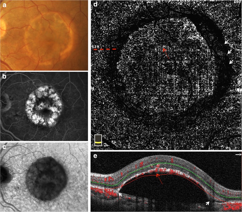
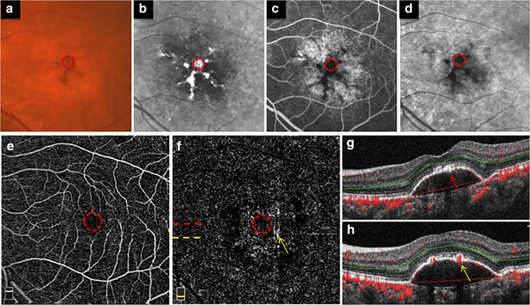
Purpose: To report the image artifacts due to retinal pigment epithelium (RPE) hyperplasia overlying retinal pigment epithelial detachment (PED) in age-related macular degeneration (AMD), which can masquerade as neovascularization on optical coherence tomography angiography (OCTA).
Methods: A hospital-based, retrospective, and cross-sectional study. Twenty-two eyes from 16 patients with non-vascularized PED related to AMD were included in this study. All patients were examined by OCTA, spectral-domain optical coherence tomography, fluorescence angiography, and indocyanine green angiography. Vascular flow signals (VFS) on both the outer retinal slab of en face OCTA and cross-sectional OCTA and their correspondence with RPE hyperplasia were evaluated.
Results: Fifteen eyes (68.2%) showed VFS on both the outer retina slab of en face OCTA and cross-sectional OCTA, all corresponding to the RPE hyperplasia overlying PED. Among them, 12 eyes with lump RPE hyperplasia outside foveal avascular zone (FAZ) all showed obvious VFS on the outer retina slab of OCTA, and 3 eyes with scattered RPE hyperplasia outside FAZ showed VFS fragments. Of note, 4 eyes had accompanied RPE hyperplasia inside FAZ, and 7 eyes without RPE hyperplasia overlying PED showed no corresponding VFS on the outer retina slab of OCTA. Additionally, a round-like dark band at the edge of PED was observed in the outer retina slab on en face OCTA in 17 eyes (77.3%).
Conclusions: RPE hyperplasia overlying PED in AMD can masquerade as neovascularization on OCTA. To avoid misdiagnosis and unnecessary treatment, this RPE hyperplasia-related image artifact should be considered when interpreting OCTA images.
Keywords: Age-related macular degeneration; Image artifacts; Optical coherence tomography angiography; Pigment epithelial detachment; Retinal pigment epithelium hyperplasia.
Conflict of interest statement
Conflict of interest
The authors declare that they have no conflict of interest.
Ethical approval
All procedures performed in studies involving human participants were in accordance with the ethical standards of the institutional and/or national research committee (the Institutional Review Board of the Zhongshan Ophthalmic Center at Sun Yat-sen University) and with the 1964 Helsinki Declaration and its later amendments or comparable ethical standards.
For this type of study, formal consent is not required.
Informed consent
Informed consent was obtained from all individual participants included in the study.
Figures


Similar articles
-
IMPROVED DETECTION AND DIAGNOSIS OF POLYPOIDAL CHOROIDAL VASCULOPATHY USING A COMBINATION OF OPTICAL COHERENCE TOMOGRAPHY AND OPTICAL COHERENCE TOMOGRAPHY ANGIOGRAPHY.Retina. 2019 Sep;39(9):1655-1663. doi: 10.1097/IAE.0000000000002228. Retina. 2019. PMID: 29927796
-
Histologic and Optical Coherence Tomographic Correlates in Drusenoid Pigment Epithelium Detachment in Age-Related Macular Degeneration.Ophthalmology. 2017 May;124(5):644-656. doi: 10.1016/j.ophtha.2016.12.034. Epub 2017 Jan 30. Ophthalmology. 2017. PMID: 28153442 Free PMC article.
-
IMAGING OF PIGMENT EPITHELIAL DETACHMENTS WITH OPTICAL COHERENCE TOMOGRAPHY ANGIOGRAPHY.Retina. 2018 Sep;38(9):1759-1769. doi: 10.1097/IAE.0000000000002016. Retina. 2018. PMID: 29293207
-
Retinal pigment epithelium tears: Classification, pathogenesis, predictors, and management.Surv Ophthalmol. 2017 Jul-Aug;62(4):493-505. doi: 10.1016/j.survophthal.2017.03.004. Epub 2017 Mar 21. Surv Ophthalmol. 2017. PMID: 28336128 Review.
-
Giant retinal pigment epithelium tears with membranous nephropathy: a case report and literature review.BMC Ophthalmol. 2024 Apr 17;24(1):177. doi: 10.1186/s12886-024-03426-5. BMC Ophthalmol. 2024. PMID: 38632537 Free PMC article. Review.
Cited by
-
Small dome-shaped pigment epithelium detachment in polypoidal choroidal vasculopathy: an under-recognized sign of polypoidal lesions on optical coherence tomography?Eye (Lond). 2022 Apr;36(4):733-741. doi: 10.1038/s41433-020-01390-0. Epub 2021 Apr 8. Eye (Lond). 2022. PMID: 33833415 Free PMC article.
-
Stem cell therapy for retinal pigment epithelium disorders.World J Stem Cells. 2025 May 26;17(5):103100. doi: 10.4252/wjsc.v17.i5.103100. World J Stem Cells. 2025. PMID: 40503367 Free PMC article. Review.
-
Artifacts in Optical Coherence Tomography Angiography.J Ophthalmic Vis Res. 2021 Apr 29;16(2):271-286. doi: 10.18502/jovr.v16i2.9091. eCollection 2021 Apr-Jun. J Ophthalmic Vis Res. 2021. PMID: 34055264 Free PMC article. Review.
-
Advances in multimodal imaging for diagnosis of pigmented ocular fundus lesions.Can J Ophthalmol. 2024 Aug;59(4):218-233. doi: 10.1016/j.jcjo.2023.07.005. Epub 2023 Jul 19. Can J Ophthalmol. 2024. PMID: 37480939 Free PMC article. Review.
-
Quantity and quality of image artifacts in optical coherence tomography angiography.PLoS One. 2019 Jan 25;14(1):e0210505. doi: 10.1371/journal.pone.0210505. eCollection 2019. PLoS One. 2019. PMID: 30682050 Free PMC article.
References
-
- Ahmed Daniel, Stattin Martin, Graf Alexandra, Forster Julia, Glittenberg Carl, Krebs Ilse, Ansari-Shahrezaei Siamak. DETECTION OF TREATMENT-NAIVE CHOROIDAL NEOVASCULARIZATION IN AGE-RELATED MACULAR DEGENERATION BY SWEPT SOURCE OPTICAL COHERENCE TOMOGRAPHY ANGIOGRAPHY. Retina. 2018;38(11):2143–2149. doi: 10.1097/IAE.0000000000001832. - DOI - PubMed
MeSH terms
Grants and funding
LinkOut - more resources
Full Text Sources
Other Literature Sources
Medical
Research Materials
