Review
. 2018 Sep 18;13(1):289.
doi: 10.1186/s11671-018-2694-z.

Graphene Family Materials in Bone Tissue Regeneration: Perspectives and Challenges

Affiliations
Review

Graphene Family Materials in Bone Tissue Regeneration: Perspectives and Challenges

Xinting Cheng et al. Nanoscale Res Lett. .

Abstract

We have witnessed abundant breakthroughs in research on the bio-applications of graphene family materials in current years. Owing to their nanoscale size, large specific surface area, photoluminescence properties, and antibacterial activity, graphene family materials possess huge potential for bone tissue engineering, drug/gene delivery, and biological sensing/imaging applications. In this review, we retrospect recent progress and achievements in graphene research, as well as critically analyze and discuss the bio-safety and feasibility of various biomedical applications of graphene family materials for bone tissue regeneration.

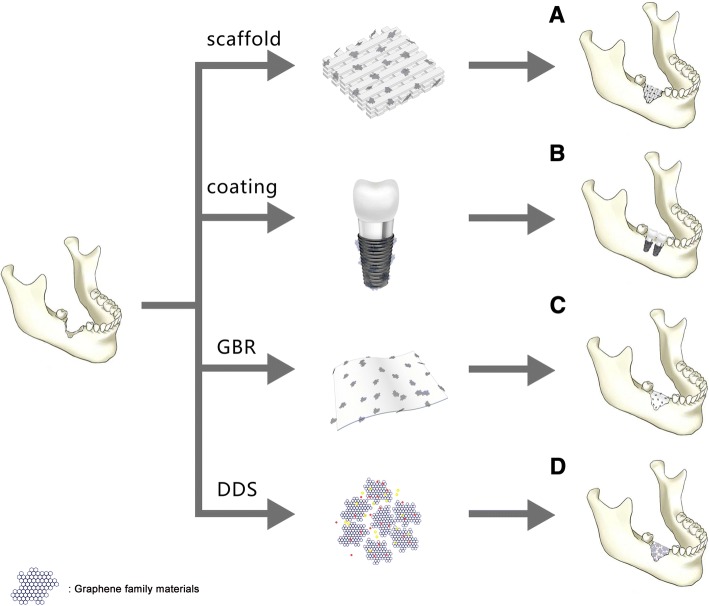
Keywords: Bone tissue regeneration; Coating; Drug delivery system; Graphene family materials; Guided bone regeneration membrane; Scaffold.


Conflict of interest statement

Competing Interests

The authors declare that they have no competing interests.

Publisher’s Note

Springer Nature remains neutral with regard to jurisdictional claims in published maps and institutional affiliations.

Figures

Fig. 1
Fig. 1
Representative HE-stained images of major organs (implantation region, liver, and kidney collected from the rats) implanted with graphene foams, GO foams, or nothing at day 14 post-implantation. No obvious organ damage or lesion was observed. Reproduced from ref. [40] with permission from the Journal of Nanoparticle Research
Fig. 2
Fig. 2
A. Graphene family materials as scaffold or a reinforcement material in scaffold for bone regeneration. B. Graphene family materials as coating transferred onto the substrate for bone regeneration. C. Graphene family as an additive in guided bone membrane. D. Graphene family materials as drug delivery system facilitates bone regeneration
Fig. 3
Fig. 3
Scheme illustration for β-TCP and β-TCP-GO scaffolds stimulated the in vivo osteogenesis. Micro-CT analysis and histological analysis of in vivo bone formation ability for the β-TCP and β-TCP-GO scaffolds after implanted in the cranial bone defects of rabbits for 8 weeks. Reproduced from ref. [85] with permission from the Journal of Carbon
Fig. 4
Fig. 4
a ALP activity in mice calvaria defects implanted with CHT/GO and b histomophometric analysis of Masson Goldner trichrome-stained sections. ###p < 0.001 vs CHT; **p < 0.01 vs control; ***p < 0.001 vs control. Reproduced from ref. [3] with permission from the Journal of Scientific Reports
Fig. 5
Fig. 5
a MTT assay after incubation of CS/Gn scaffolds and 0.25% GO/CS/Gn scaffolds with media for 48 h. The asterisk indicates a significant increase versus control, and the pound sign indicates a significant decrease versus control (p < 0.05). b, c Expression of osteogenic-related genes (RUNX2, ALP, COL-1, and OC) in mMSCs cultured on CS/Gn scaffolds and 0.25% GO/CS/Gn scaffolds for 7 and 14 days measured by quantitative RT-PCR. Reproduced from ref. [104] with permission from the Journal of International Journal of Biological Macromolecules
Fig. 6
Fig. 6
a The calvarial defects of rats were enclosed with a GO-Ti membrane. b New bone formation of the rat calvarial defects after the implantation of Ti or GO-Ti membrane at postoperative week 8. *p < 0.05 vs control; #p < 0.05 vs Ti. c Images of HE staining of the rat calvarial defects after the implantation of Ti or GO-Ti membrane at postoperative week 8. Reproduced from ref. [128] with permission from the Journal of Applied Spectroscopy Reviews
Fig. 7
Fig. 7
Schematics and scanning electron micrographs of the preparation the new GO/Ti scaffold: BMP2- and Van-loaded CGelMS were immobilized on the GO/Ti scaffold through electrostatic interactions between the functional groups of GO and CGelMS. Reproduced from ref. [149] with permission from the Journal of Biomaterials Science

Similar articles

Cited by

References

    1. Levengood SL, Zhang M. Chitosan-based scaffolds for bone tissue engineering. J Mater Chem B. 2014;2:3161–3184. doi: 10.1039/c4tb00027g. - DOI - PMC - PubMed
    1. Unnithan AR, Chan HP, Kim CS (2016) A unique scaffold for bone Tissue engineering: an osteogenic combination of graphene oxide- hyaluronic acid-chitosan with simvastatin. J Ind Eng Chem 46:182–191
    1. Hermenean A, Codreanu A, Herman H, Balta C, Rosu M, Mihali CV, et al. Chitosan-graphene oxide 3D scaffolds as promising tools for bone regeneration in critical-size mouse calvarial defects. Sci Rep. 2017;7:16641. doi: 10.1038/s41598-017-16599-5. - DOI - PMC - PubMed
    1. Holt Brian D., Wright Zoe M., Arnold Anne M., Sydlik Stefanie A. Graphene oxide as a scaffold for bone regeneration. Wiley Interdisciplinary Reviews: Nanomedicine and Nanobiotechnology. 2016;9(3):e1437. - PubMed
    1. Dimitriou R, Jones E, McGonagle D, Giannoudis PV. Bone regeneration: current concepts and future directions. BMC Med. 2011;9:66. doi: 10.1186/1741-7015-9-66. - DOI - PMC - PubMed

LinkOut - more resources