

Symplectomorphic registration with phase space regularization by entropy spectrum pathways
- PMID: 30230014
- PMCID: PMC7098261
- DOI: 10.1002/mrm.27402
Symplectomorphic registration with phase space regularization by entropy spectrum pathways
Abstract
Purpose: The ability to register image data to a common coordinate system is a critical feature of virtually all imaging studies. However, in spite of the abundance of literature on the subject and the existence of several variants of registration algorithms, their practical utility remains problematic, as commonly acknowledged even by developers of these methods.
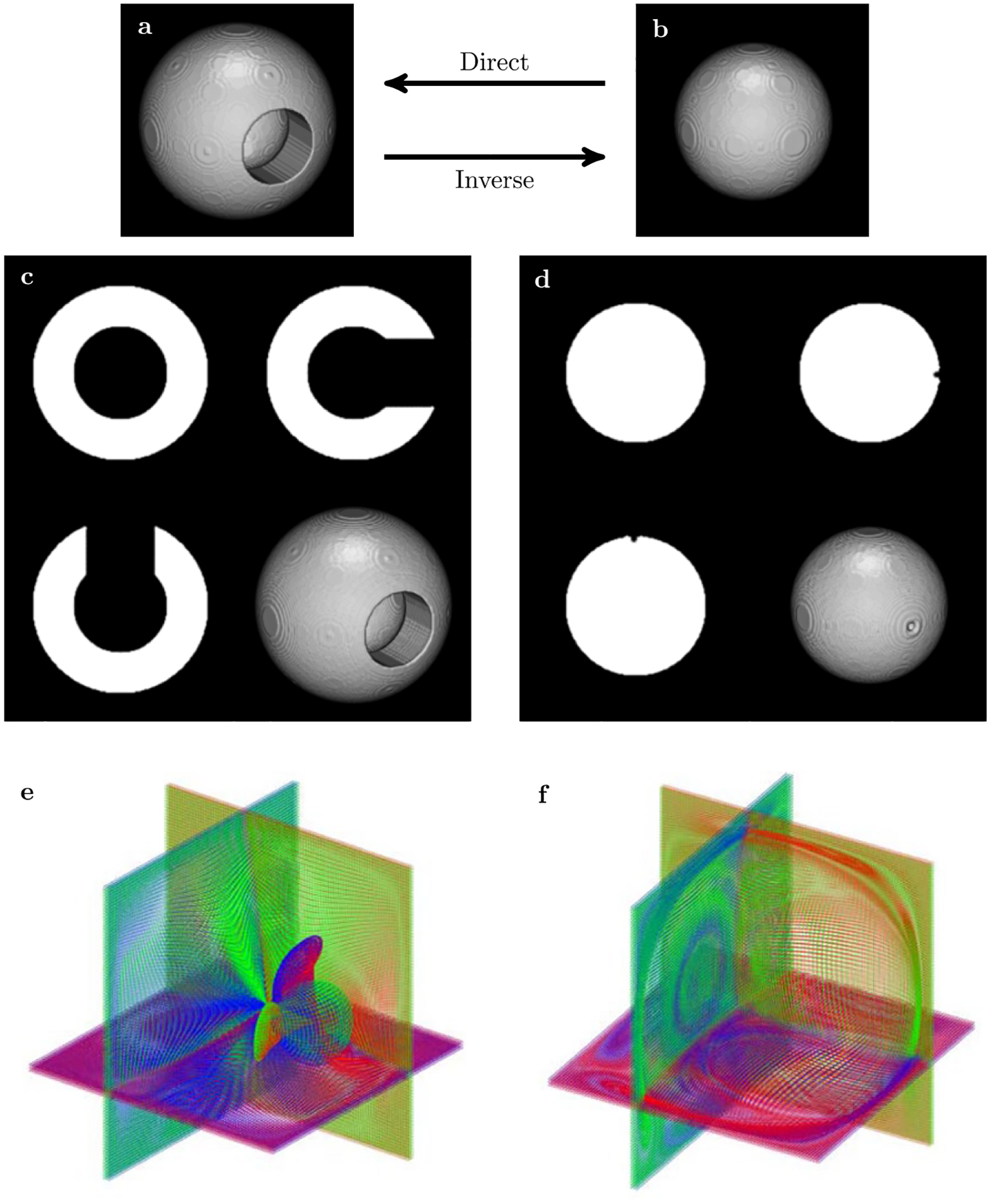
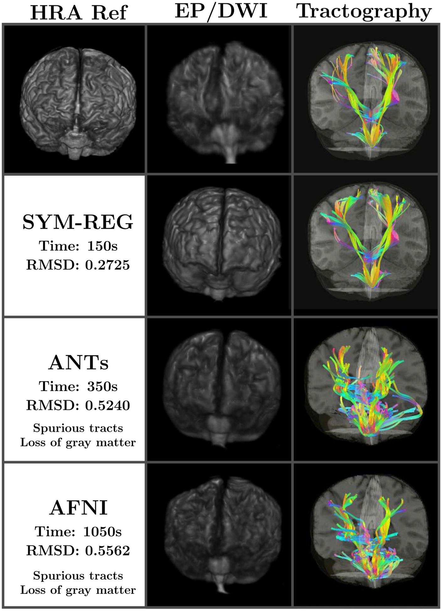
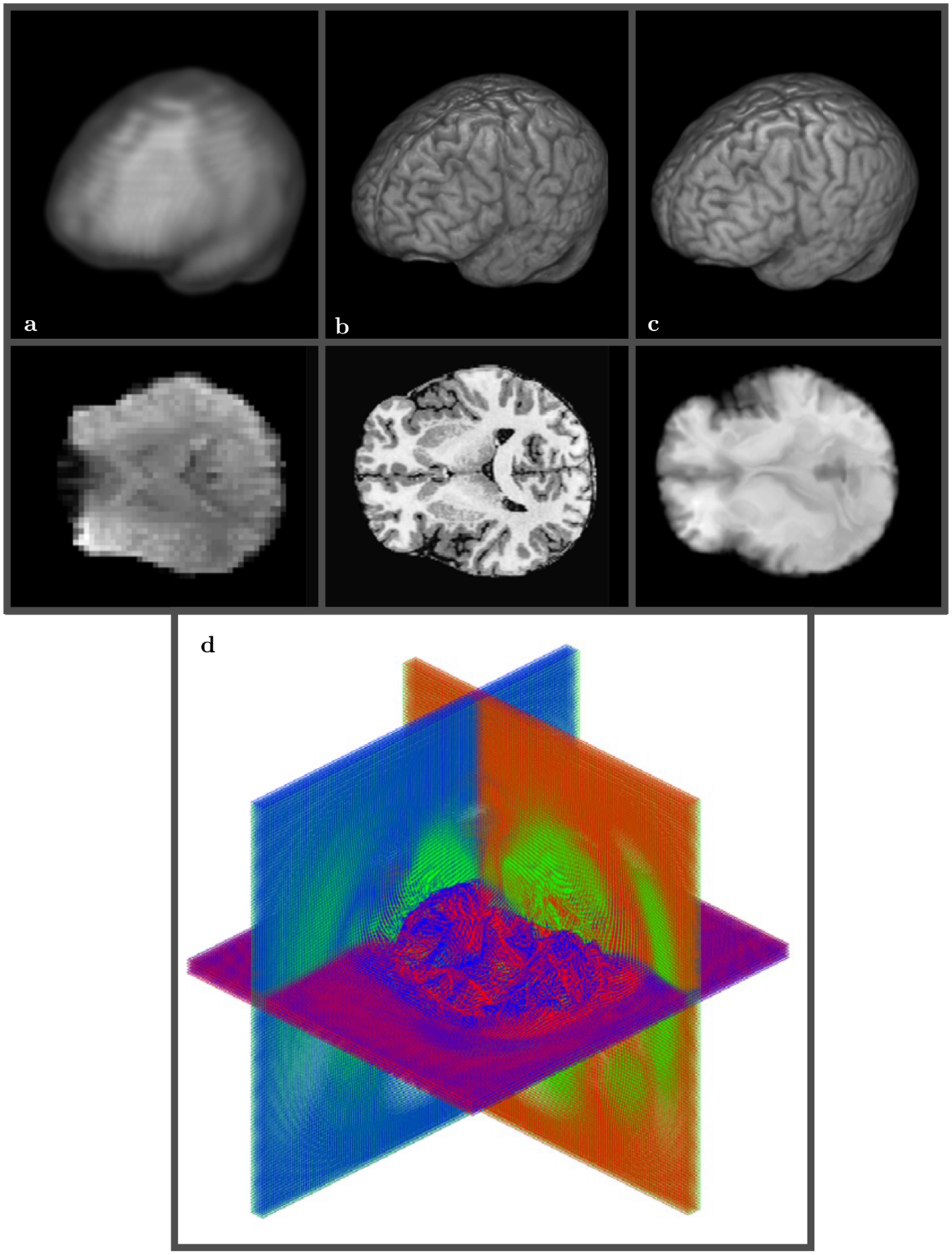
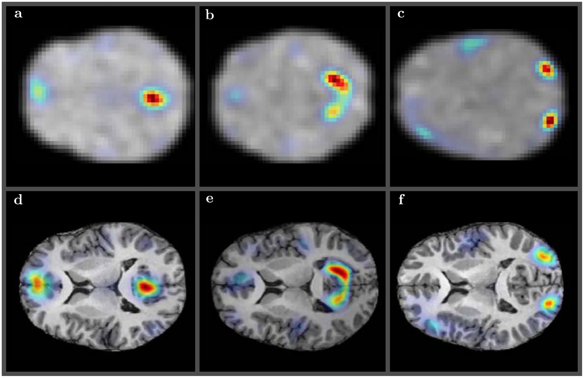
Methods: A new registration method is presented that utilizes a Hamiltonian formalism and constructs registration as a sequence of symplectomorphic maps in conjunction with a novel phase space regularization. For validation of the framework a panel of deformations expressed in analytical form is developed that includes deformations based on known physical processes in MRI and reproduces various distortions and artifacts typically present in images collected using these different MRI modalities.
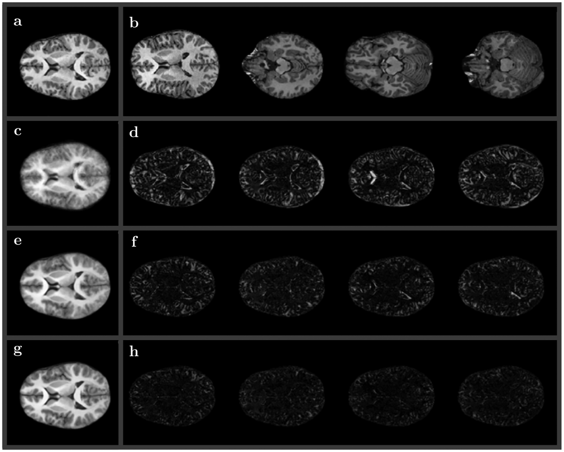
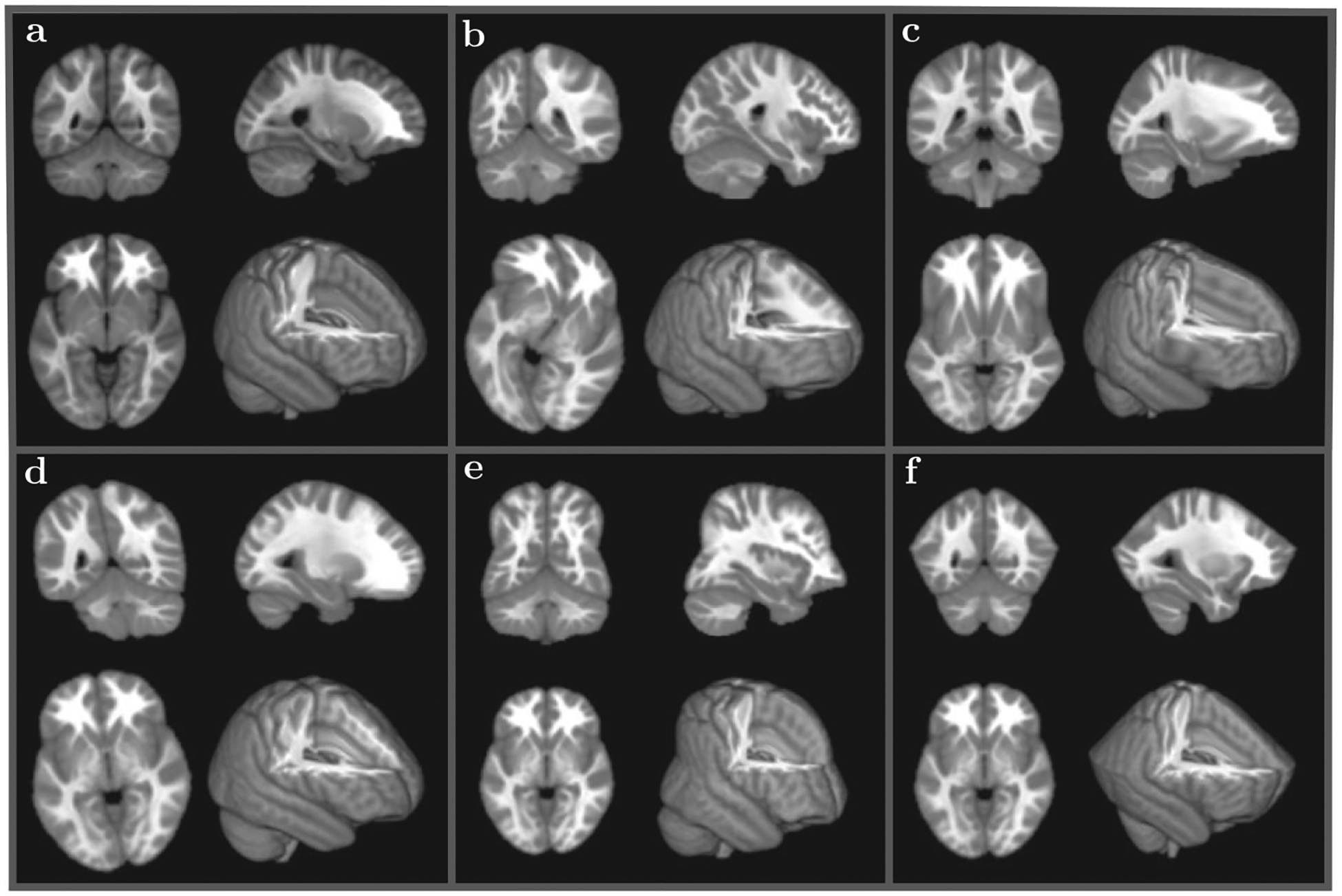
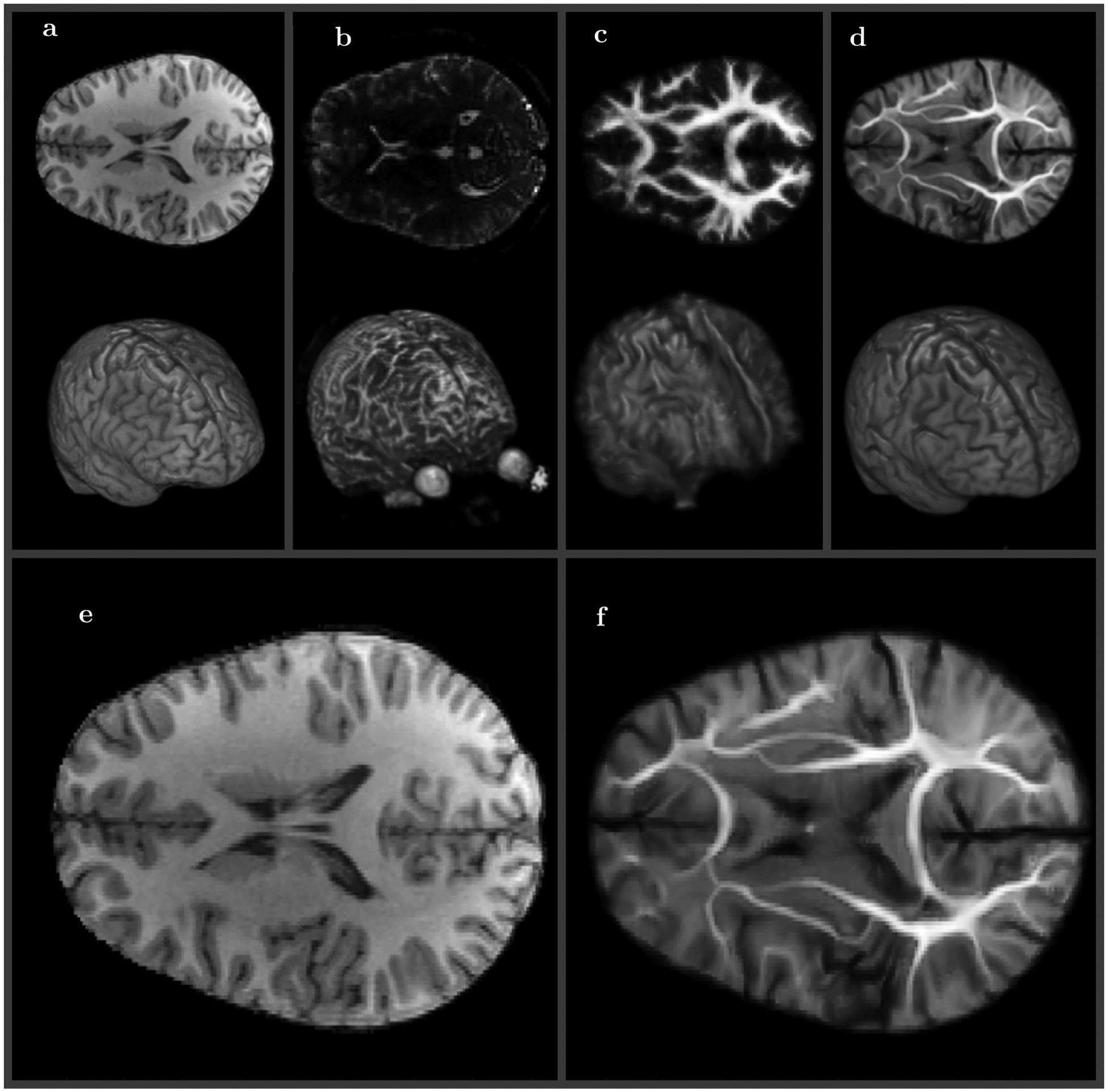
Results: The method is demonstrated on the three different magnetic resonance imaging (MRI) modalities by mapping between high resolution anatomical (HRA) volumes, medium resolution diffusion weighted MRI (DW-MRI) and HRA volumes, and low resolution functional MRI (fMRI) and HRA volumes.
Conclusions: The method has shown an excellent performance and the panel of deformations was instrumental to quantify its repeatability and reproducibility in comparison to several available alternative approaches.
Keywords: diffeomorphic mapping; diffusion tensor imaging; functional MRI; non-linear registration; symplectomorphic mapping.
© 2018 International Society for Magnetic Resonance in Medicine.
Figures
Similar articles
-
Eddy-current-induced distortion correction using maximum reconciled mutual information in diffusion MR imaging.Int J Comput Assist Radiol Surg. 2019 Mar;14(3):463-472. doi: 10.1007/s11548-018-01901-1. Epub 2019 Jan 25. Int J Comput Assist Radiol Surg. 2019. PMID: 30684107
-
In-plane "superresolution" MRI with phaseless sub-pixel encoding.Magn Reson Med. 2018 Dec;80(6):2384-2392. doi: 10.1002/mrm.27209. Epub 2018 Apr 15. Magn Reson Med. 2018. PMID: 29656440
-
Novel distortion correction method for diffusion-weighted imaging based on non-rigid image registration between low b value image and anatomical image.Magn Reson Imaging. 2019 Apr;57:277-284. doi: 10.1016/j.mri.2018.12.002. Epub 2018 Dec 10. Magn Reson Imaging. 2019. PMID: 30543851
-
Co-registration and distortion correction of diffusion and anatomical images based on inverse contrast normalization.Neuroimage. 2015 Jul 15;115:269-80. doi: 10.1016/j.neuroimage.2015.03.050. Epub 2015 Mar 27. Neuroimage. 2015. PMID: 25827811 Free PMC article.
-
Correction of susceptibility artifacts in diffusion tensor data using non-linear registration.Med Image Anal. 2007 Dec;11(6):588-603. doi: 10.1016/j.media.2007.05.004. Epub 2007 Jun 9. Med Image Anal. 2007. PMID: 17664081
Cited by
-
Diffusion MRI tractography of the locus coeruleus-transentorhinal cortex connections using GO-ESP.Magn Reson Med. 2022 Apr;87(4):1816-1831. doi: 10.1002/mrm.29088. Epub 2021 Nov 18. Magn Reson Med. 2022. PMID: 34792198 Free PMC article.
-
Universal theory of brain waves: from linear loops to nonlinear synchronized spiking and collective brain rhythms.Phys Rev Res. 2020 Apr-Jun;2(2):023061. doi: 10.1103/PhysRevResearch.2.023061. Epub 2020 Apr 21. Phys Rev Res. 2020. PMID: 33718881 Free PMC article.
-
Imaging of brain electric field networks with spatially resolved EEG.Elife. 2025 Jun 5;13:RP100123. doi: 10.7554/eLife.100123. Elife. 2025. PMID: 40472276 Free PMC article.
-
Imaging of brain electric field networks with spatially resolved EEG.Res Sq [Preprint]. 2025 Mar 12:rs.3.rs-2432269. doi: 10.21203/rs.3.rs-2432269/v3. Res Sq. 2025. Update in: Elife. 2025 Jun 05;13:RP100123. doi: 10.7554/eLife.100123. PMID: 38659785 Free PMC article. Updated. Preprint.
-
JEDI: Joint Estimation Diffusion Imaging of macroscopic and microscopic tissue properties.Magn Reson Med. 2020 Aug;84(2):966-990. doi: 10.1002/mrm.28141. Epub 2020 Jan 9. Magn Reson Med. 2020. PMID: 31916626 Free PMC article.
References
-
- Christensen GE, Rabbitt RD, Miller MI. 3D brain mapping using a deformable neuroanatomy. Phys Med Biol. 1994;39:609–618. - PubMed
-
- Ashburner J A fast diffeomorphic image registration algorithm. Neuroimage. 2007;38:95–113. - PubMed
-
- Narayanan R, Fessler JA, Park H, Meyerl CR. Diffeomorphic nonlinear transformations: a local parametric approach for image registration. Inf Process Med Imaging. 2005;19:174–185. - PubMed
-
- Vercauteren T, Pennec X, Perchant A, Ayache N. Symmetric log-domain diffeomorphic Registration: a demonsbased approach. Med Image Comput Comput Assist Interv. 2008;11:754–761. - PubMed
-
- Li X, Long X, Laurienti P, Wyatt C. Registration of images with varying topology using embedded maps. IEEE Trans Med Imaging. 2012;31:749–765. - PubMed
Publication types
MeSH terms
Grants and funding
LinkOut - more resources
Full Text Sources
Other Literature Sources
