Review
. 2018 Sep;20 Suppl 2(Suppl 2):28-50.
doi: 10.1111/dom.13378.

Biosynthesis, structure, and folding of the insulin precursor protein

Affiliations
Review

Biosynthesis, structure, and folding of the insulin precursor protein

Ming Liu et al. Diabetes Obes Metab. 2018 Sep.

Abstract

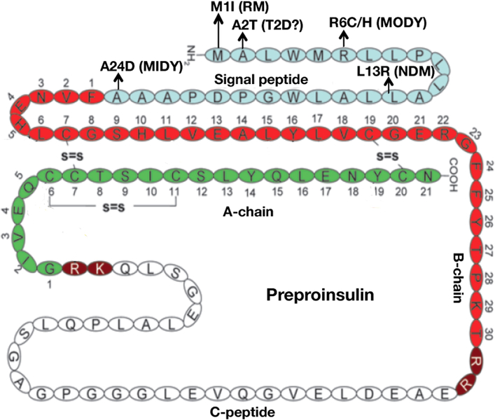
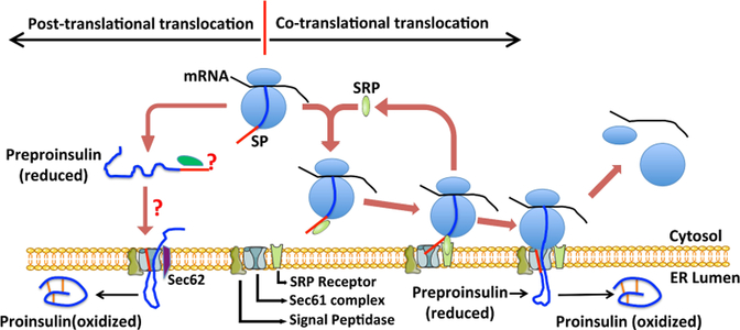
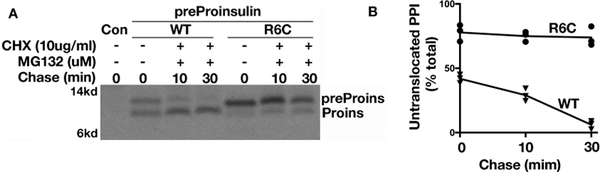
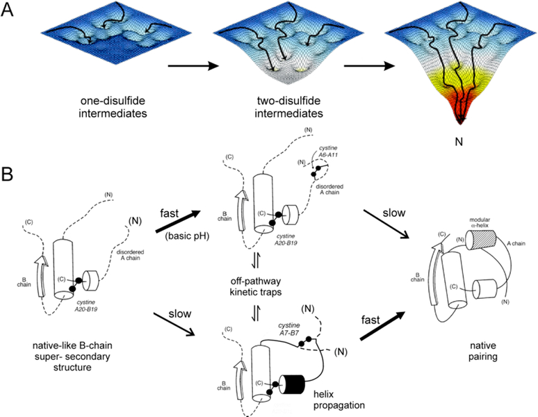
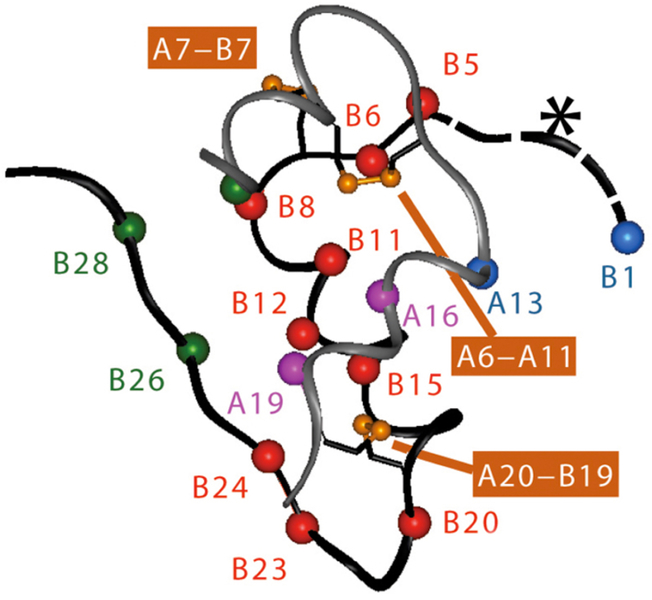
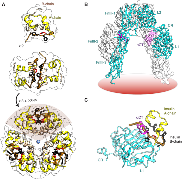
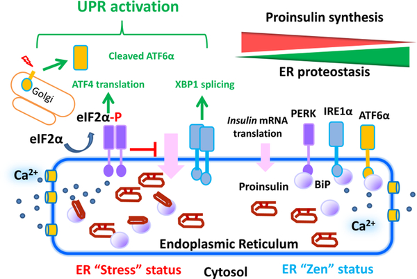
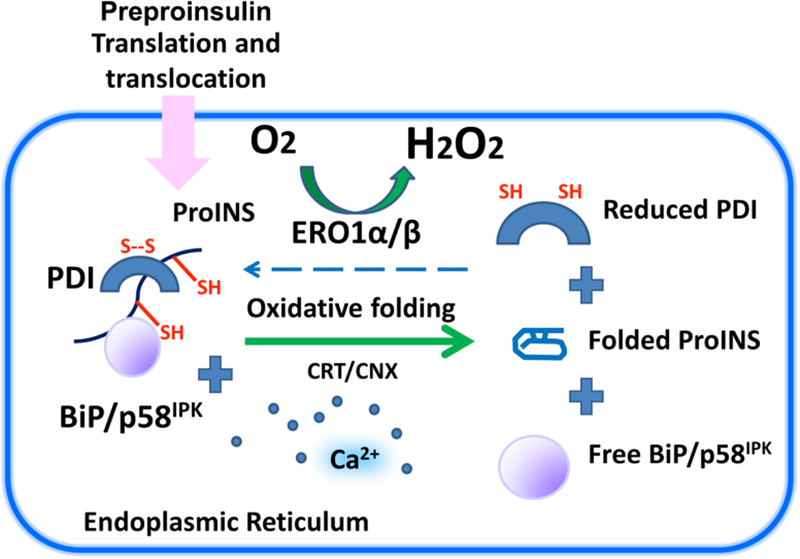
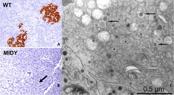
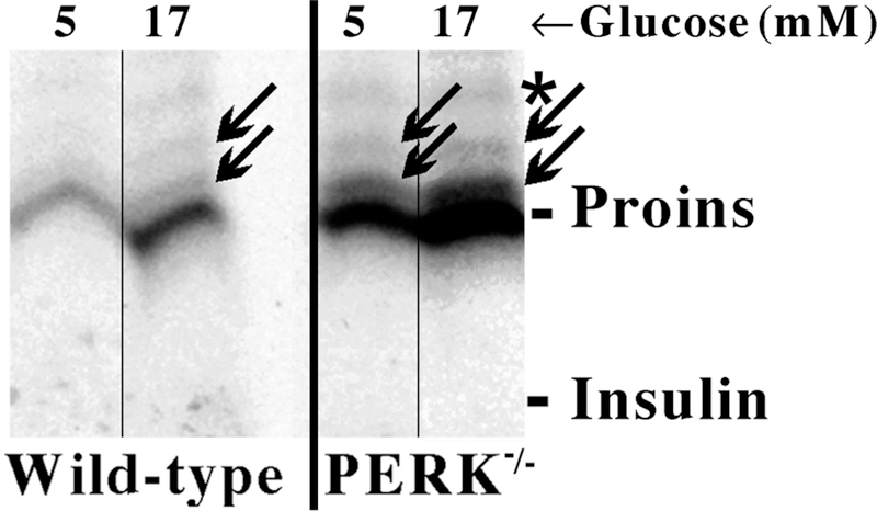
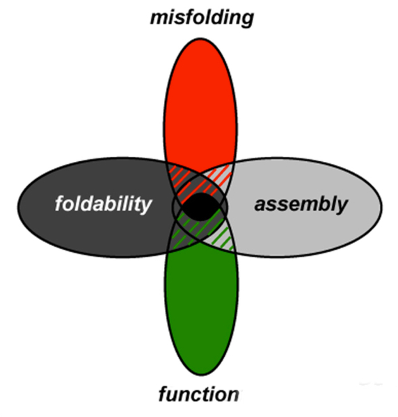
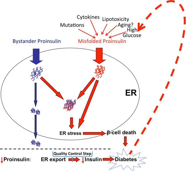
Insulin synthesis in pancreatic β-cells is initiated as preproinsulin. Prevailing glucose concentrations, which oscillate pre- and postprandially, exert major dynamic variation in preproinsulin biosynthesis. Accompanying upregulated translation of the insulin precursor includes elements of the endoplasmic reticulum (ER) translocation apparatus linked to successful orientation of the signal peptide, translocation and signal peptide cleavage of preproinsulin-all of which are necessary to initiate the pathway of proper proinsulin folding. Evolutionary pressures on the primary structure of proinsulin itself have preserved the efficiency of folding ("foldability"), and remarkably, these evolutionary pressures are distinct from those protecting the ultimate biological activity of insulin. Proinsulin foldability is manifest in the ER, in which the local environment is designed to assist in the overall load of proinsulin folding and to favour its disulphide bond formation (while limiting misfolding), all of which is closely tuned to ER stress response pathways that have complex (beneficial, as well as potentially damaging) effects on pancreatic β-cells. Proinsulin misfolding may occur as a consequence of exuberant proinsulin biosynthetic load in the ER, proinsulin coding sequence mutations, or genetic predispositions that lead to an altered ER folding environment. Proinsulin misfolding is a phenotype that is very much linked to deficient insulin production and diabetes, as is seen in a variety of contexts: rodent models bearing proinsulin-misfolding mutants, human patients with Mutant INS-gene-induced Diabetes of Youth (MIDY), animal models and human patients bearing mutations in critical ER resident proteins, and, quite possibly, in more common variety type 2 diabetes.

Keywords: Sec61 translocon; disulphide-linked protein complexes; polypeptide chain initiation; secretory protein biosynthetic pathway; unfolded protein response.


Conflict of interest statement

Conflict of interest

None declared.

Figures

Figure 1.
Figure 1.. Human preproinsulin and its signal peptide mutations associated with diabetes in humans.
Preproinsulin is comprised of the signal peptide (light blue), insulin-B chain (red), C-peptide (white), and insulin-A chain (green). Signal peptide mutations reported to cause diabetes in humans are indicated. RM: recessive mutation; T2D: type 2 diabetes; MODY: maturity onset diabetes of the young; NDM: neonatal diabetes; MIDY: mutant INS-gene induced diabetes of youth.
Figure 2.
Figure 2.. Preproinsulin translocation across the ER membrane.
Newly synthesized preproinsulin molecules can be translocated into the ER through both SRP-dependent co-translational translocation and SRP independent post-translational translocation. In co-translational translocation, SRP recognizes and binds the preproinsulin SP emerging from the ribosomes, forming SRP-ribosome-preproinsulin complexes that interact with SRP receptor on the ER membrane, thereby targeting the nascent preproinsulin to the Sec61 translocon. For post-translational translocation, although Sec62 is reportedly involved, the chaperones maintaining translocation competence of fully-synthesized preproinsulin in the cytosol, and its mechanism of delivery to the ER membrane, have not yet been identified. This figure is reprinted with modification, with permission from Vitamins and Hormones (reference 19).
Figure 3.
Figure 3.. The n-region positive charge in preproinsulin signal peptide is critical for efficient post-translational translocated of preproinsulin.
A. 293T cells transfected with plasmids encoding preproinsulin wild-type (WT) or R6C mutant were pulse-labeled with 35S-Met/Cys for 5 min followed by 0, 10, or 30 min of chase in the presence of 10 µM MG132 and 10 µg/ml cycloheximide (CHX). Post-translational translocation of preproinsulin-WT and R6C were analyzed by immunoprecipitation, SDS-PAGE, and phosphorimaging. B. The signal-uncleaved and signal-cleaved forms of preproinsulin were quantified; the signal-uncleaved fraction is shown. Reprinted with permission from the J. Biol. Chem. (reference 26).
Figure 4.
Figure 4.. Energy landscape paradigm.
(A) Successive disulfide pairing enables a sequence of folding trajectories on ever-steeper funnel-shaped free-energy landscapes. (B) Preferred pathway of disulfide pairing. Initial formation of cystine A20-B19 (left) is directed by a nascent hydrophobic core comprising the central B-domain alpha-helix (residues B9-B19), part of the C-terminal B-chain beta-strand (B24-B26), and part of the C-terminal A-domain alpha-helix (A16-A20). Alternative pathways mediate successive disulfide pairing (middle panel) leading in turn to the native state (right). The mechanism of disulfide pairing is perturbed by clinical mutations associated with a monogenic syndrome of DM due to toxic misfolding of the variant proinsulin in the ER. Figure is adapted from Ref [66]; panel A is adapted from an image kindly provided by J. Williamson.
Figure 5.
Figure 5.. Critical sites governing the foldability of proinsulin are widely distributed in insulin.
Asterisk indicates N-terminal segment of the B chain, which promotes foldability but is dispensable in the mature hormone [98]. Conserved side chains in or adjoining the C-terminal -helix of the A chain (LeuA16 and TyrA19; Ca purple spheres) and at multiple sites in the B chain (Ca red spheres) impair insulin chain combination in accord with studies of mutant proinsulins in mammalian cell lines and the distribution of clinical non-cysteine mutations in the insulin gene. Contacts between the side chains PheB1 and IleA13 (Ca blue spheres), although not well ordered in the native state, contribute to the cellular foldability of proinsulin. Residues IleA2, TyrB26, and ProB28 (Ca green spheres) contribute to the structure and stability of the native state but are not required for efficient disulfide pairing in chain combination. Disulfide bridges are as indicated (orange). Coordinates were obtained from Protein Databank file 4INS and correspond to molecule 1 of the classical 2-Zn insulin hexamer [99].
Figure 6A
Figure 6A. Structure and assembly of insulin with key roles of PheB24.
Assembly of zinc insulin hexamer. The monomeric hormone (A- and B chains, top panel) forms zinc-free dimers via anti-parallel association of B-chain α-helices and C-terminal β-strands (brown, middle panel); two zinc ions then mediate assembly of three dimers to form classical hexamer (T6, bottom panel). The A chain is shown in yellow (ribbon), and the B chain in light brown (B1-B19) or brown (B20-B30). Conserved aromatic residues PheB25 and TyrB26 are shown as black sticks whereas PheB24 is red. The Zn2+ ion is depicted in blue. The figure was in part modified from reference [109] with permission of the authors. Coordinates were obtained from 4INS. Figure 6B and6C. Structure of IR receptor ectodomain. (B) Λ-shaped IR ectodomain homodimer. One protomer is shown as a ribbon (labeled), and the other as molecular surface. Domains are: L1, first leucine-rich repeat domain; CR, cysteine-rich domain; L2, second leucine-rich repeat domain; FnIII-1–3, respective first, second and third fibronectin Type III domains; and αCT, α-chain C-terminal segment. (C) Model illustrating insulin in its classical free conformation bound to Site 1 of the microreceptor (L1-CR + αCT 704–719; designated IR) [104, 105]. L1 and part of CR are shown in cyan, and αCT in magenta. PheB24, PheB25 and TyrB26 are as in Figure 6A. The B chain is shown in dark gray (B6-B19); the position of the brown tube (residues B20-B30) would lead to steric clash between B26-B30 and αCT. The figure was in part modified from references [109] and [110] with permission of the authors. Coordinates were obtained from PDB entries 4OGA, 2DTG, and 3W11.
Figure 7.
Figure 7.. Illustration of ER stress response pathways and their activation by excessive proinsulin synthesis.
Increased synthesis and abundance of newly-made proinsulin correlates with increased BiP binding to proinsulin, which is associated with dimerization and activation of PERK and IRE1α proteins, and translocation of full-length ATF6α to the Golgi complex for proteolytic processing. Subsequent translation of ATF4 and spliced XBP1, together with the liberated cytosolic domain of ATF6α, activate major transcriptional limbs of the stress response designed to restore proteostasis in the ER.
Figure 8.
Figure 8.. Proinsulin undergoes oxidative folding upon biosynthetic delivery into the ER lumen.
Upon arrival in the ER lumen, proinsulin undergoes oxidative folding to form its three essential disulfide bonds, which is thought to be facilitated by members of the PDI family of ER oxidoreductases. The Ero1 family of upstream oxidases are considered important in shuttling the reducing equivalents (originating from proinsulin substrate) to molecular oxygen, while regenerating oxidized PDI proteins. In addition, Ca2+ dependent and –independent chaperones such as BiP and p58IPK also play indispensable roles in assisting proinsulin folding, along with key ER resident proteins for other secretory protein substrates, such as calreticulin and calnexin.
Figure 9:
Figure 9:. Pancreata and beta cells of transgenic MIDY pigs, reproduced from [226, 227].
(A) WT pig pancreas stained by immunoperoxidase with anti-insulin, counterstained with DAPI. (B) Two-year old transgenic MIDY pig pancreas stained as in panel A. (C) Transmission electron microscopy of pancreas from a 4.5-month old transgenic MIDY pig bearing only few beta cell secretory granules (arrows) and hypertrophied ER (asterisks).
Figure 10:
Figure 10:. Two distinct nonnative intramolecular disulfide isomers of proinsulin (at arrows; the native isomer position is also indicated).
Isolated mouse pancreatic islets were preincubated for 40 min at the respective glucose concentrations, and were then pulse-labeled with 35S-amino acids at these glucose concentrations. The islets were lysed, immunoprecipitated with anti-insulin, and analyzed by nonreducing Tris-tricine-urea-SDS-PAGE as in [93]. An asterisk identifies a proinsulin conversion intermediate, the processing of which takes place beyond the ER.
Figure 11.
Figure 11.. Evolution of insulin is constrained by multiple factors.
Venn diagram illustrating influence of protein misfolding, foldability and assembly as well as the traditional importance of direct biological function at the receptor level.
Figure 12:
Figure 12:. An hypothesis linking proinsulin misfolding to the pathogenesis of type 2 diabetes.
Proinsulin must fold to become exported from the endoplasmic reticulum (ER) and make insulin (pathway in blue). Many factors may cause proinsulin misfolding (see text). Misfolded proinsulin molecules (in red) recruit bystander proinsulin molecules into complexes defective for ER export. Decreased export of proinsulin decreases insulin production and secretion leading to higher blood glucose. Hyperglycemia is a factor promoting additional biosynthesis of misfolded proinsulin creating a dangerous positive feedback loop that promotes diabetes. Ultimately, high level proinsulin misfolding can trigger ER stress and beta cell death.

References

    1. Jainandunsing S, van Miert JNI, Rietveld T, Darcos Wattimena JL, Sijbrands EJG, de Rooij FWM. A stable isotope method for in vivo assessment of human insulin synthesis and secretion. Acta Diabetol 2016; 53: 935–944 - PMC - PubMed
    1. Gold G, Gishizky ML, Grodsky GM. Evidence that glucose “marks” beta cells resulting in preferential release of newly synthesize insulin. Science 1982; 218: 56–58 - PubMed
    1. Papa FR. Endoplasmic reticulum stress, pancreatic beta-cell degeneration, and diabetes. Cold Spring Harb Perspect Med 2012; 2: a007666. - PMC - PubMed
    1. Hou JC, Min L, Pessin JE. Insulin granule biogenesis, trafficking and exocytosis. Vitam Horm 2009; 80: 473–506 - PMC - PubMed
    1. Arvan P, Castle D. Sorting and storage during secretory granule biogenesis: looking backward and looking forward. Biochem J 1998; 332: 593–610 - PMC - PubMed

Publication types