. 2018 Nov;22(11):5732-5742.
doi: 10.1111/jcmm.13849. Epub 2018 Sep 19.

PKCα replaces AMPK to regulate mitophagy: Another PEDF role on ischaemic cardioprotection

Affiliations

PKCα replaces AMPK to regulate mitophagy: Another PEDF role on ischaemic cardioprotection

Haoran Miao et al. J Cell Mol Med. 2018 Nov.

Abstract

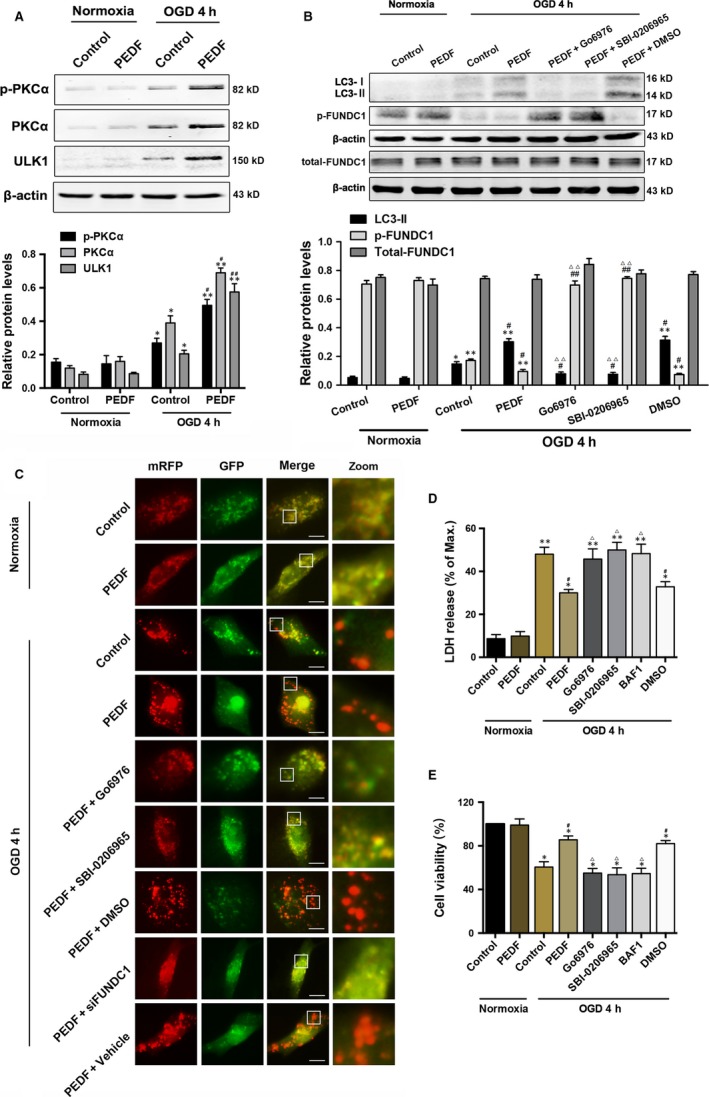
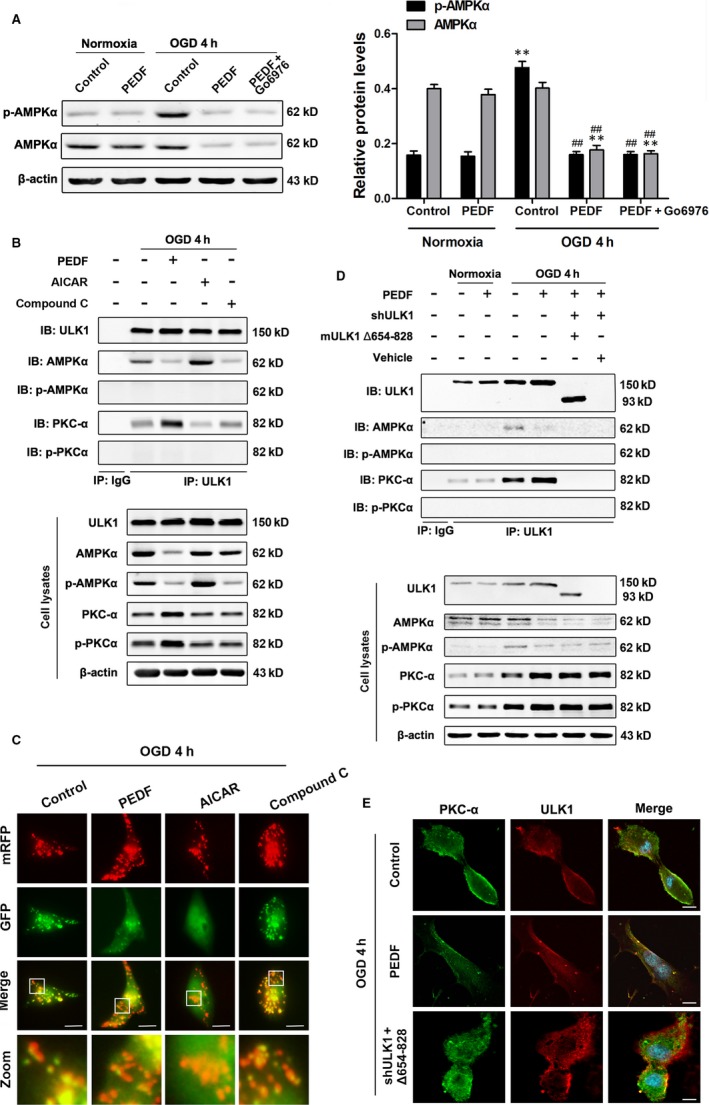
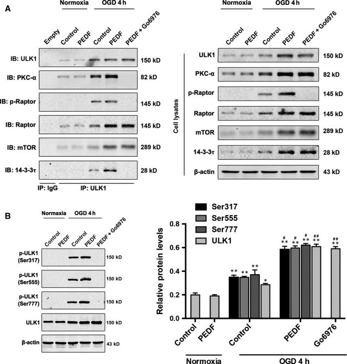
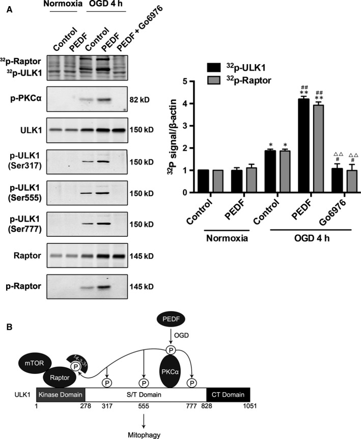
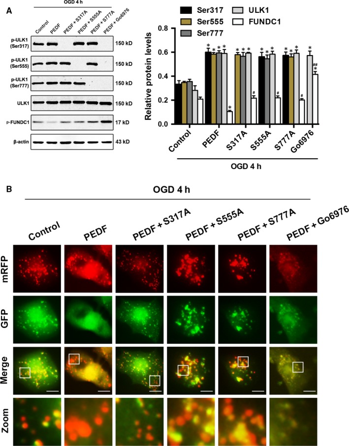
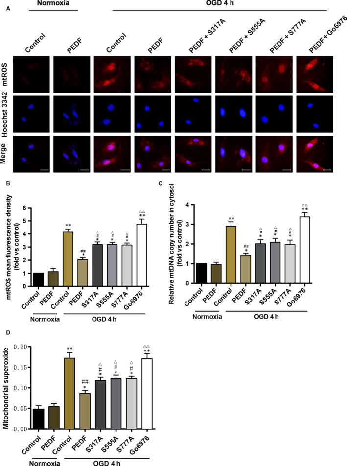
Both decreased autophagy positive regulator AMP activated protein kinase (AMPK) level and promoted mitophagy are observed in oxygen-glucose deprivation (OGD) cardiomyocytes treated with pigment epithelium-derived factor (PEDF). This contradictory phenomenon and its underlying mechanisms have not been thoroughly elucidated. Our previous study reveals that PEDF increases the protein kinase Cα (PKCα) and phospho-PKCα (p-PKCα) contents to promote mitophagy. Thus, we investigated the association between PKCα and mitophagy. Here we identify an interaction between PKCα and Unc-51-like kinase 1 (ULK1), essential component of mitophagy. Further analyses show this is a direct interaction within a domain of ULK1 that termed the serine/threonine-rich domain (S/T domain). Notably, a deletion mutant ULK1 that lacks the binding domain is defective in mediating PEDF-induced mitophagy. Furthermore, we demonstrate that ULK1 is phosphorylated at Ser317/555/777 and Raptor is also phosphorylated by phospho-PKCα. Phospho-ULK1 (p-ULK1) at these sites are all essential for PEDF-induced mitophagy and reduce the release of mitochondrial ROS and DNA. This study therefore identifies a previously uncharacterized interaction between the ULK1 and PKCα that can replace the AMPK-dependent mitophagy processes.

Keywords: Phosphorylation; Unc-51-like kinase 1; cardioprotection; mitophagy; pigment epithelium-derived factor; protein kinase Cα.


Figures

Figure 1
Figure 1
Exogenous PEDF in OGD cardiomyocytes is cardioprotective via PEDFPKCα‐ULK1‐FUNDC1 pathway. A, Protein levels of p‐PKCα, PKCα and ULK1 in cardiomyocytes treated with 10 nmol/L PEDF or not under normal conditions or subjected to oxygen‐glucose deprivation (OGD) for 4 hours, n = 6. B, Western bolt for LC3‐I, LC3‐II, p‐FUNDC1 and FUNDC1 in cardiomyocytes treated with PEDF (10 nmol/L), PKCα inhibitor Go6976 (1 μmol/L), ULK1 inhibitor SBI‐0206965(1 μmol/L) or DMSO before OGD 4 hours, n = 6. C, Representative images showing LC3 staining in different groups of cardiomyocytes infected with GFPRFPLC3 adenovirus for 24 hours, bar = 60 μm. Cardiomyocytes were treated with PEDF, Go6976, SBI‐0206965, DMSO and siFUNDC1 under normal conditions or before OGD for 4 hours, n = 30 from three independent experiments. D and E, Cell viability and cell death were assayed by (D) CCK‐8 and (E) LDH release assays. Cardiomyocytes were treated with PEDF, Go6976, SBI‐0206965, mitophagy inhibitor bafilomycin A1 (BAF1; 50 nmol/L) or DMSO under normal conditions or before OGD for 4 hours, n = 6. *P < 0.05, **P < 0.01 vs normal control, # P < 0.05, ## P < 0.01 vsOGD control, Δ P < 0.05, ΔΔ P < 0.01 vs OGD+PEDF group
Figure 2
Figure 2
PKCα interacts with the S/T domain of ULK1. A, Protein levels of p‐AMPKα, AMPKα in cardiomyocytes treated with PEDF or not under normal conditions or subjected to OGD for 4 hours, n = 6. B, Cardiomyocytes were treated with PEDF, AMPKα inhibitor (Compound C, 10 μmol/L) or AMPKα activator (AICAR, 1 mmol/L), then immunoprecipitated with ULK1 antibody. ULK1, AMPKα, p‐AMPKα, PKCα and p‐PKCα were determined using their antibodies, n = 4. IP: immunoprecipitation, IB: immunoblotting. C, Representative images showing LC3 staining in different groups of Cardiomyocytes infected with GFPRFPLC3 adenovirus for 24 hours, bar = 60 μm. Cardiomyocytes were treated with PEDF, AICAR and Compound C before OGD for 4 hours, n = 30 from three independent experiments. D, Cardiomyocytes were treated with PEDF, lentiviral short hairpin RNA targeting rat ULK1 (shULK1), deletion mutant ULK1 (mULK1 Δ654‐828) or empty vehicle, then immunoprecipitated with ULK1 antibody. ULK1, AMPKα, p‐AMPKα, PKCα and p‐PKCα were determined using their antibodies, n = 4. IP: immunoprecipitation, IB: immunoblotting. E, Confocal immunofluorescence analysis of PKCα interaction with ULK1 in cardiomyocytes, bar = 60 μm. **P < 0.01 vs normal control, ## P < 0.01 vs OGD control
Figure 3
Figure 3
PKCα induces the phosphorylation of Raptor and ULK1 at S317, S555 and S777. A, Cardiomyocytes were treated with PEDF or PEDF+Go6976 and then immunoprecipitated with ULK1 antibody. ULK1, PKCα, p‐Raptor, Raptor, mTOR and 14‐3‐3τ were determined using their antibodies, n = 5. (B) Western bolt for p‐ULK1 (Ser317, 555, 777) and ULK1 in cardiomyocytes treated with PEDF or PEDF+Go6976 under normal conditions or before OGD 4 hours, n = 6. *P < 0.05, **P < 0.01 vs relative normal control, # P < 0.05, ## P < 0.01 vs relative OGD control
Figure 4
Figure 4
Phospho‐PKCα contributes its phosphate group to phospho‐ULK1 and phospho‐Raptor. A, Cardiomyocytes were deprived of oxygen and glucose 4 hours as indicated after treated with PEDF and PEDF+Go6976. p‐ULK1 and p‐Raptor were immunoblotted and phosphorylation was examined by 32P‐autoradiogram. Proteins were resolved by SDSPAGE and visualized with autoradiogram (top) and Western blot (WB; bottom), n = 6. B, Schematic representation of PEDF‐mediated p‐PKCα induces the phosphorylation of ULK1 and suppresses the inhibitory effect of mTOR on the ULK1 autophagic complex. *P < 0.05, **P < 0.01 vs normal control, # P < .05, ## P < 0.01 vs OGD control, ΔΔ P < 0.01 vs OGD+PEDF group
Figure 5
Figure 5
Phospho‐ULK1 induced by PEDF is important for FUNDC1‐mediated mitophagy. A, Western bolt for p‐ULK1 (Ser317, 555, 777), ULK1 and p‐FUNDC1 in cardiomyocytes treated with PEDF or PEDF+Go6976 before OGD 4 for hours, n = 6. S317/555/777A mutants were transfected into cardiomyocytes as indicated. *P < 0.05 vs relative normal control, # P < 0.05, ## P < 0.01 vs relative OGD control. (B) Representative images showing LC3 staining in different groups of cardiomyocytes infected with GFPRFPLC3 adenovirus for 24 hours, bar = 60 μm. Cardiomyocytes were treated with PEDF or PEDF+Go6976before OGD 4 hours, n = 30 from three independent experiments
Figure 6
Figure 6
The release of mitochondrial ROS and DNA in cytosol is decreased by PEDF‐induced phospho‐ULK1. A and B, Fluorescence microscope of (A) Mito‐SOX Red‐labelled ROS production and release in cytosol and quantification of (B) fluorescence density, bar = 100 μm, n = 30 from 3 independent experiments. Cardiomyocytes were treated with PEDF or PEDF+Go6976 under normal conditions or before OGD 4 hours. S317/555/777A mutants were transfected into cardiomyocytes as indicated. C, Cytosolic mitochondrial DNA copy number was measured by quantitative PCR, n = 7. Wild type or S317/555/777A mutational cardiomyocytes were treated with PEDF or Go6976 under normal conditions or before OGD 4 hours. D, Quantification of mitochondrial superoxide was detected with flow cytometric, n = 3. Wild type or S317/555/777A mutational cardiomyocytes were treated with PEDF or Go6976 under normal conditions or before OGD 4 hours. *P < 0.05, **P < 0.01 vs normal control, # P < 0.05, ## P < 0.01 vs OGD control, Δ < 0.05, ΔΔ P < 0.01 vs OGD+PEDF group

References

    1. Nichols M, Townsend N, Scarborough P, Rayner M. Cardiovascular disease in Europe 2014: epidemiological update. Eur Heart J. 2014;35:2929. - PubMed
    1. Yeh RW, Sidney S, Chandra M, Sorel M, Selby JV, Go AS. Population trends in the incidence and outcomes of acute myocardial infarction. N Engl J Med. 2010;362:2155‐2165. - PubMed
    1. Roger VL, Go AS, Lloyd‐Jones DM, et al. ; Wylie‐Rosett J ; American Heart Association Statistics C ; Stroke Statistics S . Heart disease and stroke statistics–2011 update: a report from the American Heart Association. Circulation 2011;123:e18‐e209. - PMC - PubMed
    1. Matsuda N, Sato S, Shiba K, et al. PINK1 stabilized by mitochondrial depolarization recruits Parkin to damaged mitochondria and activates latent Parkin for mitophagy. J Cell Biol. 2010;189:211‐221. - PMC - PubMed
    1. Kubli DA, Gustafsson AB. Mitochondria and mitophagy: the yin and yang of cell death control. Circ Res. 2012;111:1208‐1221. - PMC - PubMed

Publication types

MeSH terms