. 2018 Sep 19;13(9):e0202590.
doi: 10.1371/journal.pone.0202590. eCollection 2018.

Therapeutic potential of extracellular vesicles derived from human mesenchymal stem cells in a model of progressive multiple sclerosis

Affiliations

Therapeutic potential of extracellular vesicles derived from human mesenchymal stem cells in a model of progressive multiple sclerosis

Fernando Laso-García et al. PLoS One. .

Abstract

Extracellular vesicles (EVs) have emerged as important mediators of intercellular communication and as possible therapeutic agents in inflammation-mediated demyelinating diseases, including multiple sclerosis (MS). In the present study, we investigated whether intravenously administered EVs derived from mesenchymal stem cells (MSCs) from human adipose tissue might mediate recovery in Theiler's murine encephalomyelitis virus (TMEV)-induced demyelinating disease, a progressive model of MS. SJL/J mice were subjected to EV treatment once the disease was established. We found that intravenous EV administration improved motor deficits, reduced brain atrophy, increased cell proliferation in the subventricular zone and decreased inflammatory infiltrates in the spinal cord in mice infected with TMEV. EV treatment was also capable of modulating neuroinflammation, given glial fibrillary acidic protein and Iba-1 staining were reduced in the brain, whereas myelin protein expression was increased. Changes in the morphology of microglial cells in the spinal cord suggest that EVs also modulate the activation state of microglia. The clear reduction in plasma cytokine levels, mainly in the Th1 and Th17 phenotypes, in TMEV mice treated with EVs confirms the immunomodulatory ability of intravenous EVs. According to our results, EV administration attenuates motor deficits through immunomodulatory actions, diminishing brain atrophy and promoting remyelination. Further studies are necessary to establish EV delivery as a possible therapy for the neurodegenerative phase of MS.


Conflict of interest statement

The authors have declared that no competing interests exist.

Figures

Fig 1
Fig 1. Experimental protocol scheme.
EVs were isolated from human adipose tissue-derived MSCs and subsequently characterised. Four- to six-week-old female SJL/J mice were inoculated intracerebrally into the right hemisphere with 2 × 10⁶ plaque-forming units of the Daniel strain of TMEV. On day 60 postinfection, treatment (saline or EVs) was administered through the tail vein. Distribution of mice: sham (n = 7), TMEV-VH (n = 10) and TMEV-EVs (n = 10). On day 75 postinfection, we evaluated mouse behaviour and performed imaging, biomarker, histological and molecular studies. Abbreviations: TMEV: Theiler’s murine encephalomyelitis virus; EVs: extracellular vesicles; TMEV-EVs: Theiler’s murine encephalomyelitis virus with EV treatment; TMEV-VH: Theiler’s murine encephalomyelitis virus with saline treatment; MRI: magnetic resonance imaging; IH: immunohistochemistry; IF: immunofluorescence; WB: western blot.
Fig 2
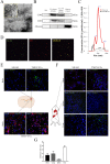
Fig 2. EV characterisation and biodistribution after intravenous administration.
EVs were characterised using various techniques: (A) Electron microscope image. EVs with size smaller than 100 nm were observed by electron microscope. (B) Western blot. Detection of EVs with specific markers (positive: CD81 and CD63; negative: albumin) by western blot assay. Negative control samples are supernatant with debris and dead cells. The gels image was cropped. (C) Characteristics of particles. Size and concentration of the particles detected in the sample EVs isolated and EVs-FBS (culture media) by NanoSight. (D) Phenotype of EVs. Immunofluorescence of EVs labelled with DiI (red) and the specific EV marker antibody anti-ALIX (green). (E) Biodistribution of the EVs in the brain. Top: after intravenous administration, EVs were observed in the brain by the TAPA-1 marker (green), using immunofluorescence. Bottom: Colabelling with EVs (green) and cellular markers (GFAP/NeuN) (red) in the brain, using immunofluorescence. (F) Biodistribution of the EVs in peripheral organs. EVs were found in various peripheral organs, such as the lungs, spleen and liver, by immunofluorescence (green). (G) EV quantification. Fluorescence intensity quantification (arbitrary units) in brain and peripheral organs. Abbreviations: EVs: extracellular vesicles; human ADMSCs: human adipose tissue-derived mesenchymal stem cells; EVs-FBS: extracellular vesicles -depleted FBS media; DiI: lipophilic Tracer CellTrackerTM CM-DiI; GFAP: glial fibrillary acidic protein; NeuN: neuronal specific nuclear protein.
Fig 3
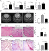
Fig 3
(A) Behavioural evaluation. Behaviour was evaluated by various tests: activity-cage, passive avoidance and object recognition (n = 7 animals in the sham group; n = 10 animals each in the TMEV-VH and TMEV-EVs groups). Data are mean ± SD, *p < .05. (B) Ventricular size. Qualitative and quantitative analysis of T2-weighted MRI images showed significant differences in ventricle sizes (coronal) between the TMEV-VH and TMEV-EVs groups (n = 4 animals per group). (C) Hematoxylin and eosin stain. Spinal cord sections, with hematoxylin and eosin stain, in which inflammatory infiltrates can be observed (black arrows) in the TMEV-VH and TMEV-EVs groups (n = 4 animals per group). (D) Cell proliferation. KI-67-positive cells in the SVZ (n = 10 animals per group). Data are mean ± SD, *p < .05. Abbreviations: EVs: extracellular vesicles; SVZ: subventricular zone; TMEV: Theiler’s murine encephalomyelitis virus; TMEV-EVs: Theiler’s murine encephalomyelitis virus with EV treatment; TMEV-VH: Theiler’s murine encephalomyelitis virus with saline treatment.
Fig 4
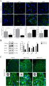
Fig 4. Brain repair-associated markers after EV treatment at 2 weeks post-treatment.
(A) Immunofluorescence of brain repair markers. Significant difference in brain repair marker expression (GFAP, Iba-1, CNPase and MBP) (green) between the TMEV-VH and TMEV-EVs groups by immunofluorescence (n = 4 animals per group). Data are mean ± SD, *p < .05. (B) Western blot. Brain repair marker expression (GFAP, Iba-1, CNPase and MBP) with their quantifications by western blot. Data are mean ± SD, *p < .05. (C) Immunofluorescence in spinal cord. Morphological changes in microglia activation in spinal cord by immunofluorescence with Iba-1 (20X, 40X) (green). EVs: extracellular vesicles; VH: vehicle; TMEV-EVs: Theiler’s murine encephalomyelitis virus with EV treatment; TMEV-VH: Theiler’s murine encephalomyelitis virus with saline treatment; CNPase: 2',3'-cyclic-nucleotide 3'-phosphodiesterase; MBP: myelin basic protein; GFAP: glial fibrillary acidic protein.
Fig 5
Fig 5. Plasma levels of cytokines analysed at the chronic phase and 2 weeks after EV treatment.
The TMEV-VH group showed higher levels of these cytokines in plasma compared with the treated group (n = 7 animals in the sham group; n = 7 animals in the TMEV-VH group and n = 12 animals in the TMEV-EVs group). Data are mean ± SEM, *p < .05 sham vs. TMEV-EVs; Δp < .05 TMEV-VH vs. TMEV-EVs; оp < .05 TMEV-VH vs. sham. Abbreviations: EVs: extracellular vesicles; TMEV-EVs: Theiler’s murine encephalomyelitis virus with EV treatment; TMEV-VH: Theiler’s murine encephalomyelitis virus with saline treatment.

Comment in

References

    1. Noseworthy JH, Lucchinetti C, Rodriguez M, Weinshenker BG. Multiple sclerosis. N Engl J Med. 2000;343:938–952. 10.1056/NEJM200009283431307 - DOI - PubMed
    1. Compston A, Coles A. Multiple sclerosis. Lancet. 2008;372:1502–17. 10.1016/S0140-6736(08)61620-7 - DOI - PubMed
    1. Dutta R, Trapp BD. Mechanisms of neuronal dysfunction and degeneration in multiple sclerosis. Prog Neurobiol. 2011;93:1–12. 10.1016/j.pneurobio.2010.09.005 - DOI - PMC - PubMed
    1. Trapp BD, Nave KA. Multiple sclerosis: an immune or neurodegenerative disorder?. Annu Rev Neurosci. 2008;31:247–69. 10.1146/annurev.neuro.30.051606.094313 - DOI - PubMed
    1. Tsunoda I, Fujinami RS. Inside-Out versus Outside-In models for virus induced demyelination: axonal damage triggering demyelination. Springer Semin Immunopathol. 2002;24:105–125. 10.1007/s00281-002-0105-z - DOI - PMC - PubMed

Publication types

MeSH terms

Substances