

Methodological Approaches to Study Extracellular Vesicle miRNAs in Epstein⁻Barr Virus-Associated Cancers
- PMID: 30231493
- PMCID: PMC6164614
- DOI: 10.3390/ijms19092810
Methodological Approaches to Study Extracellular Vesicle miRNAs in Epstein⁻Barr Virus-Associated Cancers
Abstract
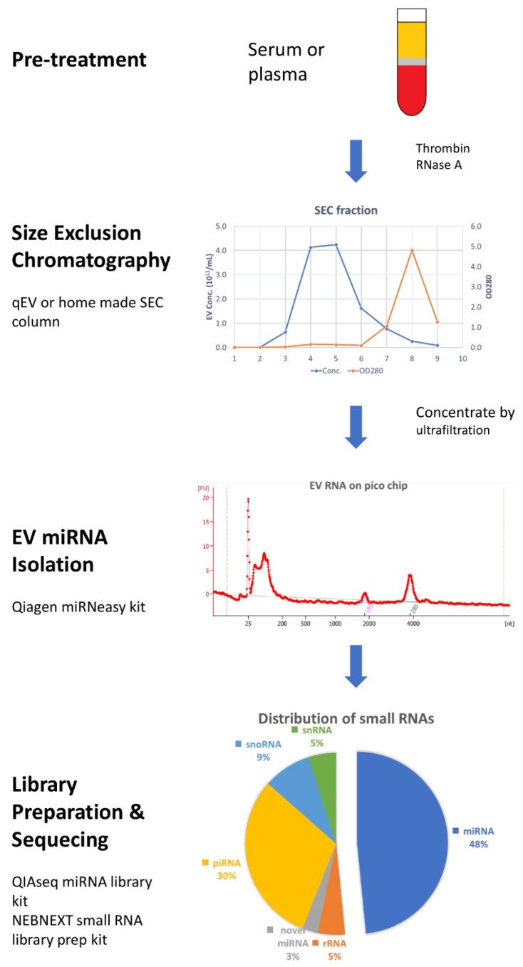
Epstein Barr-virus (EBV) was the first virus identified to be associated with human cancer in 1964 and is found ubiquitously throughout the world's population. It is now established that EBV contributes to the development and progression of multiple human cancers of both lymphoid and epithelial cell origins. EBV encoded miRNAs play an important role in tumor proliferation, angiogenesis, immune escape, tissue invasion, and metastasis. Recently, EBV miRNAs have been found to be released from infected cancer cells in extracellular vesicles (EVs) and regulate gene expression in neighboring uninfected cells present in the tumor microenvironment and possibly at distal sites. As EVs are abundant in many biological fluids, the viral and cellular miRNAs present within EBV-modified EVs may serve as noninvasion markers for cancer diagnosis and prognosis. In this review, we discuss recent advances in EV isolation and miRNA detection, and provide a complete workflow for EV purification from plasma and deep-sequencing for biomarker discovery.
Keywords: exosomes; extracellular vesicle; herpesvirus; microRNA; microvesicles; oncosomes.
Conflict of interest statement
The authors declare no conflict of interest.
Figures
Similar articles
-
Vesicle-bound EBV-BART13-3p miRNA in circulation distinguishes nasopharyngeal from other head and neck cancer and asymptomatic EBV-infections.Int J Cancer. 2019 May 15;144(10):2555-2566. doi: 10.1002/ijc.31967. Epub 2018 Dec 6. Int J Cancer. 2019. PMID: 30411781 Free PMC article.
-
The roles of EBV-encoded microRNAs in EBV-associated tumors.Crit Rev Oncol Hematol. 2019 Mar;135:30-38. doi: 10.1016/j.critrevonc.2019.01.014. Epub 2019 Jan 25. Crit Rev Oncol Hematol. 2019. PMID: 30819444 Review.
-
Epstein-Barr virus microRNAs in the pathogenesis of human cancers.Cancer Lett. 2021 Feb 28;499:14-23. doi: 10.1016/j.canlet.2020.11.019. Epub 2020 Nov 25. Cancer Lett. 2021. PMID: 33248209 Review.
-
Epstein-Barr virus-encoded microRNAs as regulators in host immune responses.Int J Biol Sci. 2018 Apr 5;14(5):565-576. doi: 10.7150/ijbs.24562. eCollection 2018. Int J Biol Sci. 2018. PMID: 29805308 Free PMC article. Review.
-
Role of exosomes as a proinflammatory mediator in the development of EBV-associated lymphoma.Blood. 2018 Jun 7;131(23):2552-2567. doi: 10.1182/blood-2017-07-794529. Epub 2018 Apr 22. Blood. 2018. PMID: 29685921
Cited by
-
Human Pluripotent Stem Cell-Derived Extracellular Vesicles: Characteristics and Applications.Tissue Eng Part B Rev. 2020 Apr;26(2):129-144. doi: 10.1089/ten.TEB.2019.0252. Epub 2020 Jan 16. Tissue Eng Part B Rev. 2020. PMID: 31847715 Free PMC article. Review.
-
Metabolomics of small extracellular vesicles derived from isocitrate dehydrogenase 1-mutant HCT116 cells collected by semi-automated size exclusion chromatography.Front Mol Biosci. 2023 Jan 11;9:1049402. doi: 10.3389/fmolb.2022.1049402. eCollection 2022. Front Mol Biosci. 2023. PMID: 36710884 Free PMC article.
-
Extracellular Vesicles in Epstein-Barr Virus Pathogenesis.Curr Clin Microbiol Rep. 2019 Sep;6(3):121-131. doi: 10.1007/s40588-019-00123-6. Epub 2019 Jul 3. Curr Clin Microbiol Rep. 2019. PMID: 32051811 Free PMC article.
-
The role of the metabolite cargo of extracellular vesicles in tumor progression.Cancer Metastasis Rev. 2021 Dec;40(4):1203-1221. doi: 10.1007/s10555-021-10014-2. Epub 2021 Dec 27. Cancer Metastasis Rev. 2021. PMID: 34957539 Free PMC article. Review.
-
Immunosuppressive Tumor Microenvironment and Immunotherapy of Epstein-Barr Virus-Associated Malignancies.Viruses. 2022 May 10;14(5):1017. doi: 10.3390/v14051017. Viruses. 2022. PMID: 35632758 Free PMC article. Review.
References
Publication types
MeSH terms
Substances
Grants and funding
LinkOut - more resources
Full Text Sources
Other Literature Sources
Research Materials
