. 2018 Dec;17(12):2767-2779.
doi: 10.1158/1535-7163.MCT-18-0348. Epub 2018 Sep 19.

Histone Deacetylase Inhibitors Synergize with Catalytic Inhibitors of EZH2 to Exhibit Antitumor Activity in Small Cell Carcinoma of the Ovary, Hypercalcemic Type

Affiliations

Histone Deacetylase Inhibitors Synergize with Catalytic Inhibitors of EZH2 to Exhibit Antitumor Activity in Small Cell Carcinoma of the Ovary, Hypercalcemic Type

Yemin Wang et al. Mol Cancer Ther. 2018 Dec.

Abstract

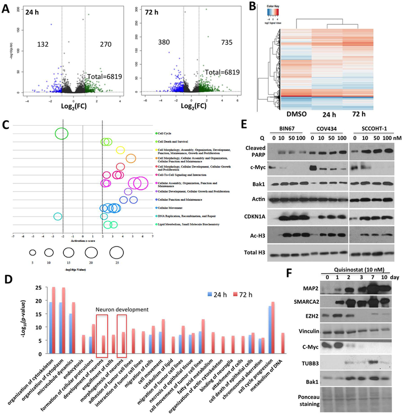
Small cell carcinoma of the ovary, hypercalcemic type (SCCOHT) is a rare but extremely lethal malignancy that mainly impacts young women. SCCOHT is characterized by a diploid genome with loss of SMARCA4 and lack of SMARCA2 expression, two mutually exclusive ATPases of the SWI/SNF chromatin-remodeling complex. We and others have identified the histone methyltransferase EZH2 as a promising therapeutic target for SCCOHT, suggesting that SCCOHT cells depend on the alternation of epigenetic pathways for survival. In this study, we found that SCCOHT cells were more sensitive to pan-HDAC inhibitors compared with other ovarian cancer lines or immortalized cell lines tested. Pan-HDAC inhibitors, such as quisinostat, reversed the expression of a group of proteins that were deregulated in SCCOHT cells due to SMARCA4 loss, leading to growth arrest, apoptosis, and differentiation in vitro and suppressed tumor growth of xenografted tumors of SCCOHT cells. Moreover, combined treatment of HDAC inhibitors and EZH2 inhibitors at sublethal doses synergistically induced histone H3K27 acetylation and target gene expression, leading to rapid induction of apoptosis and growth suppression of SCCOHT cells and xenografted tumors. Therefore, our preclinical study highlighted the therapeutic potential of combined treatment of HDAC inhibitors with EZH2 catalytic inhibitors to treat SCCOHT.


Conflict of interest statement

Conflict of interest: The authors declare no potential conflicts of interest.

Figures

Figure 1.
Figure 1.. SCCOHT cells are sensitive to HDAC inhibition.
(A) Epigenetic drug screen identifies putative therapeutic agents for SCCOHT. Averaged growth inhibition rates of SCCOHT cell lines by individual drug treatment were plotted against that of the other ovarian cell lines. (B) Epigenetic drugs preferentially suppressed the growth of SCCOHT cells vs other cell lines in the drug screen. (C) SCCOHT cells displayed significantly higher cellular sensitivity to pan-HDAC inhibitors SAHA, panobinostat and quisinostat. Cells were treated with HDAC inhibitors at various doses for 6-days before being fixed and quantitated by crystal violet assay. (D) Quisinostat increased histone H3 acetylation rapidly. (E) Quisinostat suppressed the colony formation ability of SCCOHT cells after two weeks. (F) Quisinostat blocked the formation of spheroids in BIN67 cells. Cells were exposed to quisinostat at indicated doses for 7 days immediately after being embedded in matrigel. (G) Quisinostat inhibited the growth of pre-formed SCCOHT spheroids as measured by Celltiter Glo viability assay. * P<0.05, ** P<0.01, *** P<0.001
Figure 2.
Figure 2.. Quisinostat induces cell cycle arrest, apoptosis and differentiation in SCCOHT cells.
(A) Quisinostat suppressed SCCOHT cell proliferation as measured by BrdU incorporation after 72 hours of treatment. (B) SCCOHT cell cycle distribution was determined upon quisinostat treatment for 72 hours. (C) Cell apoptosis were quantitated by quantitating caspase3/7 activation through fluorescent microscope coupled with live cell imaging (see Materials and Methods for details) in SCCOHT cells treated with quisinostat. (D) Quisinostat treatment triggered differentiation of SCCOHT cells. Phase contrast images were taken 72 hours after 50 nM quisinostat treatment. * P<0.05, ** P<0.01, *** P<0.001
Figure 3.
Figure 3.. The effect of quisinostat on the proteome of BIN67 cells.
(A) Volcano plot of the proteome of BIN67 cells exposed to 10 nM of quisinostat for 24 or 72 hours in comparison to vehicle treatment. Peptide data were subjected to PECA analysis for identification of significantly altered proteins (p.fdr<0.05 and |Log2FC|>=1). (B) Clustering analysis of proteins significantly altered by quisinostat treatment for 24 and 72 hours. (C) The enriched biological functions were predicted by IPA analysis of significantly altered proteins caused by qusinostat treatment for 72 hours. (D) Significantly increased (z-score >2) or decreased (z-score <−2) biological functions were predicted by IPA analysis upon quisinostat treatment. (E) The expression of cdk inhibitor CDKN1A and apoptotic proteins were induced upon quisinostat treatment for 24 hours by western blot analysis. (F) The expression of SMARCA2, neuronal markers (MAP2, TUBB3) and stem cell markers (EZH2, c-Myc) were analyzed by western blotting in BIN67 cells treated with 10 nM of quisinostat.
Figure 4.
Figure 4.. Re-instatement of SMARCA4 mimics the effect of HDAC inhibitors in SCCOHT cells through regulating genes involved in cell fate determination.
(A) Volcano plot of the proteome of BIN67 cells that transduced with GFP or SMARCA4 expressing lentivirus for 96 hours. Peptide data were subjected to PECA analysis for identification of significantly altered proteins (p.fdr<0.05 and |Log2FC|≥1). (B) The enriched biological functions were predicted by IPA analysis of significantly altered proteins caused by SMARCA4 re-expression. (C) Re-expression of SMARCA4 caused differentiation of SCCOHT cells. (D) SMARCA4 re-expression and quisinostat treatment regulated a common set of proteins. (E) Proteins co-regulated by SMARCA4 and quisinostat were predicted to significantly activate several biological processes, such as neuritogenesis and decrease cell cycle progression.
Figure 5.
Figure 5.. In vivo efficacy of quisinostat in SCCOHT mouse xenograft models.
(A-C) The efficacy of quisinostat was evaluated in BIN67-derived mouse subcutaneous xenograft model. Average tumor volumes of either vehicle, 10 or 20 mg/kg quisinostat-treated group were plotted against the time post cell inoculation (A). Final tumor weight was determined and compared between treated groups (B). The effect of quisinostat on downstream targets were evaluated by western blotting for tumors harvested at the end of the study (C). (D, E) Quisinostat suppressed the growth of SCCOHT-1 mouse subcutaneous xenograft tumors and increased survival of mice to humane endpoint. * P<0.05, ** P<0.01, *** P<0.001
Figure 6.
Figure 6.. Synergism between quisinostat and EZH2 inhibitor EPZ-6438 in SCCOHT cells.
(A) SCCOHT cells were exposed to combination of quisinostat and EPZ-6438 treatment at various ratio for 6 days and then processed to cell viability measurement by crystal violet assay. Combination index of drugs were calculated using the Chou-Talalay method and the CalcuSyn software. Synergism were predicted when combination index <1 at each drug combination. (B) Cells were pre-treated with DMSO or 0.25 μM EPZ-6438 for 3 days. A cell-permeable apoptosis dye (NucView™ 488 dye) was then added together with or without 2.5 nM quisinostat for monitoring induction of apoptosis. Fluorescent objects were counted for determination of apoptotic index as described in Materials and Methods. (C) Western blot analysis of histone H3K27 acetylation or tri-methylation levels and CDKN1A expression upon 10 nM quisinostat (Q), 0.5 μM EPZ-6438 (E) treatment alone or in combination. (D) The in vivo efficacy of quisinostat and EPZ-6438 drug combination in the BIN67 xenograft model. Average tumor volumes of each treatment arm were plotted against the time post cell inoculation. * P<0.05, ** P<0.01, *** P<0.001

Similar articles

Cited by

References

    1. Longy M, Toulouse C, Mage P, Chauvergne J, Trojani M. Familial cluster of ovarian small cell carcinoma: a new mendelian entity? J Med Genet 1996;33(4):333–5. - PMC - PubMed
    1. Martinez-Borges AR, Petty JK, Hurt G, Stribling JT, Press JZ, Castellino SM. Familial small cell carcinoma of the ovary. Pediatr Blood Cancer 2009;53(7):1334–6 doi 10.1002/pbc.22184. - DOI - PubMed
    1. Florell SR, Bruggers CS, Matlak M, Young RH, Lowichik A. Ovarian small cell carcinoma of the hypercalcemic type in a 14 month old: the youngest reported case. Med Pediatr Oncol 1999;32(4):304–7. - PubMed
    1. Estel R, Hackethal A, Kalder M, Munstedt K. Small cell carcinoma of the ovary of the hypercalcaemic type: an analysis of clinical and prognostic aspects of a rare disease on the basis of cases published in the literature. Arch Gynecol Obstet 2011;284(5):1277–82 doi 10.1007/s00404-011-1846-5. - DOI - PubMed
    1. Young RH, Oliva E, Scully RE. Small cell carcinoma of the ovary, hypercalcemic type. A clinicopathological analysis of 150 cases. Am J Surg Pathol 1994;18(11):1102–16. - PubMed

Publication types

MeSH terms