. 2018 Nov 12;92(23):e01360-18.
doi: 10.1128/JVI.01360-18. Print 2018 Dec 1.

Molecular Characterization of the Viroporin Function of Foot-and-Mouth Disease Virus Nonstructural Protein 2B

Affiliations

Molecular Characterization of the Viroporin Function of Foot-and-Mouth Disease Virus Nonstructural Protein 2B

D P Gladue et al. J Virol. .

Abstract

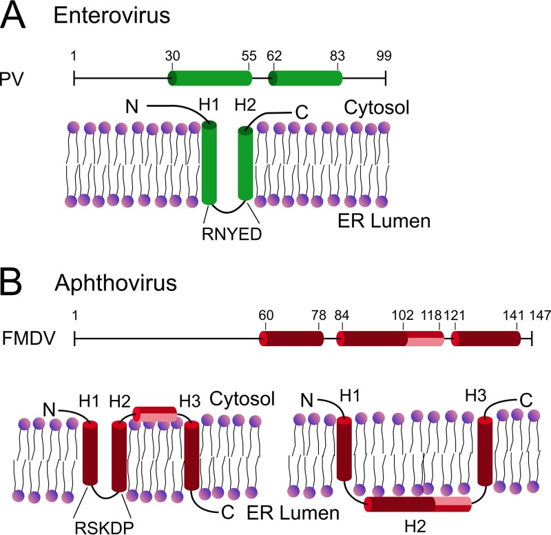
Nonstructural protein 2B of foot-and-mouth disease (FMD) virus (FMDV) is comprised of a small, hydrophobic, 154-amino-acid protein. Structure-function analyses demonstrated that FMDV 2B is an ion channel-forming protein. Infrared spectroscopy measurements using partially overlapping peptides that spanned regions between amino acids 28 and 147 demonstrated the adoption of helical conformations in two putative transmembrane regions between residues 60 and 78 and between residues 119 and 147 and a third transmembrane region between residues 79 and 106, adopting a mainly extended structure. Using synthetic peptides, ion channel activity measurements in planar lipid bilayers and imaging of single giant unilamellar vesicles (GUVs) revealed the existence of two sequences endowed with membrane-porating activity: one spanning FMDV 2B residues 55 to 82 and the other spanning the C-terminal region of 2B from residues 99 to 147. Mapping the latter sequence identified residues 119 to 147 as being responsible for the activity. Experiments to assess the degree of insertion of the synthetic peptides in bilayers and the inclination angle adopted by each peptide regarding the membrane plane normal confirm that residues 55 to 82 and 119 to 147 of 2B actively insert as transmembrane helices. Using reverse genetics, a panel of 13 FMD recombinant mutant viruses was designed, which harbored nonconservative as well as alanine substitutions in critical amino acid residues in the area between amino acid residues 28 and 147. Alterations to any of these structures interfered with pore channel activity and the capacity of the protein to permeabilize the endoplasmic reticulum (ER) to calcium and were lethal for virus replication. Thus, FMDV 2B emerges as the first member of the viroporin family containing two distinct pore domains.IMPORTANCE FMDV nonstructural protein 2B is able to insert itself into cellular membranes to form a pore. This pore allows the passage of ions and small molecules through the membrane. In this study, we were able to show that both current and small molecules are able to pass though the pore made by 2B. We also discovered for the first time a virus with a pore-forming protein that contains two independent functional pores. By making mutations in our infectious clone of FMDV, we determined that mutations in either pore resulted in nonviable virus. This suggests that both pore-forming functions are independently required during FMDV infection.

Keywords: 2B; FMD; FMDV; foot-and-mouth disease; viroporin.


Figures

FIG 1
FIG 1
(A) Comparative multisequence alignment of multiple FMDV 2B isolates and predicted structural features. Residues conserved among FMDV isolates are shown as dots. Substituted residues described in Table 2 are shown in red. (B) Amino acids of O1 Campos were used as a representative sequence to predict secondary structure (H is helical) amphiphilicity (with M showing predicted transmembrane regions), and a Kyte-Doolittle plot is shown. Predictions were done on the ExPASY server (39, 40). (C) The peptides used in this study mapped to the amino acid sequence of 2B from O1 Campos. The indicated residues in red are changes from the original amino acid sequence.
FIG 1
FIG 1
(A) Comparative multisequence alignment of multiple FMDV 2B isolates and predicted structural features. Residues conserved among FMDV isolates are shown as dots. Substituted residues described in Table 2 are shown in red. (B) Amino acids of O1 Campos were used as a representative sequence to predict secondary structure (H is helical) amphiphilicity (with M showing predicted transmembrane regions), and a Kyte-Doolittle plot is shown. Predictions were done on the ExPASY server (39, 40). (C) The peptides used in this study mapped to the amino acid sequence of 2B from O1 Campos. The indicated residues in red are changes from the original amino acid sequence.
FIG 2
FIG 2
Ion channel activity of 2B peptides. (A) Current recordings in 150 mM KCl, at different potentials, after the addition of 2B1, 2B2, 2B3, or 2B4 to ER-like lipid bilayers. (B) Histograms of the current jump amplitudes recorded for 2B2 and 2B4 with voltage set at 50 mV (left and right panels, respectively). (C) Current recordings measured in 150 mM CaCl2 at 50 mV.
FIG 3
FIG 3
Single-ER-GUV permeabilization induced by 2B peptides. (A) Micrographs depicting Rho-PE-labeled ER-GUVs (orange circumferences) immersed in a solution containing Alexa Fluor 488 (green background). Samples on top correspond to control untreated ER-GUVs (left) or ER-GUVs treated with the p7C peptide derived from CSFV p7 viroporin (11). Bottom samples correspond to ER-GUVs treated with different 2B peptides at the doses displayed on the panels. Bars correspond to 25 μm in all micrographs. (B) Distribution of ER-GUVs according to their percentage of permeabilization to Alexa Fluor 488 after treatment with the different 2B peptides (left) and mean permeabilization values in the samples (right). Peptides were applied at the doses displayed on the panels. (C) Stability of the membrane permeabilization state induced by 2B2 and 2B4. (Top) ER-GUVs permeabilized to Alexa Fluor 488 after incubation for 2 h with 2B2 or 2B4 (green) were supplemented externally with Alexa Fluor 647 (red) and additionally incubated for 2 h before image processing. The presence of both probes inside vesicles can be observed in the merged images (bottom right panels). The “Control” panel displays an intact vesicle incubated in the absence of peptide. (Bottom) Bars represent the degree of filling of individual ER-GUVs with Alexa Fluor 488 (dark green) and Alexa Fluor 647 (light green) after a 4-h incubation with the 2B peptides.
FIG 4
FIG 4
Mapping of membrane-porating activity within the C-terminal end of FMDV-2B. (A) Sequences and range covered by the 2B4-derived overlapping peptides 2B4a, 2B4b, and 2B4c. (B) Ion channel activity of the peptides. Shown are electrophysiological recordings after the addition of 2B4a, 2B4b, and 2B4c (left) and a histogram of current jump amplitudes recorded in lipid bilayers treated with 2B4c (right). Conditions are otherwise as described in the legend of Fig. 2. (C and D) Single-ER-GUV permeabilization. Shown are micrographs of ER-GUVs treated with 2B4-derived peptides (C) and their distributions (D) according to the percentage of permeabilization per vesicle (left) and calculated mean permeabilization values (right). Conditions are otherwise as described in the legend of Fig. 3.
FIG 5
FIG 5
Structures and orientations adopted by 2B peptides in ER-like membranes. (A) Comparison of ATR-IR spectra of 2B2 and 2B3 peptides in the amide I region. Peaks at 1,656 and 1,630 cm−1 are indicative of α-helical and extended secondary structures, respectively. The main contained orientation is inferred from the ratio of peak areas recorded with incident light polarized parallel (║) and perpendicular (┬) to the membrane normal. a.u., arbitrary units. (B) Models for the orientation of the membrane-associated structures adopted by 2B-derived peptides, which are based on the values of the order parameters and tilt angles displayed in Table 1. Models considering single, continuous helices are accompanied by models considering kinked helical structures.
FIG 6
FIG 6
Assessment of the presence of virus replication after transfection. (A) The presence of virus RNA was detected by real-time RT-PCR. Total RNA was isolated from either transfected BHK-21 cells or LFBK-V6 cells used for the third (p.3) and sixth (p.6) blind passages of transfected extracts. Data are expressed as CT (cycle threshold) values for all infectious clone constructs described in Table 2. (B) Western blot analysis of FMDV 2B protein expression in cell extracts obtained from either transfected BHK-21 cells (1) or LFBK-V6 cells used for the second blind passages of transfected extracts (2). Expression of 2B in five blind passages of cell transfected with construct 83P is shown (3). C+, positive control, which is an extract of cells infected with FMDV O1 Campos.
FIG 7
FIG 7
Functional effects of lethal mutations targeting 2B2 and 2B4 transmembrane regions. (A) Current recordings in 150 mM KCl, at a potential of 50 mV, after addition to ER-like lipid bilayers of 2B2 or 2B4 peptide variants incorporating the mutations (recordings on the left and right, respectively). (B) Mean permeabilization values for ER-GUVs treated with 2B2 or 2B4 peptide variants (left and right, respectively). (C) Locations of native and mutated forms of 2B in the ER by confocal microscopy. Micrographs on the left illustrate individual cells coexpressing several GFP constructs and the ER marker mCh-Sec61. Control GFP (i.e., devoid of membrane anchors) labeled the complete cell but was excluded from the ER (top panels), whereas GFP-2B constructs were excluded from the nucleus and colocalized with mCh-Sec61. The panel on the right displays colocalization of mCh-Sec61 and GFP in the samples, including the different 2B mutants, as calculated with the ImageJ plug-in Coloc 2 (http://imagej.net/Coloc_2). Measurements were carried out in at least 6 cells, as for those displayed in the micrograph. Bars represent mean values ± standard errors (SEs). Maximal colocalization with mCh-Sec61 was observed for the fusions, including the 2B.WT protein and the 2B.2 and 2B.12 mutants.
FIG 8
FIG 8
Effects of 2B.2 and 2B.12 lethal mutations on thapsigargin-induced calcium release from the endoplasmic reticulum. (A) Kinetic traces of Ca2+ efflux from the ER into the cytosol in cells after thapsigargin addition (final concentration of 10 μM; addition time is indicated by the arrow). Cells were transfected with GFP (black) or the GFP fusions GFP-2B.WT (red), GFP-2B.2 (blue), and GFP-2B.12 (purple). (B) Maximal extents of rhod-2/AM intensity change upon thapsigargin addition to the above-described samples. The analyzed cells transfected with the GFP-2B fusions displayed expression levels comparable to those of the GFP controls. Intensity value distributions were determined for at least 12 single cells, and mean values for the different samples are depicted as box-and-whisker plots (the ends of the whiskers represent SDs).
FIG 9
FIG 9
Models for the membrane topologies adopted by viroporins 2B derived from picornaviruses. (A) Enterovirus 2B proteins contain membrane-integral hairpins formed by two poorly hydrophobic helices connected through a short polar turn. (B) Alternative models for FMDV 2B. (Left) Assuming the existence of a membrane-integral hairpin analogous to that of enteroviruses, transmembrane helix 2 (H2) and helix 3 (H3) would be connected through an amphipathic short helix. The C terminus would be facing the lumen of the ER. (Right) Provided that the N and C termini are oriented toward the cytosol, only H1 and H3 would transverse the lipid bilayer.

Similar articles

Cited by

References

    1. Domingo E, Baranowski E, Escarmis C, Sobrino F. 2002. Foot-and-mouth disease virus. Comp Immunol Microbiol Infect Dis 25:297–308. doi:10.1016/S0147-9571(02)00027-9. - DOI - PubMed
    1. de Jong AS, Wessels E, Dijkman HB, Galama JM, Melchers WJ, Willems PH, van Kuppeveld FJ. 2003. Determinants for membrane association and permeabilization of the coxsackievirus 2B protein and the identification of the Golgi complex as the target organelle. J Biol Chem 278:1012–1021. doi:10.1074/jbc.M207745200. - DOI - PubMed
    1. Nieva JL, Agirre A, Nir S, Carrasco L. 2003. Mechanisms of membrane permeabilization by picornavirus 2B viroporin. FEBS Lett 552:68–73. doi:10.1016/S0014-5793(03)00852-4. - DOI - PubMed
    1. van Kuppeveld FJ, Galama JM, Zoll J, van den Hurk PJ, Melchers WJ. 1996. Coxsackie B3 virus protein 2B contains cationic amphipathic helix that is required for viral RNA replication. J Virol 70:3876–3886. - PMC - PubMed
    1. Aldabe R, Irurzun A, Carrasco L. 1997. Poliovirus protein 2BC increases cytosolic free calcium concentrations. J Virol 71:6214–6217. - PMC - PubMed

Publication types

MeSH terms

LinkOut - more resources