. 2018 Nov 12;92(23):e01308-18.
doi: 10.1128/JVI.01308-18. Print 2018 Dec 1.

Redundant Late Domain Functions of Tandem VP2 YPX3L Motifs in Nonlytic Cellular Egress of Quasi-enveloped Hepatitis A Virus

Affiliations

Redundant Late Domain Functions of Tandem VP2 YPX3L Motifs in Nonlytic Cellular Egress of Quasi-enveloped Hepatitis A Virus

Olga González-López et al. J Virol. .

Abstract

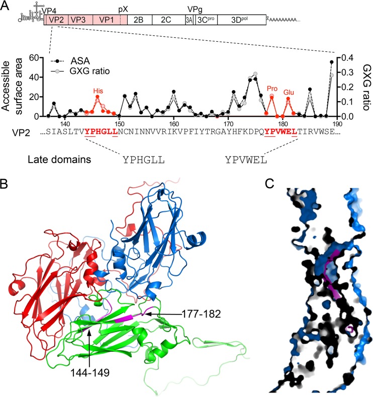
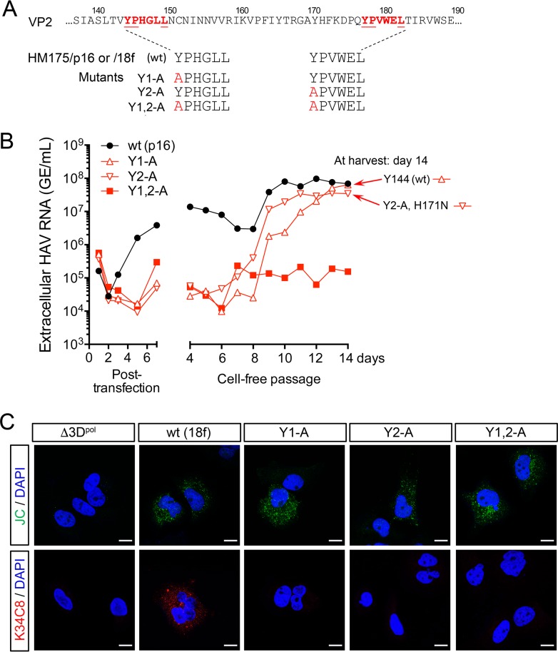
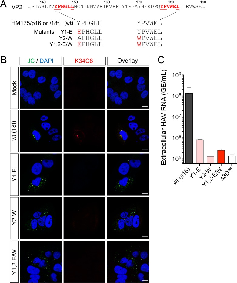
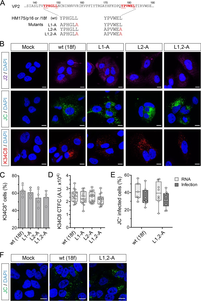
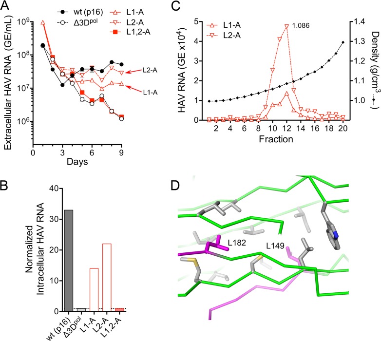
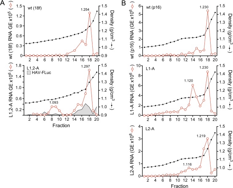
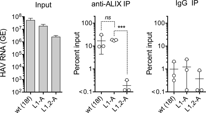
The quasi-envelopment of hepatitis A virus (HAV) capsids in exosome-like virions (eHAV) is an important but incompletely understood aspect of the hepatovirus life cycle. This process is driven by recruitment of newly assembled capsids to endosomal vesicles into which they bud to form multivesicular bodies with intraluminal vesicles that are later released at the plasma membrane as eHAV. The endosomal sorting complexes required for transport (ESCRT) are key to this process, as is the ESCRT-III-associated protein, ALIX, which also contributes to membrane budding of conventional enveloped viruses. YPX1or3L late domains in the structural proteins of these viruses mediate interactions with ALIX, and two such domains exist in the HAV VP2 capsid protein. Mutational studies of these domains are confounded by the fact that the Tyr residues (important for interactions of YPX1or3L peptides with ALIX) are required for efficient capsid assembly. However, single Leu-to-Ala substitutions within either VP2 YPX3L motif (L1-A and L2-A mutants) were well tolerated, albeit associated with significantly reduced eHAV release. In contrast, simultaneous substitutions in both motifs (L1,2-A) eliminated virus release but did not inhibit assembly of infectious intracellular particles. Immunoprecipitation experiments suggested that the loss of eHAV release was associated with a loss of ALIX recruitment. Collectively, these data indicate that HAV YPX3L motifs function as redundant late domains during quasi-envelopment and viral release. Since these motifs present little solvent-accessible area in the crystal structure of the naked extracellular capsid, the capsid structure may be substantially different during quasi-envelopment.IMPORTANCE Nonlytic release of hepatitis A virus (HAV) as exosome-like quasi-enveloped virions is a unique but incompletely understood aspect of the hepatovirus life cycle. Several lines of evidence indicate that the host protein ALIX is essential for this process. Tandem YPX3L "late domains" in the VP2 capsid protein could be sites of interaction with ALIX, but they are not accessible on the surface of an X-ray model of the extracellular capsid, raising doubts about this putative late domain function. Here, we describe YPX3L domain mutants that assemble capsids normally but fail to bind ALIX and be secreted as quasi-enveloped eHAV. Our data support late domain function for the VP2 YPX3L motifs and raise questions about the structure of the HAV capsid prior to and following quasi-envelopment.

Keywords: ESCRT; exosome; picornavirus; quasi-envelope.


Figures

FIG 1
FIG 1
HAV VP2 YPX3L late domain motifs. (A) Schematic representation of the two tandem late domain motifs in the HAV VP2 capsid protein. The organization of the HAV genome is shown at the top with the polyprotein coding region displayed as an extended box. Below is shown a segment of the VP2 protein with the two conserved YPX3L motifs in red font and with the accessible surface area (ASA) of each residue plotted above along with the ratio to calculated GXG value (surface area of the residue relative to that in a peptide in which it is flanked on each side by Gly) based on the X-ray model of the naked HAV capsid (17). (B) Protomer subunit of the HAV capsid showing the adjacent, antiparallel orientation of the two late domain motifs (L1, residues 144 to 149, and L2, residues 177 to 182, highlighted in magenta) within the VP2 (green) β-barrel (17). VP1 is shown in blue, and VP3 is shown in red. (C) Section through the HAV capsid wall, showing the buried position of residues within the late domain motifs (magenta).
FIG 2
FIG 2
Tyr-to-Ala mutations in the HAV VP2 YPX3L motifs ablate capsid assembly. (A) YPX3L late domain Tyr-to-Ala mutants constructed in the background of p16 and 18f virus (Fig. 1A). (B) Extracellular virus release following electroporation of Huh-7.5 cells with wt (HM175/p16) or related Y1-A, Y2-A, and Y1,2-A late domain mutants. Medium was replaced daily, and virus was quantified by reverse transcription-quantitative PCR (RT-qPCR). Cell-free virus was passaged from lysates harvested on day 7 postelectroporation, with supernatant fluid virus titers followed for an additional 14 days. Sequencing of viruses at day 14 postpassage revealed reversion of the Y1-A mutation to wt (Tyr144) and the presence of a second-site substitution in Y2-A (VP2 H171N). (C) Laser scanning confocal fluorescence microscopy of Huh-7.5 cells electroporated with the indicated wt (HM175/18f) or mutant RNA and fixed and stained 48 h later with either JC polyclonal human (top row) or K34C8 murine monoclonal anticapsid (bottom row) antibodies. The absence of K34C8 fluorescence despite abundant JC fluorescence in cells transfected with the mutant RNAs is consistent with the absence of capsid assembly. Nuclear counterstaining was with DAPI (blue). Δ3Dpol is a genome-length HAV RNA in which Gly-Ala-Ala has replaced the Gly-Asp-Asp motif in the 3Dpol RNA-dependent RNA polymerase. Bars, 10 µm.
FIG 3
FIG 3
JC polyclonal human convalescent antibody labels only HAV structural proteins in viral RNA-transfected cells examined by confocal fluorescence microscopy. (A) Schematic representation of the subgenomic 18f-FLuc RNA derived from the HM175/18f virus used in this study, in which the firefly luciferase sequence has replaced most of the VP2-VP1 coding sequence. Gly-Ala-Ala has replaced the 3Dpol Gly-Asp-Asp sequence in the replication-incompetent 18f-FLuc/Δ3Dpol mutant. (B) Cells were either mock electroporated or electroporated with wt (HM175/18f) virus RNA, the replicon 18f-FLuc, or 18f-FLuc/Δ3Dpol and then fixed and stained 72 h later with either J2, a murine monoclonal antibody that binds dsRNA in a sequence-independent fashion (23), or the JC polyclonal antibody. The absence of JC fluorescence in cells transfected with 18f-FLuc RNA, despite abundant J2 fluorescence indicative of RNA replication, indicates that JC fails to detect nonstructural proteins of the virus under the conditions used. Bars, 10 µm.
FIG 4
FIG 4
Late domain mutants with Y1-E, Y2-W, and Y1,2-E/W substitutions are unable to efficiently assemble capsids and thus severely handicapped in replication. (A) YPX3L late domain mutants constructed in the background of p16 and 18f viruses (Fig. 1A). (B) Confocal fluorescence microscopic images of cells mock electroporated or electroporated 48 h previously with wt (HM175/18f) or related mutant RNAs as shown. Absence of K34C8 fluorescence despite readily detectable expression of structural proteins labeled with JC polyclonal antibody suggests a failure of capsid assembly. Bars, 10 µm. (C) Extracellular HAV RNA in supernatant of cells 9 days after electroporation with wt (HM175/p16) or related Y1-E, Y2-W, or Y1,2-E/W RNAs. Cells transfected with each of the mutants released less than 1% of the amount of virus released by cells transfected with the wt control. GE, genome equivalents.
FIG 5
FIG 5
Capsid assembly is not impaired by carboxy-terminal Leu-to-Ala substitutions within the VP2 YPX3L motifs of HAV. (A) YPX3L late domain Leu-to-Ala mutants constructed in the background of p16 and 18f viruses (Fig. 1A). (B) Confocal immunofluorescence microscopy of Huh-7.5 cells mock electroporated or electroporated with wt (HM175p16) or related L1-A, L2-A, or L1,2-A double mutant RNAs. Cells were fixed 48 h postelectroporation and stained with J2 (monoclonal anti-dsRNA), JC (polyclonal human anti-HAV), or K34C8 (monoclonal HAV anticapsid) antibodies. Strong K34C8 fluorescence in cells transfected with each of the mutants is indicative of efficient capsid assembly (22). Nuclear counterstaining was with DAPI (blue). Bars, 10 µm. (C) Percentage of cells stained with the K34C8 anticapsid monoclonal antibody following transfection with each of the mutants. Data shown are means ± standard deviations from 4 independent experiments. (D) Intensity of K34C8 fluorescence (CTFC, corrected total fluorescence intensity per cell) in cells transfected with wt or the indicated mutant viral RNA. All comparisons between cells transfected with mutant RNAs and those transfected with wt control RNAs were nonsignificant statistically (P > 0.29 by one-way ANOVA). A.U., arbitrary units. (E) Assay for infectious virus produced in cells transfected with wt (18f) or L1,2-A RNA. Results shown represent percent cells staining positively with JC antibody 48 h after RNA electroporation (RNA) or 48 h after inoculation of fresh cells with lysates of the electroporated cells (Infection). Data are from 7 to 10 low-power microscopy fields of cells under each condition and are representative of two independent experiments. (F) Confocal microscopic images of Huh-7.5 cells inoculated with cell-free lysates prepared from cells 48 h after electroporation with wt or L1,2-A RNA or no RNA (mock). Cells were stained with polyclonal JC antibody to HAV 48 h after inoculation.
FIG 6
FIG 6
Nonlytic viral egress is reduced by Ala substitutions of the carboxy-terminal Leu of the VP2 YPX3L late domain motifs. (A) Extracellular virus quantified by RT-qPCR following electroporation of wild-type (HM175/p16) or the related L1-A, L2-A, L1,2-A, or replication-incompetent Δ3Dpol mutant RNAs. See legend to Fig. 2B for details. (B) Intracellular viral RNA abundance 9 days after transfection of the indicated wt or mutant virus. Abundance was normalized to that present in cells transfected with the replication-incompetent Δ3Dpol RNA. (C) Isopycnic iodixanol gradients were loaded with virus present in supernatant fluids of cultures 9 days following electroporation of the single L1-A or L2-A motif mutants, and fractions were assayed after centrifugation for HAV RNA by RT-qPCR. Virus present in the peak fractions (fraction 12, 1.086 g/cm3) was sequenced, and the presence of each mutation was confirmed. (D) Orientation of the side chains of Leu149 (L149) and Leu182 (L182) (magenta) within the HAV capsid, showing the lack of a direct interaction between these residues.
FIG 7
FIG 7
Isopycnic iodixanol gradient profiles showing density distributions of intracellular wt and mutant viral RNAs. (A) Gradients loaded with lysates from cells electroporated 72 h previously with wt (HM175/18f) (top) or the related L1,2-A mutant (bottom). Also shown in the bottom panel is the density distribution of subgenomic replicon 18f-FLuc RNA (HAV-FLuc). A unique peak of viral RNA is present in the L1,2-A gradient (1.093 g/cm3). (B) Gradients loaded with lysates from cells electroporated 9 days previously with wt (HM175/p16) (top) or the related L1-A (middle) and L2-A (bottom) mutants. Unique, low-density RNA peaks are present in gradients loaded with lysates of cells transfected with either mutant.
FIG 8
FIG 8
Immunoprecipitation of encapsidated viral RNA present in lysates of cells prepared 48 h following electroporation with wt (HM175/18f), L1-A, or L1,2-A mutant HAV RNAs. Input HAV RNA present in each lysate is shown on the left, quantified by RT-qPCR, followed (left to right) by the percentage of each RNA precipitated by anti-ALIX antibody and nonspecific immunoglobulin (IgG). Data shown represent means ± standard deviations from 3 independent experiments. Dashed lines indicate statistical comparisons by two-sided paired t test: ***, P = 0.0007; ns, nonsignificant.

Similar articles

Cited by

References

    1. Jacobsen KH. 2018. Globalization and the changing epidemiology of hepatitis A virus. Cold Spring Harb Perspect Med doi:10.1101/cshperspect.a031716. - DOI - PMC - PubMed
    1. Lanford RE, Feng Z, Chavez D, Guerra B, Brasky KM, Zhou Y, Yamane D, Perelson AS, Walker CM, Lemon SM. 2011. Acute hepatitis A virus infection is associated with a limited type I interferon response and persistence of intrahepatic viral RNA. Proc Natl Acad Sci U S A 108:11223–11228. doi:10.1073/pnas.1101939108. - DOI - PMC - PubMed
    1. Shin EC, Jeong SH. 2018. Natural history, clinical manifestations, and pathogenesis of hepatitis A. Cold Spring Harb Perspect Med 8:a031708. doi:10.1101/cshperspect.a031708. - DOI - PMC - PubMed
    1. Lemon SM. 1985. Type A viral hepatitis: new developments in an old disease. N Engl J Med 313:1059–1067. doi:10.1056/NEJM198510243131706. - DOI - PubMed
    1. Zhou Y, Callendret B, Xu D, Brasky KM, Feng Z, Hensley LL, Guedj J, Perelson AS, Lemon SM, Lanford RE, Walker CM. 2012. Dominance of the CD4+ T helper cell response during acute resolving hepatitis A virus infection. J Exp Med 209:1481–1492. doi:10.1084/jem.20111906. - DOI - PMC - PubMed

Publication types

MeSH terms

Substances

LinkOut - more resources