. 2018 Nov 12;92(23):e01486-18.
doi: 10.1128/JVI.01486-18. Print 2018 Dec 1.

Cellular Protein Kinase D Modulators Play a Role during Multiple Steps of Herpes Simplex Virus 1 Egress

Affiliations

Cellular Protein Kinase D Modulators Play a Role during Multiple Steps of Herpes Simplex Virus 1 Egress

Élisabeth Roussel et al. J Virol. .

Abstract

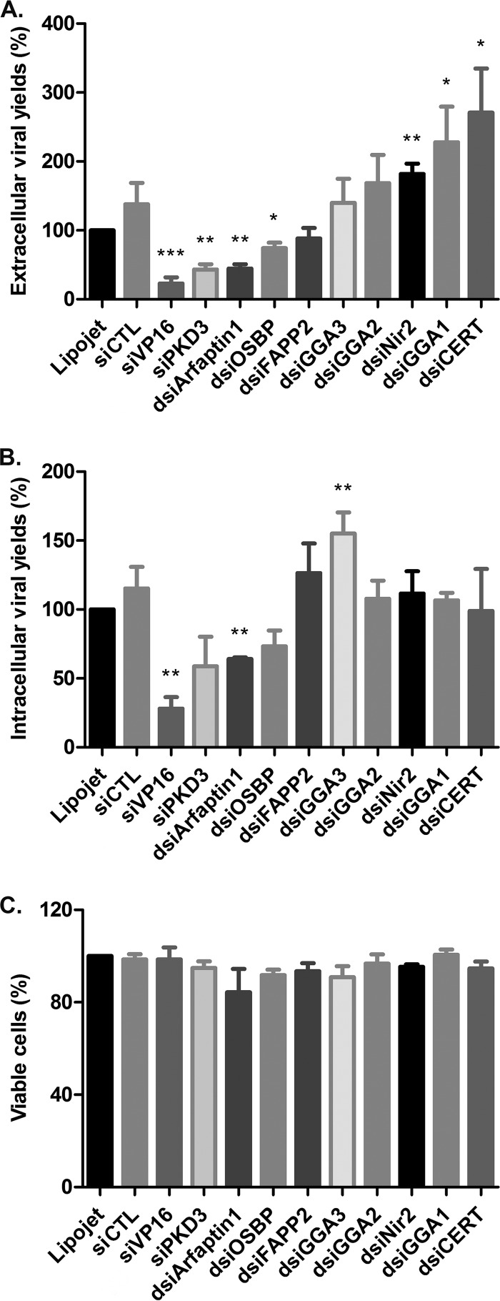
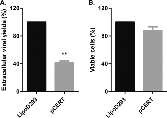
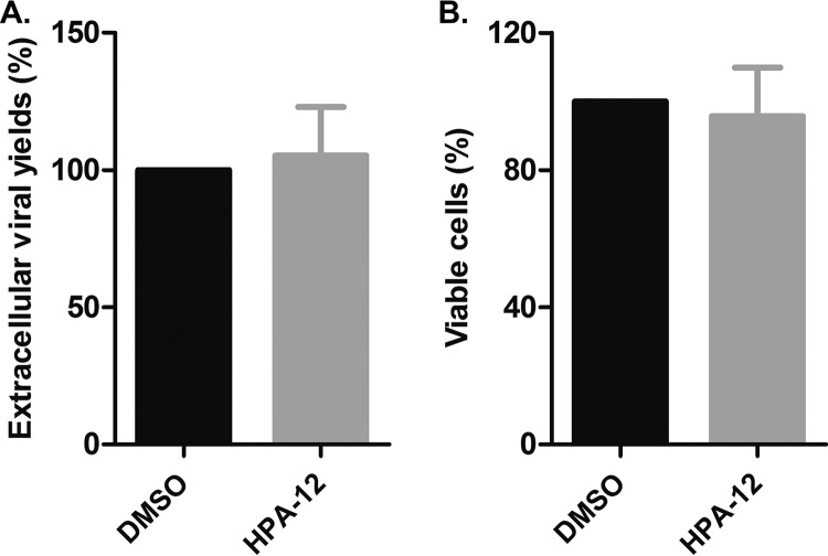
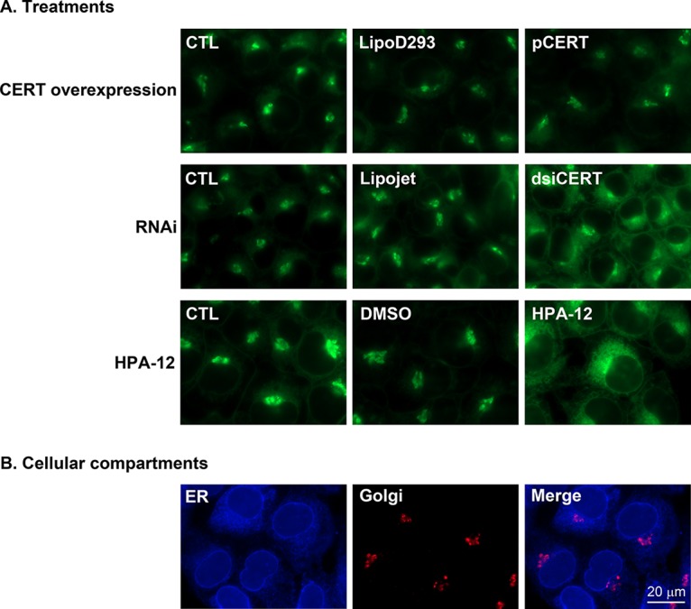
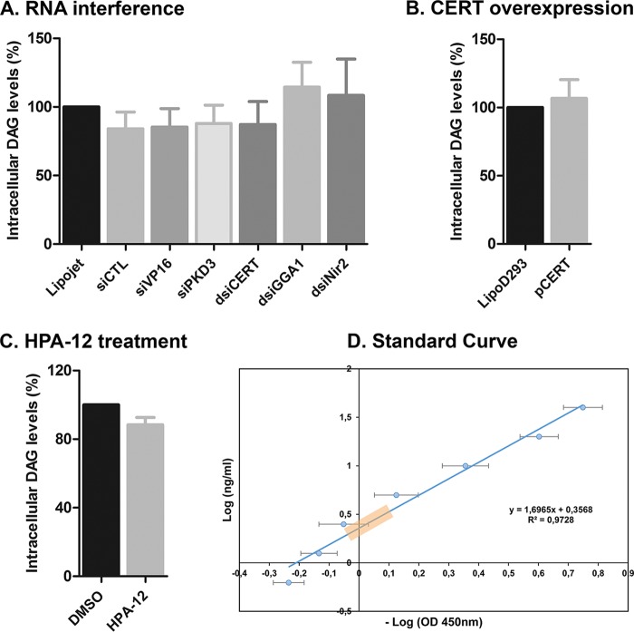
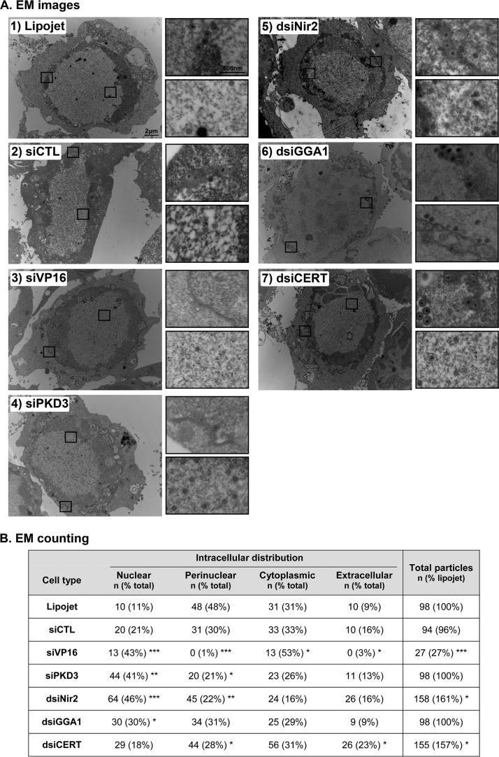
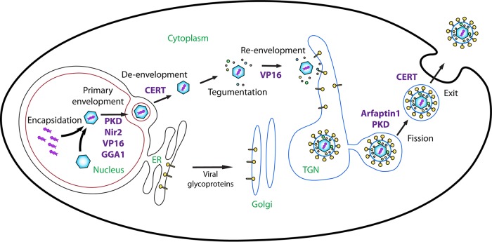
The assembly of new herpes simplex virus 1 (HSV-1) particles takes place in the nucleus. These particles then travel across the two nuclear membranes and acquire a final envelope from a cellular compartment. The contribution of the cell to the release of the virus is, however, little known. We previously demonstrated, using a synchronized infection, that the host protein kinase D and diacylglycerol, a lipid that recruits the kinase to the trans-Golgi network (TGN), promote the release of the virus from that compartment. Given the role this cellular protein plays in the herpes simplex virus 1 life cycle and the many molecules that modulate its activity, we aimed to determine to what extent this virus utilizes the protein kinase D pathway during a nonsynchronized infection. Several molecular protein kinase D (PKD) regulators were targeted by RNA interference and viral production monitored. Surprisingly, many of these modulators negatively impacted the extracellular release of the virus. Overexpression studies, the use of pharmacological reagents, and assays to monitor intracellular lipids implicated in the biology of PKD suggested that these effects were oddly independent of total intracellular diacylglycerol levels. Instead, mapping of the viral intermediates by electron microscopy suggested that some of these modulators could regulate distinct steps along the viral egress pathway, notably nuclear egress. Altogether, this suggests a more complex contribution of PKD to HSV-1 egress than originally anticipated and new research avenues to explore.IMPORTANCE Viruses are obligatory parasites that highjack numerous cellular functions. This is certainly true when it comes to transporting viral particles within the cell. Herpesviruses share the unique property of traveling through the two nuclear membranes by subsequent budding and fusion and acquiring their final envelope from a cellular organelle. Albeit disputed, the overall evidence from many laboratories points to the trans-Golgi network (TGN) as the source of that membrane. Moreover, past findings revealed that the host protein kinase D (PKD) plays an important role at that stage, which is significant given the known implication of that protein in vesicular transport. The present findings suggest that the PKD machinery not only affects the late stages of herpes simplex virus I egress but also modulates earlier steps, such as nuclear egress. This opens up new means to control these viruses.

Keywords: Arf; CERT; GGA1; HSV; Nir2; PKD; arfaptin; egress; protein kinase D; transport.


Figures

FIG 1
FIG 1
Role of PKD and its modulators in vesicular cargo transport at the TGN. Step 1. Cytoplasmic PKD is recruited to the TGN by DAG and Arf1. Step 2. DAG activates nPKCs that in turn activate PKD by phosphorylation. Active PKD then phosphorylates PI4KIIIβ, which has been recruited to the TGN beforehand by Arf1. This enables the conversion by PI4KIIIβ of PI to PI4P. Step 3. PI4P acts as a scaffold and recruits multiple proteins to the TGN, including Arfaptin1, whose role is to inhibit Arf1 and prevent premature fission. PI4P also recruits FAPP2, OSBP, and CERT. These modulators regulate, respectively, the transport of glucosylceramide between the TGN and PM, of cholesterol between the ER and Golgi compartment/TGN, and of ceramide between the ER and TGN. PKD additionally activates PI5P4K, which converts PI5P to PI(4,5)P2. All this ultimately leads to an accumulation of DAG and PKD at the nascent vesicle. Step 4. When the optimal local concentration of DAG is reached, PKD phosphorylates Arfaptin1, leading to its detachment from the TGN and the activation of Arf1. The latter then activates PLD, which metabolizes DAG into PA, which is subsequently converted into LPA by other enzymes. Nir2 negatively regulates the conversion of DAG into LPA. Step 5. The accumulation of LPA at the neck of the vesicle induces strong curvature of the membrane and, ultimately, its fission from the TGN. Concomitantly, Arf1 recruits adaptor proteins, such as GGA1 to -3, to help the formation of the vesicle, cargo selection, and the coating of the vesicles. Step 6. PKD phosphorylates FAPP2, OSBP, and CERT, thereby promoting their detachment from the TGN, leading to a decrease in DAG concentration and, ultimately, the release of PKD from the TGN. Green boundaries indicate activation, and red boundaries indicate inactivation. See the text for details and abbreviations.
FIG 2
FIG 2
Impacts of PKD modulators on HSV-1 yields. (A, B) 143B cells were transfected for 48 h with the LipoJet reagent alone or with different siRNAs and dsiRNAs before being infected for 24 h. The supernatants (A) and cell fractions (B) were then harvested, and the virus produced quantified in plaque assays. (C) The viability of 143B cells treated in parallel as described above was measured using alamarBlue. The mean values and SEM from five independent experiments each performed in duplicate are shown in all panels. The data are normalized to the average obtained with samples transfected with LipoJet alone. Significant differences between the results for cells subjected to RNAi and those treated with LipoJet alone were evaluated with Student’s bilateral tests (*, P < 0.05; **, P < 0.01; ***, P < 0.001).
FIG 3
FIG 3
CERT overexpression hinders viral production. (A) 143B cells were transfected for 24 h with the LipoD293 reagent alone or plasmid expressing hemagglutinin (HA)-tagged wild-type CERT (pCERT). Cells transfected for 24 h were subsequently infected for 24 h, and the supernatants harvested for plaque assays. The mean values and SEM are derived from two independent experiments each performed in duplicate. (B) Cells treated in parallel in 96-well plates were monitored for their viability using alamarBlue. In this case, the bars and error bars show the mean values and SEM from three independent experiments each performed in triplicate. In all cases, the data are normalized to the average value obtained with samples transfected with LipoD293 alone. Student’s bilateral tests showed the significant differences between results for cells transfected with pCERT and cells treated with LipoD293 alone (**, P < 0.01).
FIG 4
FIG 4
Inhibition of CERT with the HPA-12 drug does not affect the egress of the virus. (A) 143B cells were infected in the presence of DMSO or 2.5 μM HPA-12 for 16 h. The supernatants were then harvested and used for plaque assays. The mean values and SEM from three independent experiments performed in duplicate are shown. (B) 143B cells were treated in parallel with 2.5 μM HPA-12 for 16 h, and their viability measured using alamarBlue. Error bars show the SEM of three independent experiments performed in triplicate. In both panels, the data are normalized to the average value obtained with samples treated with DMSO alone. Student’s bilateral tests did not hint at any significant differences between the results obtained using HPA-12 and DMSO alone.
FIG 5
FIG 5
CERT reagents function as expected. (A) 143B cells were treated with pCERT, dsiCERT, or HPA-12, and the localization of the fluorescent ceramide analog BODIPY FL C5-ceramide monitored under a fluorescence microscope. (B) Cells were also probed for ER (anti-calnexin antibody) and Golgi (anti-Golgin-97 antibody) markers. Representative cells out of three independent experiments are shown for each condition. The scale bar applies to all images.
FIG 6
FIG 6
Total intracellular DAG levels are not affected by RNAi, CERT overexpression, or HPA-12. (A to C) 143B cells were transfected for 48 h with the LipoJet reagent alone or with different siRNAs and dsiRNAs (A), transfected for 24 h with the LipoD293 reagent alone or with pCERT (B), or treated for 16 h with DMSO alone or 2.5 μM HPA-12 (C). The cell fractions were collected, and the total DAG levels were measured using an ELISA kit. Data are normalized to the average value obtained with samples treated with the respective control. (D) A standard curve was established with exogenous DAG and the above-named kit. Note that values for all samples described above were within the orange box and, thus, in the linear range of the assay. The mean values and SEM from three independent experiments performed in duplicate are depicted. No statistically significant differences between the results for treated and control samples were noted.
FIG 7
FIG 7
The PKD pathway can modulate distinct steps along the viral egress pathway. (A) Differential impacts of PKD modulators on viral egress. 143B cells were transfected for 48 h with the LipoJet reagent alone or with different siRNAs and dsiRNAs before being infected for 24 h. The cells were then fixed, embedded, and prepared for electron microscopy. Representative cells are shown for each condition. Boxed areas are enlarged to the right of each panel. The top zoom represents a perinuclear section, and the bottom one a nuclear section. The scale bars of 2 μm for the entire cell and 500 nm for the enlarged section apply for all images. (B) Counting of viral particles. Values are the average numbers and percentages of viral particles counted from 8 to 15 different randomly selected cells per condition. Student’s bilateral tests detected some significant differences between the results for cells subjected to RNAi and cells transfected with LipoJet alone (*, P < 0.05; **, P < 0.01; ***, P < 0.001).
FIG 8
FIG 8
Involvement of PKD modulators in HSV-1 egress. The main steps of viral egress are depicted and include the primary envelopment of the newly made viral capsids through the inner nuclear membrane, their deenvelopment via the fusion of the perinuclear virions with the outer nuclear membrane, the main tegumentation step (mostly taking place in the cytoplasm, albeit some has already occurred in the nucleus), the reenvelopment of the naked cytoplasmic particles, fission of HSV-1-laden transport carriers from the TGN, and finally, the release of the virus at the cell surface by fusion. The diagram also depicts at which steps the PKD modulators might promote or interfere with viral egress.

Similar articles

Cited by

References

    1. Fu Y, Rubin CS. 2011. Protein kinase D: coupling extracellular stimuli to the regulation of cell physiology. EMBO Rep 12:785–796. doi:10.1038/embor.2011.139. - DOI - PMC - PubMed
    1. Roy A, Ye J, Deng F, Wang QJ. 2017. Protein kinase D signaling in cancer: a friend or foe? Biochim Biophys Acta Rev Cancer 1868:283–294. doi:10.1016/j.bbcan.2017.05.008. - DOI - PMC - PubMed
    1. Malhotra V, Campelo F. 2011. PKD regulates membrane fission to generate TGN to cell surface transport carriers. Cold Spring Harb Perspect Biol 3:a005280. doi:10.1101/cshperspect.a005280. - DOI - PMC - PubMed
    1. Valverde AM, Sinnett-Smith J, Van Lint J, Rozengurt E. 1994. Molecular cloning and characterization of protein kinase D: a target for diacylglycerol and phorbol esters with a distinctive catalytic domain. Proc Natl Acad Sci U S A 91:8572–8576. doi:10.1073/pnas.91.18.8572. - DOI - PMC - PubMed
    1. Maeda Y, Beznoussenko GV, Van Lint J, Mironov AA, Malhotra V. 2001. Recruitment of protein kinase D to the trans-Golgi network via the first cysteine-rich domain. EMBO J 20:5982–5990. doi:10.1093/emboj/20.21.5982. - DOI - PMC - PubMed

Publication types

MeSH terms

Grants and funding

LinkOut - more resources