. 2018 Nov 12;92(23):e01255-18.
doi: 10.1128/JVI.01255-18. Print 2018 Dec 1.

Robust Human and Murine Hepatocyte Culture Models of Hepatitis B Virus Infection and Replication

Affiliations

Robust Human and Murine Hepatocyte Culture Models of Hepatitis B Virus Infection and Replication

Luhua Qiao et al. J Virol. .

Abstract

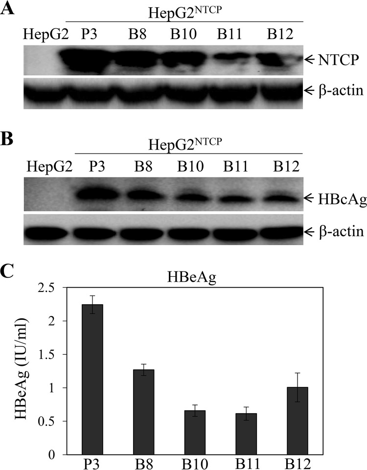
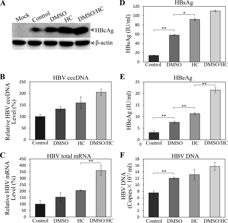
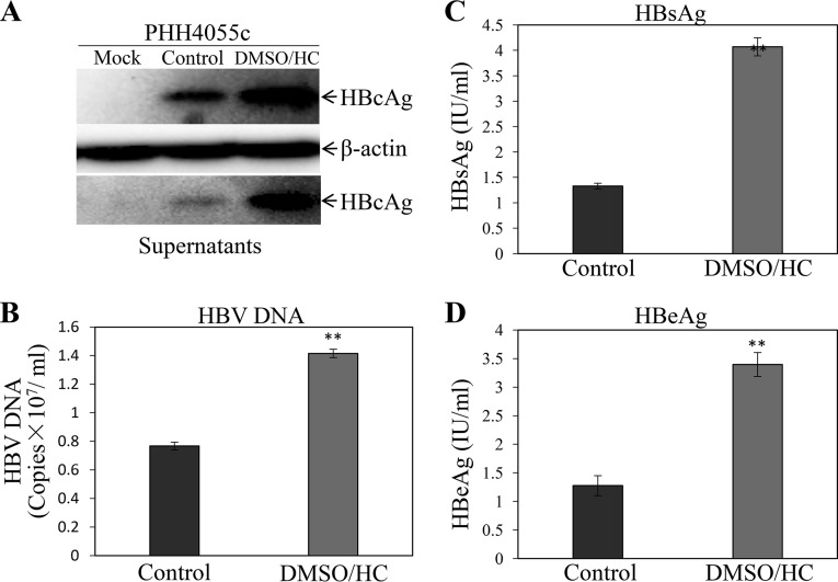
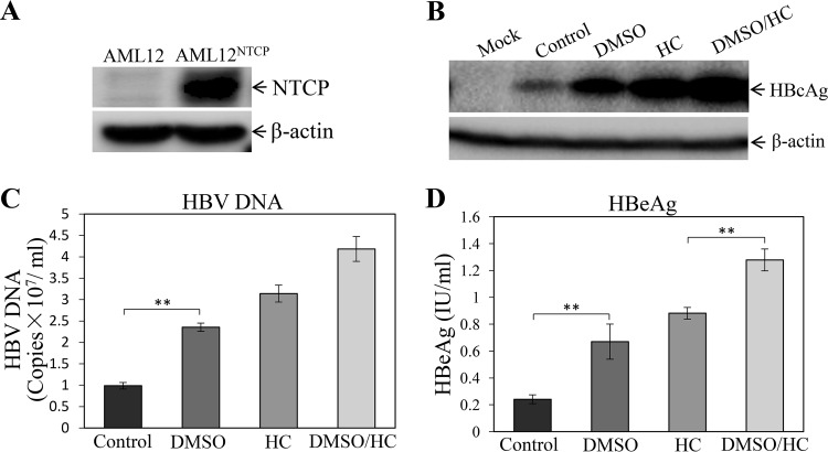
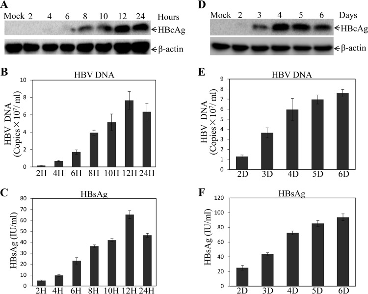
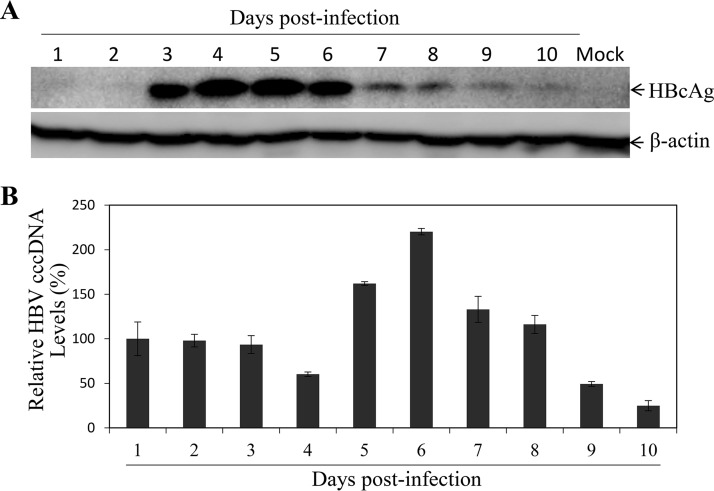
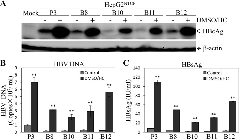
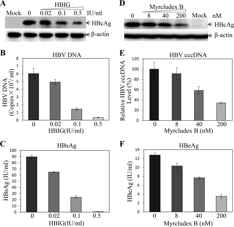
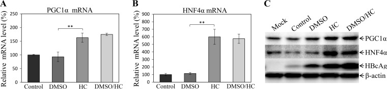
Hepatitis B virus (HBV) is a major cause of chronic liver diseases, including hepatitis, cirrhosis, and hepatocellular carcinoma. HBV research has been hampered by the lack of robust cell culture and small animal models of HBV infection. The discovery of sodium taurocholate cotransporting polypeptide (NTCP) as an HBV receptor has been a landmark advance in HBV research in recent years. Ectopic expression of NTCP in nonpermissive HepG2, Huh7, and AML12 cell lines confers HBV susceptibility. However, HBV replication in these human and murine hepatocyte cell lines appeared suboptimal. In the present study, we constructed stable NTCP-expressing HepG2 and AML12 cell lines and found that HBV permissiveness is correlated with NTCP expression. More significantly, we developed robust HBV cell culture models by treating the HBV-infected cells with dimethyl sulfoxide (DMSO) and hydrocortisone, which significantly promoted HBV replication and production. Mechanistic studies suggested that hydrocortisone significantly enhanced the transcription and expression of PGC1α and HNF4α, which are known to promote HBV transcription and replication. These new human and murine hepatocyte culture systems of HBV infection and replication will accelerate the determination of molecular aspects underlying HBV infection, replication, and morphogenesis in human and murine hepatocytes. We anticipate that our HBV cell culture models will also facilitate the discovery and development of antiviral drugs towards the ultimate eradication of chronic hepatitis B virus infection.IMPORTANCE HBV research has been greatly hampered by the lack of robust cell culture and small animal models of HBV infection and propagation. The discovery of NTCP as an HBV receptor has greatly impacted the field of HBV research. Although HBV infection of NTCP-expressing human and murine hepatocyte cell lines has been demonstrated, its replication in cell culture appeared inefficient. To further improve cell culture systems of HBV infection and replication, we constructed NTCP-expressing HepG2 and AML12 cell lines that are highly permissive to HBV infection. More significantly, we found that DMSO and hydrocortisone markedly enhanced HBV transcription and replication in human and murine hepatocytes when added to the cell culture medium. These new cell culture models of HBV infection and replication will facilitate HBV research and antiviral drug discovery towards the ultimate elimination of chronic hepatitis B virus infection.

Keywords: AML12; DMSO; HBV; HepG2; NTCP; PHH; hepatitis B virus; hydrocortisone; infection; morphogenesis; replication.


Figures

FIG 1
FIG 1
Correlation of NTCP expression and HBV susceptibility. (A) HepG2 cells were transfected with pLiv7/Blast/NTCP and pLiv7/Puro/NTCP, respectively. Upon selection with blasticidin (B8, B10, B11, and B12) or puromycin (P3), cell clones were picked up and expanded. The expression of NTCP was detected by Western blotting using an NTCP-specific monoclonal antibody. (B) HepG2NTCP cell lines were infected with HBV in the presence of 4% PEG 8000 for 12 h. Upon removal of unbound HBV and PEG by washing with PBS, the HBV-infected cells were incubated with fresh DME-F12 medium containing 3% FBS. After 4 days postinfection (p.i.), cells were lysed in a RIPA buffer. HBcAg in cell lysates was detected by Western blotting using an HBc-specific monoclonal antibody. (A, B) The housekeeping gene β-actin was used as an internal control. (C) The levels of HBeAg in the supernatants were quantified by chemiluminescence immunoassay. Average values (±SD) derived from three experiments are plotted.
FIG 2
FIG 2
Enhancement of HBV replication by DMSO and hydrocortisone. HepG2NTCP-P3 cells were infected with HBV at an MOI of 100 genome copies (GC) in the presence of 4% PEG 8000 for 12 h. Upon removal of unbound HBV and PEG 8000, HBV-infected cells were incubated with DME-F12 medium containing 1% DMSO, 5 μg/ml hydrocortisone (HC), or both (DMSO/HC). (A) After culturing for 4 days, cell lysates were collected for detection of HBcAg by Western blotting. (B) HBV cccDNA in the cells was quantified by qPCR, using mitochondrial DNA as an internal control for normalization. (C) Total RNA was extracted from HBV-infected cells with TRIzol reagent and was reverse transcribed with superscript III. Resulting cDNAs were quantified by qPCR. (D to F) The levels of HBsAg (D) and HBeAg (E) in the supernatants were quantified by chemiluminescence immunoassay. HBV DNA in the supernatants was quantified by qPCR (F). (B to F) Average values (±SD) derived from three experiments are plotted. *, P < 0.05; **, P < 0.01.
FIG 3
FIG 3
Enhancement of HBV transcription and replication in PHHs by DMSO and hydrocortisone. PHHs were infected with HBV at an MOI of 100 GC in the presence of 4% PEG 8000 for 12 h. The HBV-infected PHHs were cultured in the PHH maintenance medium without (Control) or with 1% DMSO and 5 μg/ml HC (DMSO/HC) for 4 days by changing medium every day. At 4 days p.i., the HBV-infected PHHs were lysed in a RIPA buffer and the cell culture supernatants were collected. (A) The levels of HBcAg in cell lysates were determined by Western blotting. (B to D) The levels of HBV DNA (B), HBsAg (C), and HBeAg (D) in the supernatants were quantified by qPCR or chemiluminescence immunoassay. Average values (±SD) derived from three experiments are plotted. **, P < 0.01.
FIG 4
FIG 4
Promotion of HBV transcription and replication in the AML12NTCP cells by DMSO and hydrocortisone. AML12NTCP cells were infected with HBV at an MOI of 100 GC in the presence of 4% PEG 8000 for 12 h. Upon removal of unbound HBV and PEG 8000, the HBV-infected AML12NTCP cells were cultured in DME-F12 medium containing 3% FBS and 1% DMSO, 5 μg/ml HC, or both (DMSO/HC). (A, B) After 4 days p.i., the HBV-infected AML12NTCP cells were lysed in a RIPA buffer and the levels of NTCP (A) and HBcAg (B) in cell lysates were measured by Western blotting. (C, D) The levels of HBV DNA (C) and HBeAg (D) in the cell culture supernatants were quantified by qPCR and chemiluminescence immunoassay, respectively. Average values (±SD) derived from three experiments are plotted. **, P < 0.01.
FIG 5
FIG 5
Optimization of conditions for HBV infection and replication. (A to C) HepG2NTCP-P3 cells were infected with HBV at an MOI of 100 GC in the presence of 4% PEG 8000 for various times (2, 4, 6, 8, 10, 12, and 24 h). HBV-infected cells were cultured in DME-F12 medium containing 3% FBS, 1% DMSO, and 5 μg/ml hydrocortisone. (A) After 4 days p.i., the HBV-infected cells were lysed and the levels of HBcAg were determined by Western blotting. (B) HBV DNA in the supernatants was quantified by qPCR. (C) The levels of HBsAg in the supernatants were quantified by chemiluminescence immunoassay. (D to F) Optimal time of HBV replication was determined by HBV infection with an MOI of 100 GC in the presence of 4% PEG 8000 for 12 h. The HBV-infected cells were lysed in a RIPA buffer at different time points (2, 3, 4, 5, and 6 days) after HBV infection. The levels of HBcAg in the infected cells (D) and of HBV DNA (E) and HBsAg (F) in the supernatants were determined by Western blotting, qPCR, and chemiluminescence immunoassay, respectively. (B, C, E, and F) Average values (±SD) derived from three experiments are plotted.
FIG 6
FIG 6
Kinetics of HBcAg and HBV cccDNA synthesis upon induction with DMSO and hydrocortisone. HepG2NTCP-P3 cells were infected with HBV at an MOI of 100 GC in the presence of 4% PEG 8000 for 12 h. HBV-infected cells were cultured in DME-F12 medium containing 3% FBS, 1% DMSO, and 5 μg/ml hydrocortisone. (A) At every day postinfection, the HBV-infected cells were lysed and the levels of HBcAg were determined by Western blotting. (B) HBV cccDNA in the cells was subjected to Hirt extraction, followed by digestion with T5 exonuclease and quantification by qPCR using cccDNA-specific primers and probe. Average values (±SD) derived from three experiments are plotted.
FIG 7
FIG 7
Comparison of HBV transcription and replication efficiencies with and without DMSO and hydrocortisone (DMSO/HC) treatment among different HepG2NTCP cell lines. Different HepG2NTCP cell lines were infected with HBV at an MOI of 100 GC in the presence of 4% PEG 8000 for 12 h and then cultured in DME-F12 medium without (−) or with 1% DMSO and 5 μg/ml HC (+). After 4 days p.i., the HBV-infected cells were lysed in a RIPA buffer and cell culture supernatants were collected. (A) HBcAg in the cell lysates was detected by Western blotting. (B, C) The levels of HBV DNA (B) and HBsAg (C) in the supernatants were quantified by qPCR and chemiluminescence immunoassay, respectively. Average values (±SD) derived from three experiments are plotted. **, P < 0.01.
FIG 8
FIG 8
Inhibition of HBV infection by HBIG and myrcludex B. HepG2NTCP-P3 cells were infected with HBV at an MOI of 100 GC in the presence of 4% PEG 8000 and increasing concentrations of HBIG (0, 0.02, 0.1, and 0.5 IU) or myrcludex B (0, 80, 40, and 200 nM) for 12 h. The HBV-infected cells were cultured with DME-F12 medium containing 3% FBS, 1% DMSO, and 5 μg/ml HC. After 4 days p.i., the HBV-infected cells were lysed in a RIPA buffer. (A, D, and E) The levels of HBcAg (A, D) and HBV cccDNA (E) in the HBV-infected cells were determined by Western blotting and qPCR, respectively. (B, C, and F) The levels of HBV DNA (B), HBsAg (C), and HBeAg (F) in the cell culture supernatants were quantified by qPCR or chemiluminescence immunoassay. Average values (±SD) derived from three experiments are plotted.
FIG 9
FIG 9
Induction of PGC1α and HNF4α gene transcription and expression by hydrocortisone. HepG2NTCP-P3 cells were infected with HBV at an MOI of 100 GC in the presence of 4% PEG 8000 for 12 h. The HBV-infected cells were cultured in DME-F12 medium containing 1% DMSO, 5 μg/ml HC, or both (DMSO/HC). (A, B) After 4 days p.i., the levels of PGC1α mRNA (A) and HNF4α mRNA (B) in the HBV-infected cells were quantified by a real-time RT-PCR method. Average values (±SD) derived from three experiments are plotted. **, P < 0.01. (C) The levels of PGC1α and HNF4α expression were determined by Western blotting using PGC1α- and HNF4α-specific monoclonal antibodies. β-Actin was used as an internal control.

Similar articles

Cited by

References

    1. Ganem D, Prince AM. 2004. Hepatitis B virus infection—natural history and clinical consequences. N Engl J Med 350:1118–1129. doi:10.1056/NEJMra031087. - DOI - PubMed
    1. Lok AS, McMahon BJ. 2009. Chronic hepatitis B: update 2009. Hepatology 50:661–662. doi:10.1002/hep.23190. - DOI - PubMed
    1. Seeger C, Mason WS. 2015. Molecular biology of hepatitis B virus infection. Virology 479-480:672–686. doi:10.1016/j.virol.2015.02.031. - DOI - PMC - PubMed
    1. Zeisel MB, Lucifora J, Mason WS, Sureau C, Beck J, Levrero M, Kann M, Knolle PA, Benkirane M, Durantel D, Michel ML, Autran B, Cosset FL, Strick-Marchand H, Trepo C, Kao JH, Carrat F, Lacombe K, Schinazi RF, Barre-Sinoussi F, Delfraissy JF, Zoulim F. 2015. Towards an HBV cure: state-of-the-art and unresolved questions—report of the ANRS workshop on HBV cure. Gut 64:1314–1326. doi:10.1136/gutjnl-2014-308943. - DOI - PubMed
    1. Gao W, Hu J. 2007. Formation of hepatitis B virus covalently closed circular DNA: removal of genome-linked protein. J Virol 81:6164–6174. doi:10.1128/JVI.02721-06. - DOI - PMC - PubMed

Publication types

MeSH terms

LinkOut - more resources