. 2018 Nov 12;92(23):e00826-18.
doi: 10.1128/JVI.00826-18. Print 2018 Dec 1.

Natural Secretory Immunoglobulins Promote Enteric Viral Infections

Affiliations

Natural Secretory Immunoglobulins Promote Enteric Viral Infections

Holly Turula et al. J Virol. .

Abstract

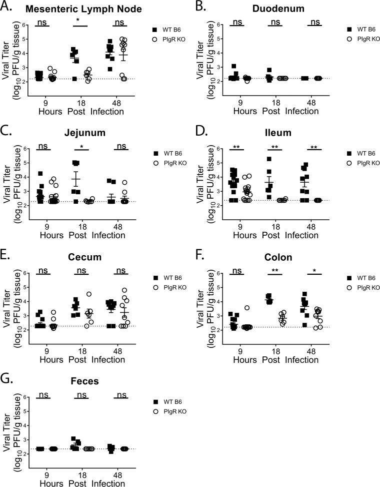
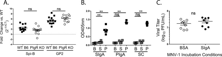
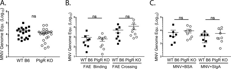
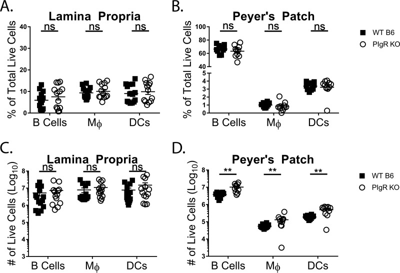
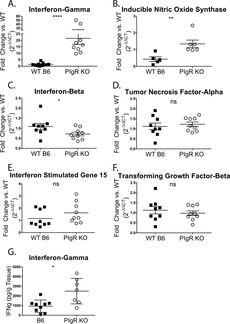
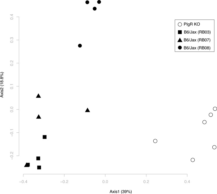
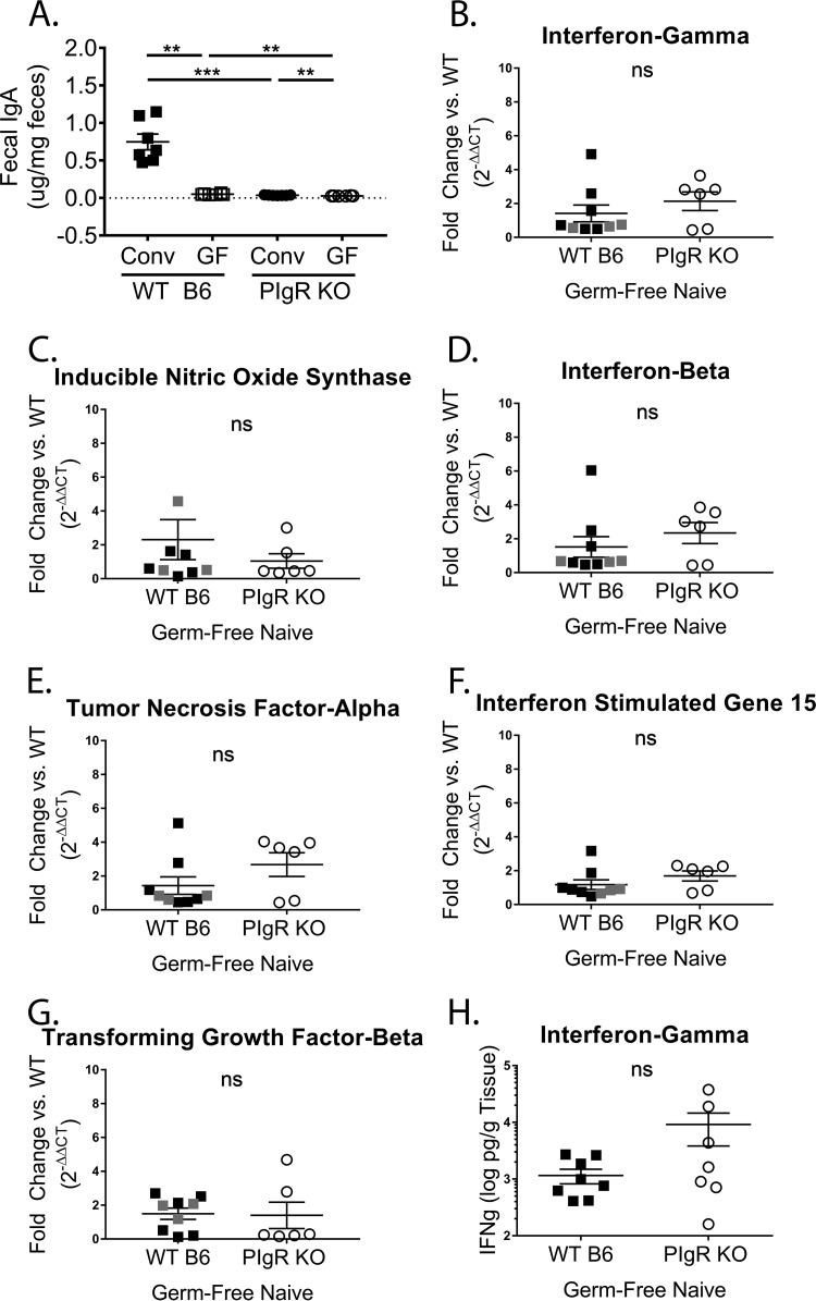
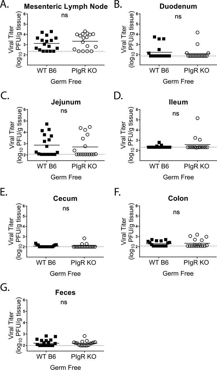
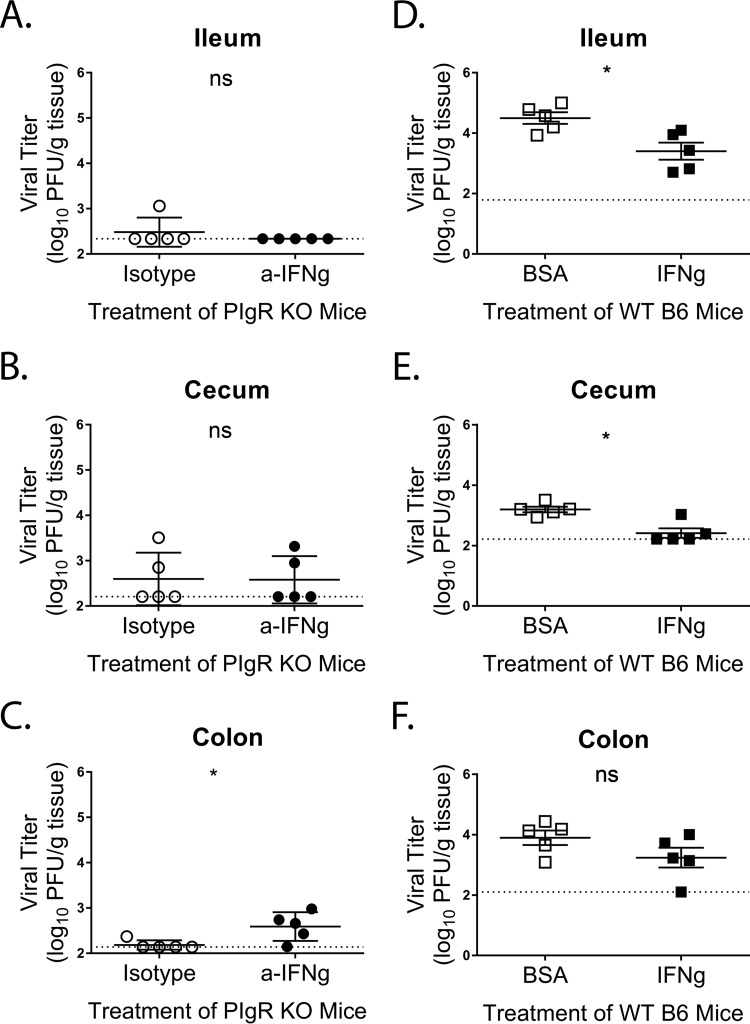
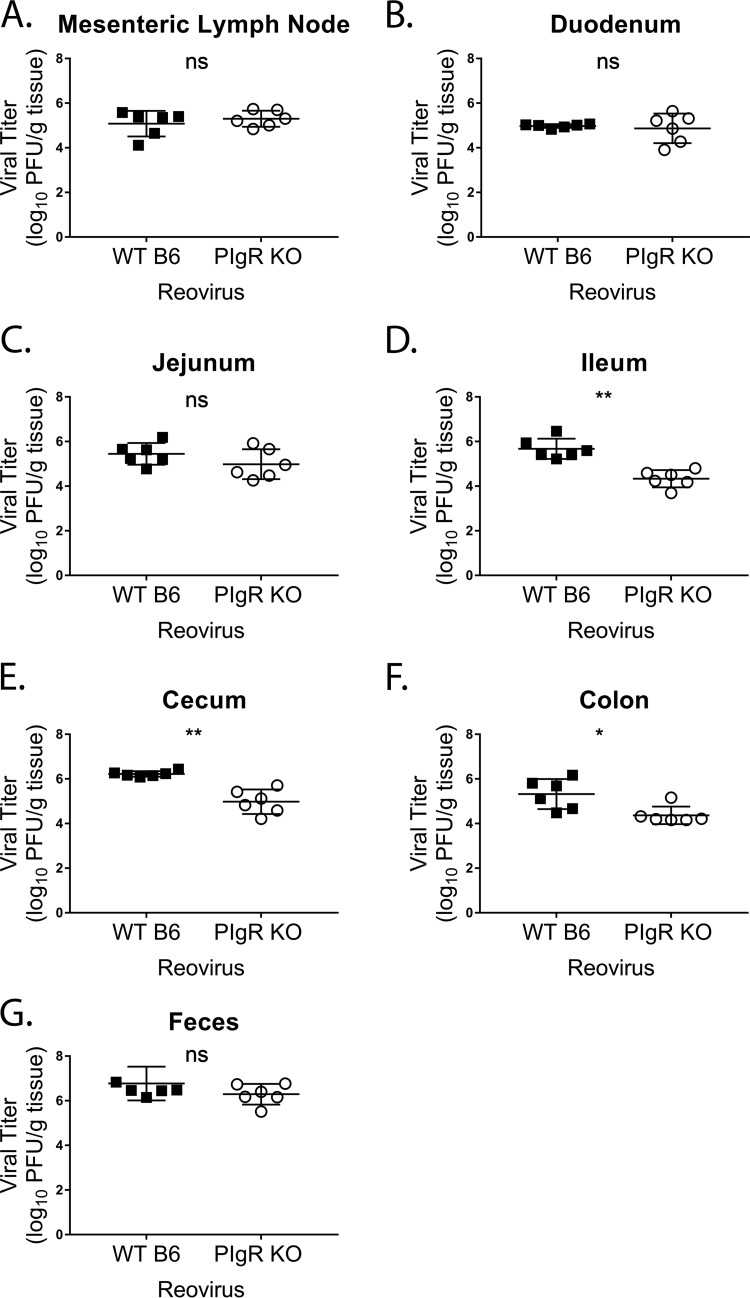
Noroviruses are enteric pathogens causing significant morbidity, mortality, and economic losses worldwide. Secretory immunoglobulins (sIg) are a first line of mucosal defense against enteric pathogens. They are secreted into the intestinal lumen via the polymeric immunoglobulin receptor (pIgR), where they bind to antigens. However, whether natural sIg protect against norovirus infection remains unknown. To determine if natural sIg alter murine norovirus (MNV) pathogenesis, we infected pIgR knockout (KO) mice, which lack sIg in mucosal secretions. Acute MNV infection was significantly reduced in pIgR KO mice compared to controls, despite increased MNV target cells in the Peyer's patch. Natural sIg did not alter MNV binding to the follicle-associated epithelium (FAE) or crossing of the FAE into the lymphoid follicle. Instead, naive pIgR KO mice had enhanced levels of the antiviral inflammatory molecules interferon gamma (IFN-γ) and inducible nitric oxide synthase (iNOS) in the ileum compared to controls. Strikingly, depletion of the intestinal microbiota in pIgR KO and control mice resulted in comparable IFN-γ and iNOS levels, as well as MNV infectious titers. IFN-γ treatment of wild-type (WT) mice and neutralization of IFN-γ in pIgR KO mice modulated MNV titers, implicating the antiviral cytokine in the phenotype. Reduced gastrointestinal infection in pIgR KO mice was also observed with another enteric virus, reovirus. Collectively, our findings suggest that natural sIg are not protective during enteric virus infection, but rather, that sIg promote enteric viral infection through alterations in microbial immune responses.IMPORTANCE Enteric virus, such as norovirus, infections cause significant morbidity and mortality worldwide. However, direct antiviral infection prevention strategies are limited. Blocking host entry and initiation of infection provides an established avenue for intervention. Here, we investigated the role of the polymeric immunoglobulin receptor (pIgR)-secretory immunoglobulin (sIg) cycle during enteric virus infections. The innate immune functions of sIg (agglutination, immune exclusion, neutralization, and expulsion) were not required during control of acute murine norovirus (MNV) infection. Instead, lack of pIgR resulted in increased IFN-γ levels, which contributed to reduced MNV titers. Another enteric virus, reovirus, also showed decreased infection in pIgR KO mice. Collectively, our data point to a model in which sIg-mediated microbial sensing promotes norovirus and reovirus infection. These data provide the first evidence of the proviral role of natural sIg during enteric virus infections and provide another example of how intestinal bacterial communities indirectly influence MNV pathogenesis.

Keywords: RNA virus; enteric viruses; gastrointestinal infection; pathogenesis.


Figures

FIG 1
FIG 1
Kinetics of MNV-1-NR infection in WT mice. WT mice were infected by oral gavage with 3.8 × 105 PFU/animal of neutral-red-labeled MNV-1. Mesenteric lymph nodes (A), stomach (B), duodenum (C), jejunum (D), ileum (E), cecum (F), colon (G), and feces (H) of five mice per time point were harvested at the indicated times in a darkened room using a red photolight. The tissue homogenate was serially diluted and exposed to white light for 30 min. Replicated viral titers in the indicated tissues were assessed via plaque assay. The detection thresholds are indicated by dotted lines. The data are from two independent experiments. The error bars represent standard errors of the mean (SEM).
FIG 2
FIG 2
Characterization of pIgR KO mice. Sera and feces were collected from naive pIgR KO and WT mice, and IgA concentrations were measured via ELISA. (A) Serum diluted 1:500. (B) Ten percent fecal slurries diluted 1:200. The limit of detection (LOD) was set at 0. Each symbol is a data point from an individual animal. The error bars represent SEM. The data were analyzed using the Mann-Whitney U test. ***, P < 0.001; ****, P < 0.0001.
FIG 3
FIG 3
Acute norovirus infection is reduced in pIgR KO mice. Mice were infected by oral gavage with 3.8 × 105 PFU/animal neutral-red-labeled MNV-1, and tissues were harvested at 9, 18, and 48 hpi in a darkened room using a red photolight. The tissue homogenates were serially diluted and exposed to white light for 30 min. Replicated viral titers in the indicated tissues of WT and pIgR KO mice were assessed via plaque assay. The sensitivity threshold for each graph, indicated by the dotted line, is as follows (log PFU per gram of tissue): 2.17 (A), 2.21 (B), 2.3 (C), 2.37 (D), 2.27 (E), 2.22 (F), and 2.37 (G). The data are pooled from at least two independent experiments, and each symbol is a data point from an individual animal. The error bars represent SEM. The data were analyzed using the Mann-Whitney U test. *, P < 0.05; **, P < 0.01; ns, not statistically significant.
FIG 4
FIG 4
Natural sIgA binding to MNV-1 fails to neutralize infection in vitro. (A) Transcript levels of M cell-related Spi-B and GP2 genes from WT and pIgR KO Peyer's patches over GAPDH transcript levels are shown as 2−ΔΔCT in relation to the WT. ΔCT values were analyzed for statistical significance. (B) ELISA was performed by coating microtiter plates with 0.1 mg/ml of recombinant murine secretory sIgA, murine pIgA, or murine secretory component (SC) and incubated with bacterially expressed P or S domain of the MNV-1 capsid or ELISA coating buffer. Protein domains were detected using an MNV-1 capsid antibody, followed by a peroxidase-conjugated secondary antibody. Absorbance (optical density [OD]) was read at 405 nm. Six replicates are shown. (C) MNV-1 (2.8 × 107 PFU) was preincubated with 0.25 mg/ml recombinant sIgA or bovine serum albumin for 1 h in a 37°C water bath, and neutralization was assessed via plaque assay. The error bars represent SEM. The data were analyzed using the Mann-Whitney U test. **, P < 0.01; ns, not statistically significant.
FIG 5
FIG 5
Natural secretory immunoglobulins do not aid in MNV access to the Peyer’s patch. Intestinal ileal loop assays were performed in WT and pIgR KO mice. MNV-1 (100 μl; 3.8 × 105 PFU) was injected into the closed loop and incubated for 25 min. Viral genome copy equivalents (Equ.) were measured by qRT-PCR in Peyer’s patches. (A) MNV-1 bound to and was internalized in PP. The data were pooled from four independent experiments. (B) MNV-1 genomes bound to the follicle-associated epithelium (FAE Binding) and were internalized in the PP lamina propria (FAE Crossing). The data were pooled from four independent experiments. (C) MNV-1 was preincubated with rotavirus-specific sIgA or BSA for 1 h at 37°C before injecting the complex into the loop. The data are pooled from two independent experiments. Each symbol is a data point from an individual PP. The data mean is indicated for each group. The error bars represent SEM. The data were analyzed using the Mann-Whitney U test. ns, not statistically significant.
FIG 6
FIG 6
pIgR KO mice have increased small-intestinal APC subsets. Peyer’s patches and small-intestinal lamina propria cells were isolated from naive pIgR KO and WT mice. Live cells were analyzed via flow cytometry. The gating strategy was as follows. APCs were defined as singlets, live, CD45+ I-A/I-E (MHC-II)+. APCs were further gated into B cells (CD19+ CD11b), macrophages (CD19 CD11b+ CD64+) (Mφ), and DC (CD19 CD11b+ CD64 CD11c+). (A and B) Percentages of live lamina propria (A) and Peyer’s patch (B) cells. (C and D) Absolute numbers of live lamina propria (C) and Peyer’s patch (D) cells. The data shown are pooled from two or three independent experiments, and each symbol is a data point from an individual animal. The data mean is indicated for each group. The error bars represent SEM. The data were analyzed using the Mann-Whitney U test. **, P < 0.01; ns, not statistically significant.
FIG 7
FIG 7
IFN-γ and iNOS levels are enhanced in pIgR KO mice. (A to F) Total ileal RNA was isolated from naive conventionally housed pIgR KO and WT mice to determine host gene levels relative to gapdh. The data are shown relative to WT mice and are displayed as 2−ΔΔCT. ΔCT values were analyzed for significance. (G) Ilea from naive pIgR KO and WT mice were harvested to determine host IFN-γ protein levels via ELISA. Each symbol is the data point from an individual animal. The data mean is indicated for each group. The error bars represent SEM. *, P < 0.05; **, P < 0.01; ****, P < 0.0001; ns, not statistically significant.
FIG 8
FIG 8
Intestinal microbial communities are altered in pIgR KO mice. Ileal contents were isolated from naive pIgR KO and WT mice, and intestinal microbial communities were analyzed via 16S rRNA gene sequencing. A PCoA plot of θYC distances between bacterial communities is shown. Each symbol represents the bacterial community from the ileal content of an individual animal. The solid symbols represent WT mice, and the shapes distinguish between WT breeding rooms. The open circles represent pIgR KO mice.
FIG 9
FIG 9
IFN-γ and iNOS levels are reduced in germfree pIgR KO mice. (A) Ten percent fecal slurries diluted 1:200 from naive conventional (Conv) and germfree (GF) pIgR KO and WT mice were analyzed for IgA concentrations via ELISA. The conventional samples were distinct from those in Fig. 2B. The LOD was set at 0. (B to G) Total ileal RNA was isolated from naive germfree pIgR KO and germfree WT mice. Data points from WT mice housed at University of Michigan are indicated in black, and at Emory University in gray. Host gene levels were normalized to gapdh. The data are shown relative to WT mice and displayed as 2−ΔΔCT. ΔCT values were analyzed for significance. (H) Ilea from naive germfree pIgR KO and WT mice were harvested to determine host IFN-γ protein levels via ELISA. Each symbol is a data point from an individual animal. The error bars represent SEM. The data were analyzed using the Mann-Whitney U test. **, P < 0.01; ***, P < 0.001; ns, not statistically significant.
FIG 10
FIG 10
MNV-1 infections are similar in germfree pIgR KO and WT mice. Germfree mice were infected by oral gavage with 3.8 × 105 PFU/animal of neutral-red-labeled MNV-1, and tissues were harvested at 18 hpi in a darkened room using a red photolight. The tissue homogenate was serially diluted and exposed to white light for 30 min. Replicated viral titers in the indicated tissues of germfree WT and pIgR KO mice were assessed via plaque assay. The sensitivity threshold for each graph, indicated by a dotted line, is as follows (log PFU per gram of tissue): 2.356 (A), 2.881 (B), 2.057 (C), 2.317 (D), 2.014 (E), 2.077 (F), and 1.993 (G). The data were pooled from four independent experiments, and each symbol is a data point from an individual animal. The error bars represent SEM. The values were analyzed using the Mann-Whitney U test. ns, not statistically significant.
FIG 11
FIG 11
IFN-γ modulates MNV-1 infection in vivo. (A to C) PIgR KO mice were treated with 500 mg of anti-IFN-γ antibody (XMG1.2) or isotype control antibody via intraperitoneal injection 18 h prior to infection. Mice were infected by oral gavage with 3.8 × 10⁵ PFU/animal of neutral-red-labeled MNV-1. Tissues were harvested at 18 hpi in a darkened room using a red photolight. The tissue homogenates were serially diluted and exposed to white light for 30 min. Replicated viral titers in the indicated tissues were assessed via plaque assay. (D to F) WT mice were treated with 104 units/animal of IFN-γ or equivalent protein (BSA) via intraperitoneal injection 18 h prior to infection. Infection was performed as before. The sensitivity threshold for each graph, indicated by a dotted line, is as follows (log PFU per gram of tissue): 2.34 (A), 2.21 (B), 2.14 (C), 1.79 (D), 2.22 (E), and 2.1 (F). The data were pooled from two independent experiments, and each symbol is a data point from an individual animal. The error bars represent SEM. The values were analyzed using the Mann-Whitney U test. *, P < 0.05; ns, not statistically significant.
FIG 12
FIG 12
Acute reovirus infection is reduced in pIgR KO mice. Mice were infected by oral gavage with 2 × 106 PFU/animal of reovirus T1L, and indicated tissues, i.e., mesenteric lymph node (A), duodenum (B), jejunum (C), ileum (D), cecum (E), colon (F), and feces (G), were harvested at 24 hpi. Viral titers in the indicated tissues of WT and pIgR KO mice were assessed via plaque assay. The data were pooled from two independent experiments, and each symbol is a data point from an individual animal. The error bars represent SEM. The data were analyzed using the Mann-Whitney U test. *, P < 0.05; **, P < 0.01; ns, not statistically significant.

Similar articles

Cited by

References

    1. Kaetzel CS. 2014. Cooperativity among secretory IgA, the polymeric immunoglobulin receptor, and the gut microbiota promotes host-microbial mutualism. Immunol Lett 162:10–21. doi:10.1016/j.imlet.2014.05.008. - DOI - PMC - PubMed
    1. Corthesy B. 2009. Secretory immunoglobulin A: well beyond immune exclusion at mucosal surfaces. Immunopharmacol Immunotoxicol 31:174–179. doi:10.1080/08923970802438441. - DOI - PubMed
    1. Gorrell RJ, Wijburg OL, Pedersen JS, Walduck AK, Kwok T, Strugnell RA, Robins-Browne RM. 2013. Contribution of secretory antibodies to intestinal mucosal immunity against Helicobacter pylori. Infect Immun 81:3880–3893. doi:10.1128/IAI.01424-12. - DOI - PMC - PubMed
    1. Davids BJ, Palm JE, Housley MP, Smith JR, Andersen YS, Martin MG, Hendrickson BA, Johansen FE, Svard SG, Gillin FD, Eckmann L. 2006. Polymeric immunoglobulin receptor in intestinal immune defense against the lumen-dwelling protozoan parasite Giardia. J Immunol 177:6281–6290. doi:10.4049/jimmunol.177.9.6281. - DOI - PubMed
    1. Wijburg OL, Uren TK, Simpfendorfer K, Johansen FE, Brandtzaeg P, Strugnell RA. 2006. Innate secretory antibodies protect against natural Salmonella typhimurium infection. J Exp Med 203:21–26. doi:10.1084/jem.20052093. - DOI - PMC - PubMed

Publication types

MeSH terms