. 2018 Sep 19;10(459):eaao5620.
doi: 10.1126/scitranslmed.aao5620.

Axonal organization defects in the hippocampus of adult conditional BACE1 knockout mice

Affiliations

Axonal organization defects in the hippocampus of adult conditional BACE1 knockout mice

Ming-Hsuan Ou-Yang et al. Sci Transl Med. .

Abstract

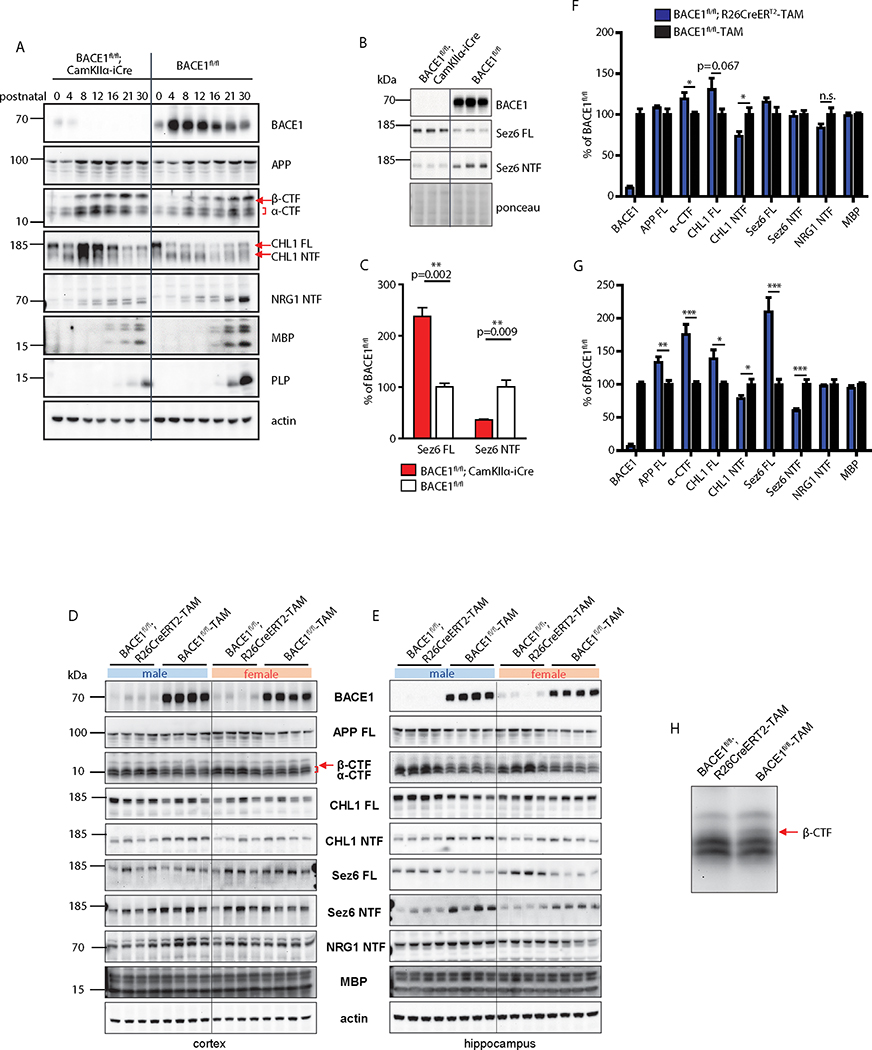
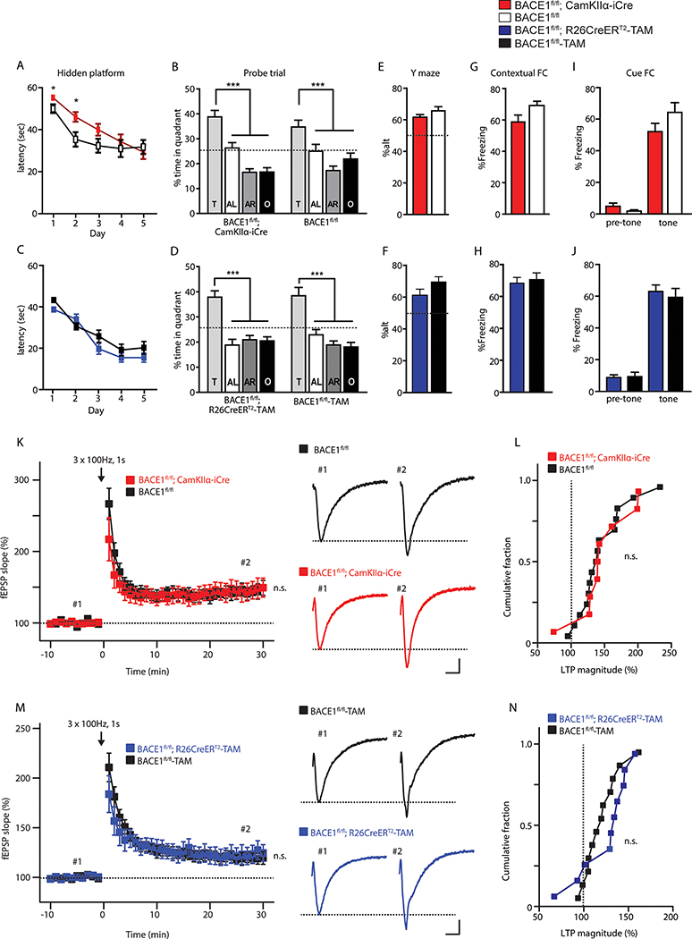
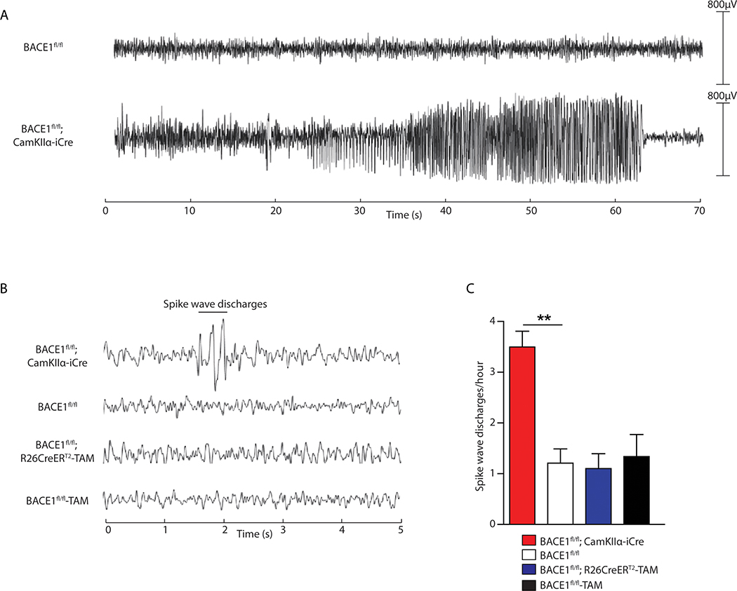
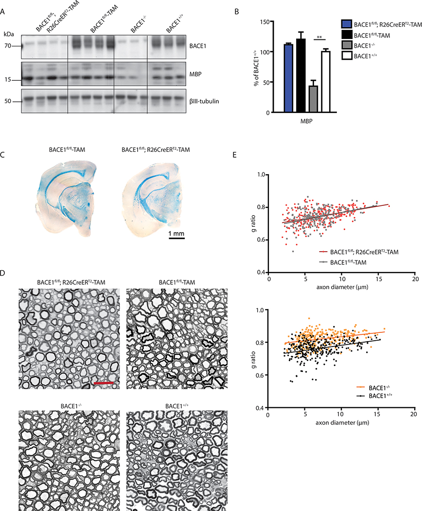
β-Site APP (amyloid precursor protein) cleaving enzyme 1 (BACE1) is the β-secretase enzyme that initiates production of the toxic amyloid-β peptide that accumulates in the brains of patients with Alzheimer's disease (AD). Hence, BACE1 is a prime therapeutic target, and several BACE1 inhibitor drugs are currently being tested in clinical trials for AD. However, the safety of BACE1 inhibition is unclear. Germline BACE1 knockout mice have multiple neurological phenotypes, although these could arise from BACE1 deficiency during development. To address this question, we report that tamoxifen-inducible conditional BACE1 knockout mice in which the Bace1 gene was ablated in the adult largely lacked the phenotypes observed in germline BACE1 knockout mice. However, one BACE1-null phenotype was induced after Bace1 gene deletion in the adult mouse brain. This phenotype showed reduced length and disorganization of the hippocampal mossy fiber infrapyramidal bundle, the axonal pathway of dentate gyrus granule cells that is maintained by neurogenesis in the mouse brain. This defect in axonal organization correlated with reduced BACE1-mediated cleavage of the neural cell adhesion protein close homolog of L1 (CHL1), which has previously been associated with axon guidance. Although our results indicate that BACE1 inhibition in the adult mouse brain may avoid phenotypes associated with BACE1 deficiency during embryonic and postnatal development, they also suggest that BACE1 inhibitor drugs developed for treating AD may disrupt the organization of an axonal pathway in the hippocampus, an important structure for learning and memory.


Conflict of interest statement

Competing interests: The authors declare no competing interests.

Figures

Fig. 1.
Fig. 1.. Conditional BACE1 knockout mice exhibit robust reduction of BACE1 and cleavage of BACE1 substrates.
(A) Immunoblots of total cortical homogenates from postnatal BACE1fl/fl;CamKIIα-iCre and BACE1fl/fl mice were probed with antibodies against BACE1 and the indicated full-length BACE1 substrates, BACE1-cleaved substrate fragments and myelin-associated proteins. (B) Immunoblots of cortical homogenates of 3 month-old BACE1fl/fl;CamKIIα-iCre mice and BACE1fl/fl mice were probed with antibodies against BACE1 and the BACE1 substrate Sez6 in membrane (Sez6 full length, FL) and soluble (Sez6 amino-terminal fragment, NTF) fractions. (C) Immunosignals shown in (B) were normalized to ponceau staining and represented as percentage of immunosignal in BACE1fl/fl mice. (D, E) Immunoblots of cortical (D) and hippocampal (E) soluble or membrane homogenates from BACE1fl/fl and BACE1fl/fl;R26CreERT2 mice that were treated with tamoxifen (TAM) at 3 months and analyzed at 1 year of age. Panels represent immunoblots of membrane (BACE1, APP FL, α/β-carboxy-terminal fragment, CTF, CHL1 FL, Sez6 FL, myelin basic protein, MBP) and soluble (CHL1 NTF, Sez6 NTF, NRG1 NTF) fractions of cortical or hippocampal homogenates. (F, G) Quantification of cortical (F) and hippocampal (G) immunoblot signals for BACE1, BACE1 substrates, and MBP in (D) and (E), respectively. Protein concentrations for tamoxifen-treated BACE1fl/fl and tamoxifen-treated BACE1fl/fl;R26CreERT2 mice were compared using unpaired two-way student’s t tests. p values for cortex are: α-CTF, p=0.036; CHL1 NTF, p=0.024. p values for hippocampus are: APP FL, p= 0.0065; α-CTF: p= 0.00032; CHL1 FL, p= 0.015; CHL1 NTF, p= 0.039; Sez6 FL, p= 0.0003; Sez6 NTF, p= 0.0003. (H) Enlarged APP β-CTF immunoblot bands from lanes 12 and 13 of (D). FL, full-length; CTF: C-terminal fragment; NTF, N-terminal fragment.
Fig. 2.
Fig. 2.. Conditional BACE1 knockout mice lack memory deficits and LTP impairment.
BACE1fl/fl;CamKIIα-iCre, tamoxifen-treated BACE1fl/fl;R26CreERT2, and respective control mice were assessed in the Morris water maze, Y-maze, fear conditioning, and longterm potentiation (LTP) tests. (A-D) Testing of conditional BACE1 knockout mice in the Morris Water Maze. (A-B) 9 month-old BACE1fl/fl;CamKIIα-iCre (n=23) mice and BACE1fl/fl (n=18) mice were trained using (A) a hidden platform (Repeated two-way ANOVA, p=0.074, F=3.382), followed by (B) a probe trial test (One-way ANOVA, FCKO=26.75, FCONT=9.86). (C-D) BACE1fl/fl;R26CreERT2-TAM (n=17) mice and control BACE1fl/fl -TAM (n=14) mice treated with tamoxifen (TAM) at 3 months were assessed in the Morris water maze at 9 months of age in (C) the hidden platform (Repeated 2-way ANOVA, p=0.089, F=3.096) followed by the (D) probe trial (One-way ANOVA, FCKO=18.68, FCONT=17.03). (E, G, I) BACE1fl/fl;CamKIIα-iCre and BACE1fl/fl mice were tested in a Y-maze (E), and in tests measuring contextual fear conditioning (FC) and cued fear conditioning (G, I, respectively). N= (23, 18) for Y-Maze and (24, 16) for fear conditioning for BACE1fl/fl;CamKIIα-iCre and BACE1fl/fl mice, respectively. (F, H, J) BACE1fl/fl;R26CreERT2-TAM and BACE1fl/fl-TAM mice treated with tamoxifen were tested in a Y-maze (F), and in tests measuring contextual (H) and cued fear conditioning (J). N= (17, 13) for Y-maze and (17, 14) for fear conditioning for BACE1fl/fl;R26CreERT2-TAM and BACE1fl/fl-TAM mice, respectively. (K-N) LTP in the CA1 region of the hippocampus. (K) Grouped data showing time-course of LTP for 1 year-old BACE1fl/fl;CamKIIα-iCre mice (red, n=9 slices from 5 animals) and control BACE1fl/fl mice (white, n=15 slices from 5 animals). (L) Cumulative probability distribution of all LTP recordings in (K). (M-N) Grouped data showing time-course of LTP for 9 month-old BACE1fl/fl;R26CreERT2-TAM (blue, n=10 slices from 3 animals) and control BACE1fl/fl-TAM mice (black, n=12 slices from 3 animals) that were injected with tamoxifen (TAM) at 3 months of age. (N) Cumulative probability distribution of all LTP recordings in (M). LTP was induced at time=0 with 3 trains of 100Hz, 1s tetanic stimulation (arrows in K and M). LTP statistics were analyzed by unpaired student’s t test.
Fig. 3.
Fig. 3.. Seizure activity was reduced or absent in BACE1 conditional knockout mice.
Seizure activity was measured in postnatal forebrain excitatory neuron BACE1 conditional knockout mice and in adult whole-body BACE1 conditional knockout mice. (A) Electrographic seizure with buildup of generalized fast activity in BACE1fl/fl;CamKIIα-iCre mice at 1 year of age (lower panel) compared to age-matched BACE1fl/fl control mice (upper panel). (B) EEG in awake animals showed epileptiform abnormalities, including spike-wave discharges, in BACE1fl/fl;CamKIIα-iCre mice (n=6) compared to normal EEG in BACE1fl/fl mice (n=4), BACE1fl/fl-TAM mice treated with tamoxifen (n=5) and BACE1fl/fl;R26CreERT2-TAM mice treated with tamoxifen (n=6). (C) Comparison of the frequency of spike-wave discharges in mice with the four indicated genotypes: BACE1fl/fl;CamKIIα-iCre, BACE1fl/fl, BACE1fl/fl;R26CreERT2-TAM and BACE1fl/fl-TAM. P values are 0.0003 by one-way ANOVA and 0.0011 by Tukey post-hoc test between BACE1fl/fl;CamKIIα-iCre mice and BACE1fl/fl mice.
Fig. 4.
Fig. 4.. Central and peripheral hypomyelination are absent in adult whole-body BACE1 conditional knockout mice.
(A) Immunoblot analysis of sciatic nerve homogenates showing BACE1 and myelin basic protein (MBP) in BACE1fl/fl;R26CreERT2-TAM, BACE1fl/fl-TAM, BACE1−/−, and BACE1+/+ mice. (B) Quantification of MBP immunoblot signals from (A), normalized to βIII-tubulin immunosignal (One-way ANOVA on MBP, p=0.0004, F=15.9. P value of 0.006 from unpaired two-way student’s t-test). (C) Luxol fast blue staining of myelinated fibers in BACE1fl/fl;R26CreERT2-TAM tamoxifen treated and control BACE1fl/fl mouse brains. (D) Images of toluidine blue stained sciatic nerve thin sections showing myelination in BACE1fl/fl;R26CreERT2-TAM compared to BACE1fl/fl-TAM tamoxifen treated mice and BACE1−/− compared to BACE1+/+ mice. Scale bar = 20 μm. (E) Quantification of myelination via g-ratio between BACE1fl/fl;R26CreERT2-TAM tamoxifen treated (n=3) and BACE1fl/fl-TAM tamoxifen treated (n=2) mice (upper panel; Linear regression analysis on slope: F=0.27, p=0.6; on intercepts: F=1.03, p=0.31), and BACE1−/− (n=3) mice and BACE1+/+ (n=3) mice (lower panel). 100 axons per animal; Linear regression analysis on slope: F=0.98, p=0.32; on intercepts: F=233.6, p<0.0001.
Fig. 5.
Fig. 5.. Adult conditional BACE1 knockout mice show disorganization of the hippocampal mossy fiber pathway.
(A) Shown are coronal brain sections from mice with the indicated genotypes (BACE1−/−, BACE1fl/fl, BACE1fl/fl;CamKIIα-iCre, BACE1fl/fl-TAM, BACE1fl/fl;R26CreERT2-TAM) revealing the hippocampus co-labeled for the mossy fiber marker synaptoporin (SPO, green, upper panel) and BACE1 (red, lower panel). Arrows delineate the boundaries of the infrapyramidal bundle (IPB). BACE1fl/fl-TAM and BACE1fl/fl;R26CreERT2-TAM mice were treated with tamoxifen at 3 months of age and analyzed at 1 year of age. BACE1−/− and BACE1fl/fl mice at 9 month of age were used as positive and negative controls, respectively. SPB, suprapyramidal bundle; slu, stratum lucidum. Scale bar = 200μm. (B) Coronal brain sections from BACE1fl/fl-TAM and BACE1fl/fl;R26CreERT2-TAM tamoxifen treated mice in (A) were co-immunostained with antibodies against CHL1 (red),the mossy fiber bouton and presynaptic terminal markers calbindin (green), and synaptophysin (blue). (C) Infrapyramidal bundle (IPB) lengths were normalized to the lengths of the suprapyramidal bundle (SPB) plus stratum lucidum (slu) and displayed as ratios, that is,IPB/(SPB+slu). The number of mice for each genotype is indicated in each bar of the graph. Error bars indicate standard error of the mean; p-values for indicated comparisons (lines) using unpaired Student’s t test are shown above the bars. (D) Both soluble and membrane fractions were prepared from the hippocampi of representative BACE1fl/fl-TAM and BACE1fl/fl;R26CreERT2-TAM tamoxifen treated mice and then were subjected to immunoblot analysis for full length (FL) CHL1 and CHL1 N-terminal fragment (β-NTF). Middle panel represents a longer exposure of the upper panel. βIII-tubulin was the loading control. (E) Ratio of CHL1 FL to CHL1 β-NTF intensities in membrane fractions (CHL1 FL/ β-NTF) and the intensity of the CHL1 β-NTF band in soluble fractions normalized to βIII-tubulin (sCHL1) displayed as arbitrary units (a.u.) for BACE1fl/fl;R26CreERT2-TAM and BACE1fl/fl-TAM mice treated with tamoxifen, mean ± SEM. (F) Correlation of CHL1 FL/ β-NTF from (E) plotted against IPB/(SPB+slu) for BACE1fl/fl;R26CreERT2-TAM and BACE1fl/fl-TAM tamoxifen treated mice (R2=0.7014, p=0.0002). (G) Correlation of sCHL1 from (E) plotted against IPB/(SPB+slu) for BACE1fl/fl;R26CreERT2-TAM and BACE1fl/fl-TAM tamoxifen treated mice (R2=0.4117, p=0.01).

Similar articles

Cited by

References

    1. Tanzi RE, The genetics of Alzheimer disease. Cold Spring Harb Perspect Med 2, (2012). - PMC - PubMed
    1. Selkoe DJ, Hardy J, The amyloid hypothesis of Alzheimer’s disease at 25 years. EMBO Mol Med 8, 595–608 (2016). - PMC - PubMed
    1. Tanzi RE, Gusella JF, Watkins PC, Bruns GA, St George-Hyslop P, Van Keuren ML, Patterson D, Pagan S, Kurnit DM, Neve RL, Amyloid beta protein gene: cDNA, mRNA distribution, and genetic linkage near the Alzheimer locus. Science 235, 880–884 (1987). - PubMed
    1. Haass C, Kaether C, Thinakaran G, Sisodia S, Trafficking and proteolytic processing of APP. Cold Spring Harb Perspect Med 2, a006270 (2012). - PMC - PubMed
    1. Vassar R, Bennett BD, Babu-Khan S, Kahn S, Mendiaz EA, Denis P, Teplow DB, Ross S, Amarante P, Loeloff R, Luo Y, Fisher S, Fuller J, Edenson S, Lile J, Jarosinski MA, Biere AL, Curran E, Burgess T, Louis JC, Collins F, Treanor J, Rogers G, Citron M, Beta-secretase cleavage of Alzheimer’s amyloid precursor protein by the transmembrane aspartic protease BACE. Science 286, 735–741 (1999). - PubMed

Publication types

MeSH terms

Substances

LinkOut - more resources