. 2018 Oct 2;115(40):E9381-E9390.
doi: 10.1073/pnas.1810133115. Epub 2018 Sep 19.

Substance P and IL-33 administered together stimulate a marked secretion of IL-1β from human mast cells, inhibited by methoxyluteolin

Affiliations

Substance P and IL-33 administered together stimulate a marked secretion of IL-1β from human mast cells, inhibited by methoxyluteolin

Alexandra Taracanova et al. Proc Natl Acad Sci U S A. .

Abstract

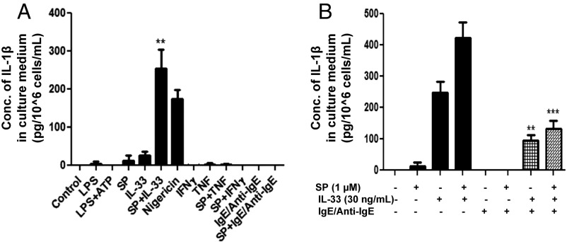
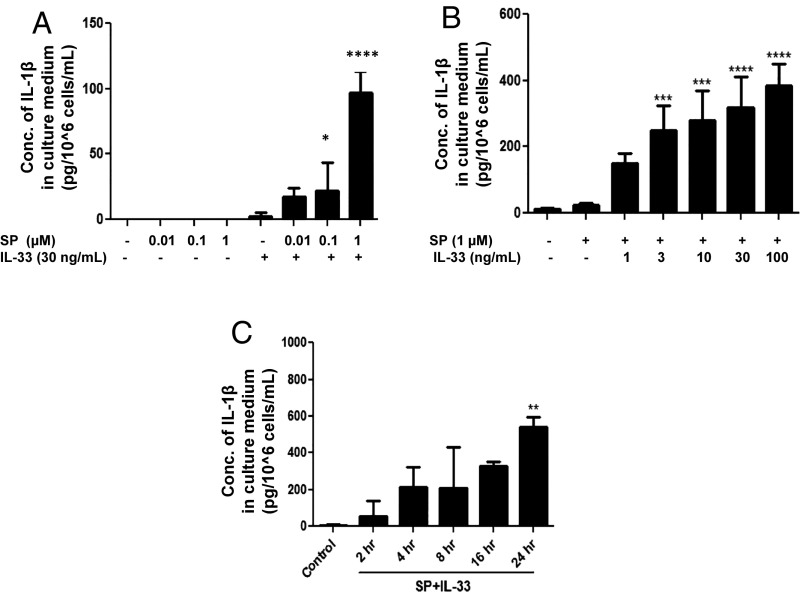
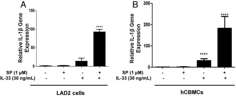
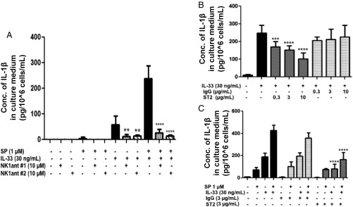
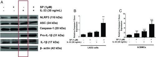
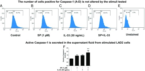
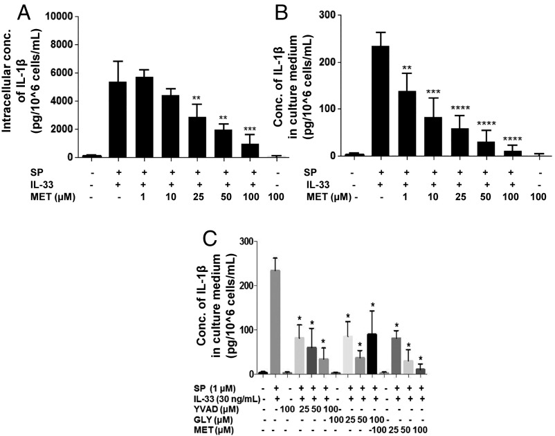
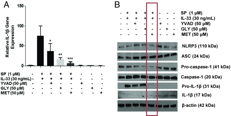
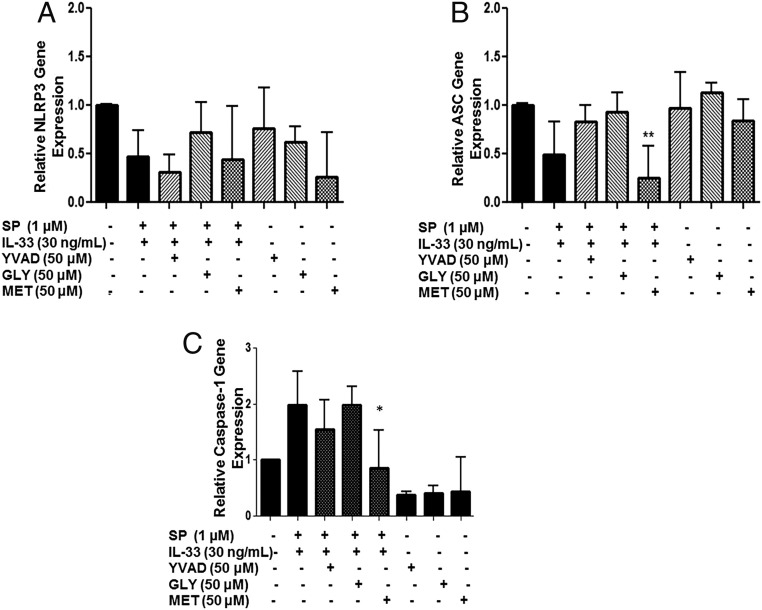
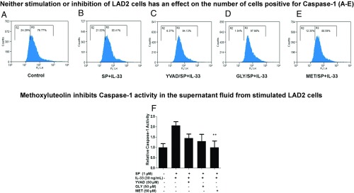
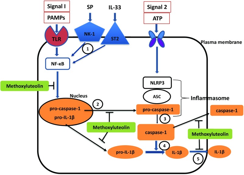
Mast cells are critical for allergic and inflammatory responses in which the peptide substance P (SP) and the cytokine IL-33 are involved. SP (0.01-1 μM) administered together with IL-33 (30 ng/mL) to human cultured LAD2 mast cells stimulates a marked increase (P < 0.0001) in secretion of the proinflammatory cytokine IL-1β. Preincubation of LAD2 (30 min) with the SP receptor (NK-1) antagonists L-733,060 (10 μM) or CP-96345 (10 µM) inhibits (P < 0.001) secretion of IL-1β stimulated by either SP (1 μM) or SP together with IL-33 (30 ng/mL). Surprisingly, secretion of IL-1β stimulated by IL-33 is inhibited (P < 0.001) by each NK-1 antagonist. Preincubation with an antibody against the IL-33 receptor ST2 inhibits (P < 0.0001) secretion of IL-1β stimulated either by IL-33 or together with SP. The combination of SP (1 μM) with IL-33 (30 ng/mL) increases IL-1β gene expression by 90-fold in LAD2 cells and by 200-fold in primary cultured mast cells from human umbilical cord blood. The combination of SP and IL-33 increases intracellular levels of IL-1β in LAD2 by 100-fold and gene expression of IL-1β and procaspase-1 by fivefold and pro-IL-1β by twofold. Active caspase-1 is present even in unstimulated cells and is detected extracellularly. Preincubation of LAD2 cells with the natural flavonoid methoxyluteolin (1-100 mM) inhibits (P < 0.0001) secretion and gene expression of IL-1β, procaspase-1, and pro-IL-1β. Mast cell secretion of IL-1β in response to SP and IL-33 reveals targets for the development of antiinflammatory therapies.

Keywords: IL-1β; IL-33; inflammation; mast cells; substance P.


Conflict of interest statement

Conflict of interest statement: T.C.T. is the recipient of US patent no. 7,906,153 covering the use of flavonoids in neuroinflammatory conditions.

Figures

Fig. 1.
Fig. 1.
(A) SP and IL-33 stimulate IL-1β secretion. LAD2 cells (1 × 10⁵ cells per well) were seeded in a 96-well culture plate and stimulated with LPS (100 ng/mL), ATP (5 μM), SP (1 μΜ), IL-33 (30 ng/mL), nigericin (10 µM), TNF (50 ng/mL), IFN-γ (100 U), IgE (1 µg/mL)/anti-IgE (5 µg/mL), or their combinations as shown for 24 h. Control cells were treated with culture medium only (n = 3, **P < 0.01 compared with unstimulated controls). (B) IgE/anti-IgE decrease the SP- and IL-33–stimulated secretion of IL-1β. LAD2 cells (1 × 10⁵ cells per well) were seeded in a 96-well culture plate, were preincubated with human IgE (1 µg/mL) overnight, and were stimulated the next day with anti-IgE (10 ng/mL) for 2 h and/or SP (1 µM) and IL-33 (30 ng/mL) for 24 h. Supernatant fluids were collected at the end of the incubation period and were assayed for IL-1β using ELISA (n = 3, **P < 0.01 and ***P < 0.001 compared to SP alone or to SP+IL-33, respectively). Conc, concentration.
Fig. 2.
Fig. 2.
Selection of the optimal doses to study IL-1β secretion stimulated by SP and IL-33 when administered in combination. (A and B) LAD2 cells (1 × 10⁵ cells per well) were stimulated with SP (0.01–1 μΜ) (n = 3, *P < 0.05 and ****P < 0.0001 compared with SP alone) (A) or IL-33 (1–100 ng/mL) (B) and their combination as shown for 24 h (n = 3, ***P < 0.001 and ****P < 0.0001 compared with SP alone). (C) Time-dependent study of IL-1β secretion. LAD2 cells (1 × 10⁵ cells per well) were stimulated with the combination of SP (1 μΜ) and IL-33 (30 ng/mL) for 2–24 h. Supernatant fluids were collected at the end of the incubation period. IL-1β secretion was assayed using ELISA (n = 3, **P < 0.01). Conc, concentration.
Fig. 3.
Fig. 3.
SP and IL-33 markedly enhance IL-1β gene expression and secretion. LAD2 cells (1 × 10⁶ cells per well) (A) and hCBMCs (0.3 × 10⁶ cell per well) (B) were seeded in a 12-well culture plate and were stimulated with SP (1 μM), IL-33 (30 ng/mL), or their combination for 6 h. IL-1β mRNA expression levels were measured by qRT-PCR and were normalized to human GAPDH endogenous control (n = 3, ****P < 0.0001).
Fig. 4.
Fig. 4.
NK-1 receptor antagonists inhibit IL-1β secretion. (A) LAD2 cells were pretreated with NK-1R antagonists L-733,060 (10 μM) (n = 3, **P < 0.01 and ****P < 0.0001 compared with IL-33 alone) and CP-96345 (10 µM) (n = 3, ****P < 0.0001 compared with SP and IL-33) for 30 min and then were stimulated with SP (1 μM), IL-33 (30 ng/mL), or their combination for 24 h. Antibody against ST2 inhibits IL-1β secretion. (B) LAD2 cells (1 × 10⁵ cells per well) were seeded in a 96-well culture plate and were preincubated with an antibody against ST2, the IL-33 receptor (ST2, 0.3–10 µg/mL), or a nonspecific antibody not recognizing ST2 (IgG control) (0.3–10 µg/mL) for 2 h and then were stimulated with IL-33 (30 ng/mL) for 24 h (n = 3, ***P < 0.01, ****P < 0.0001 compared with IL33 alone). (C) LAD2 cells were preincubated with anti-ST2 neutralizing antibody (3 ng/mL) or IgG control (3 ng/mL) for 2 h and then were stimulated with SP (1 μΜ), IL-33 (30 ng/mL), or their combination for 24 h. Collected supernatant fluids were assayed by IL-1β ELISA (n = 3, ****P < 0.0001 compared with SP and IL-33). Conc, concentration.
Fig. 5.
Fig. 5.
Expression of NLRP3 inflammasome components and the mature form of IL-1β. (A) LAD2 cells (1 × 10⁶ cells per well) were seeded in a 12-well culture plate and were stimulated with SP (1 μΜ), IL-33 (30 ng/mL), or their combination for 24 h. Cell lysates were collected after 24 h, and protein levels of the NLRP3 inflammasome components (NLRP3, ASC, caspase-1), pro-IL-1β, and active IL-1β (p17) were measured by Western blot, using β-actin as loading control (shown in a representative gel of n = 3). SP and IL-33 increase caspase-1 gene expression. (B) LAD2 cells (1 × 10⁶ cells per well), (n = 3, **P < 0.01 compared with unstimulated cells). (C) hCMBCs (0.3 × 10⁶ cell per well) were seeded in a 12-well culture plate and were stimulated by SP (1 µM), IL-33 (30 ng/mL), or their combination for 6 h. Caspase-1 gene expression was measured by qRT-PCR and normalized to human GAPDH endogenous control (n = 3, ***P < 0.001 compared with unstimulated cells).
Fig. 6.
Fig. 6.
Active caspase-1 is constitutively present. (AE) LAD2 cells (0.25 × 10⁶ cells per well) were stimulated with SP (1 μΜ), IL-33 (30 ng/mL), or their combination for 24 h. Stimulated cells were incubated with the FLICA (FL-1H) caspase-1 probe for 1 h, and active caspase-1+ cells (counts are on the vertical axis) were assessed by flow cytometry. Each panel is representative of three experiments. SP and IL-33 increase caspase-1 activity. (F) LAD2 cells (0.5 × 10⁵ cells per well) were stimulated with SP (1 μΜ), IL-33 (30 ng/mL), or their combination for 24 h; after stimulation, 50 μM YVAD-AFC substrate was added to cells and incubated for 2 h. Caspase-1 activity in the supernatant fluids was determined by measuring fluorescence at the 405-nm wavelength. The fold-increase in caspase-1 activity was determined by comparing the values to the untreated control samples (n = 3, **P < 0.01 compared with SP and IL-33).
Fig. 7.
Fig. 7.
Methoxyluteolin inhibits the secretion of IL-1β. LAD2 cells (1 × 10⁵ cells per well) were seeded in a 96-well culture plate, were pretreated with methoxyluteolin (MET, 1–100 μM) for 2 h, and then were stimulated with the combination of SP (1 μM) and IL-33 (30 ng/mL) for 24 h. LAD2 cells were also pretreated with AC-YVAD-CMK (YVAD 25–100 μM) or glybenclamide (GLY 25–100 μM). Control cells were treated with 0.1% DMSO, the highest concentration corresponding to that of 100 μM methoxyluteolin. Collected lysates (A) and supernatant fluids (B and C) were assayed for IL-1β using ELISA (n = 3, *P < 0.05, **P < 0.01, ***P < 0.001, and ****P < 0.0001 compared with SP and IL-33). Conc, concentration.
Fig. 8.
Fig. 8.
Methoxyluteolin inhibits gene and protein expression of IL-1β as well as protein expression of procaspase I and pro-IL-1β. LAD2 cells (1 × 10⁶ cells per well) were seeded in a 12-well culture plate, were preincubated with methoxyluteolin (MET, 50 μM), AC-YVAD-CMK (YVAD, 50 μM), or glybenclamide (GLY, 50 μM), and then were stimulated with the combination of SP (1 μM) and IL-33 (30 ng/mL) for 6 h. (A) Gene expression of IL-1β was measured by qRT-PCR and normalized to human GAPDH endogenous control. (n = 3, *P < 0.05, **P < 0.01, *** and P < 0.001 compared to SP and IL-33). (B) Protein expression of NLRP3, ASC, procaspase I, pro-IL-1β, and IL-1β were assayed by Western blot analysis using β-actin as the loading control. The red rectangle identifies the effect of methoxyluteolin.
Fig. 9.
Fig. 9.
Methoxyluteolin decreases caspase-1 gene expression. LAD2 cells (1 × 10⁶ cells per well) were seeded in a 12-well culture plate, were preincubated with AC-YVAD-CMK (YVAD 50 μM), glybenclamide (GLY 50 μM), or methoxyluteolin (MET 50 μM), and then were stimulated with the combination of SP (1 µM) and IL-33 (30 ng/mL) for 6 h. The gene expression of the inflammasome components NLRP3 (A) and ASC (B) and caspase-1 gene expression (C) were measured by qRT-PCR and normalized to human GAPDH endogenous control (n = 3, *P < 0.05 and **P < 0.01 compared to SP and IL-33).
Fig. 10.
Fig. 10.
(AE) Methoxyluteolin and inflammasome inhibitors do not decrease the percentage of active caspase-1–expressing human mast cells. LAD2 cells (0.25 × 10⁶ cells per well) were preincubated with AC-YVAD-CMK (YVAD, 50 μM), glybenclamide (GLY, 50 μM), or methoxyluteolin (MET, 50 μM) and then were stimulated with SP (1 μΜ) and IL-33 (30 ng/mL) for 24 h. Stimulated cells were incubated with the FLICA caspase-1 probe for 1 h, and active capsase-1+ cells were assessed by flow cytometry. Each panel is representative of three experiments. (F) Methoxyluteolin decreases caspase-1 activity. LAD2 cells (0.5 × 10⁵ cells per well) were preincubated with AC-YVAD-CMK (YVAD, 50 μM), glybenclamide (GLY, 50 μM), or methoxyluteolin (MET, 50 μM) and then were stimulated with SP (1 μΜ) and IL-33 (30 ng/mL) for 24 h. After stimulation, 50 μM YVAD-AFC substrate was added to cells and incubated for 2 h. Caspase-1 activity in the supernatant fluids was determined by measuring fluorescence at 405 nm. Fold-increase in caspase-1 activity was determined by comparing the values to the untreated control samples (n = 3, **P < 0.01 compared with SP and IL-33).
Fig. 11.
Fig. 11.
Diagrammatic representation of the stimulatory effect of SP and IL-33 on IL-1β synthesis and secretion and the proposed point of inhibition of methoxyluteolin. Our evidence indicates that (1) SP and IL-33 activate their respective receptors and stimulate synthesis of procaspase-1 and pro-IL-1β, possibly via NF-κB, activation, which is inhibited by methoxyluteolin. (2) Procaspase-1 and pro-IL-1β are released from the nucleus, a process that could also be inhibited by methoxyluteolin. (3) In the cytoplasm, caspase-1, which is already active, converts pro-IL-1β to active IL-1β. (4) Some of the procaspase-1 is converted to caspase-1, but this may be a minor contribution since the NLRP3 inflammasome does not seem to be involved. (5) IL-1β and active caspase-1 are then secreted extracellularly, a process that is also inhibited by methoxyluteolin. Orange boxes and ovals indicate facts supported by our findings. Open boxes and ovals indicate pathways not supported by our data. The (T) attached to methoxyluteolin indicates possible points of inhibition.

Similar articles

Cited by

References

    1. Rodewald HR, Dessing M, Dvorak AM, Galli SJ. Identification of a committed precursor for the mast cell lineage. Science. 1996;271:818–822. - PubMed
    1. Kitamura Y, Ito A. Mast cell-committed progenitors. Proc Natl Acad Sci USA. 2005;102:11129–11130. - PMC - PubMed
    1. Schmetzer O, Valentin P, Church MK, Maurer M, Siebenhaar F. Murine and human mast cell progenitors. Eur J Pharmacol. 2016;778:2–10. - PubMed
    1. Galli SJ, Tsai M. Mast cells in allergy and infection: Versatile effector and regulatory cells in innate and adaptive immunity. Eur J Immunol. 2010;40:1843–1851. - PMC - PubMed
    1. Theoharides TC, Valent P, Akin C. Mast cells, mastocytosis, and related disorders. N Engl J Med. 2015;373:163–172. - PubMed

Publication types

LinkOut - more resources