. 2018 Sep 20;3(18):e122188.
doi: 10.1172/jci.insight.122188.

Necroptosis of infiltrated macrophages drives Yersinia pestis dispersal within buboes

Affiliations

Necroptosis of infiltrated macrophages drives Yersinia pestis dispersal within buboes

Mohammad Arifuzzaman et al. JCI Insight. .

Abstract

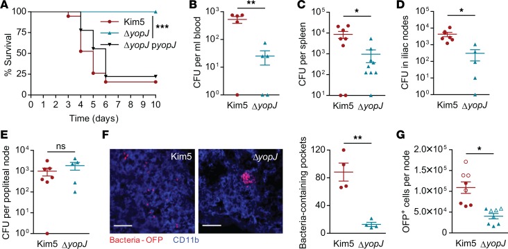
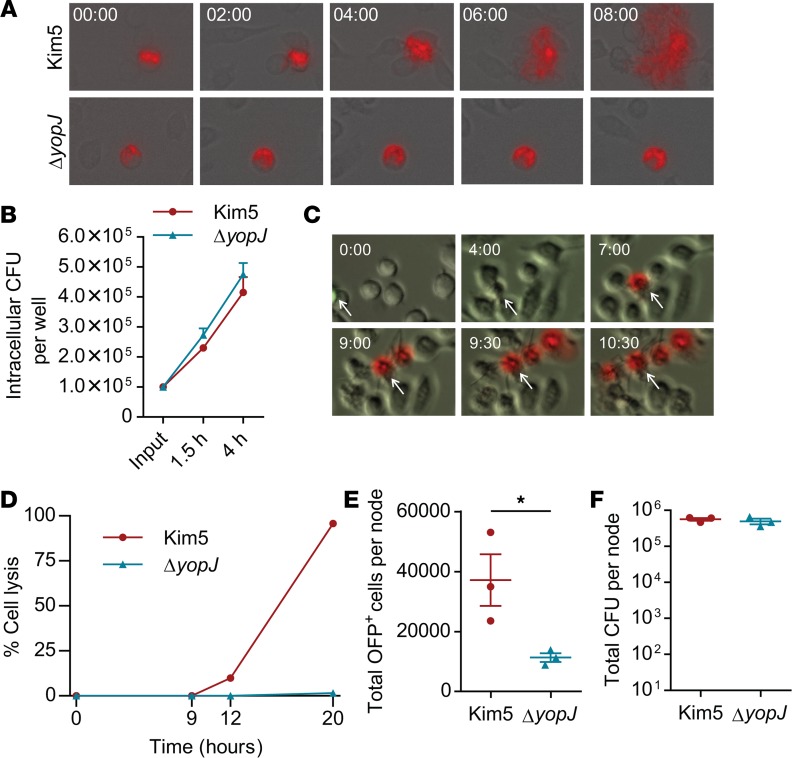
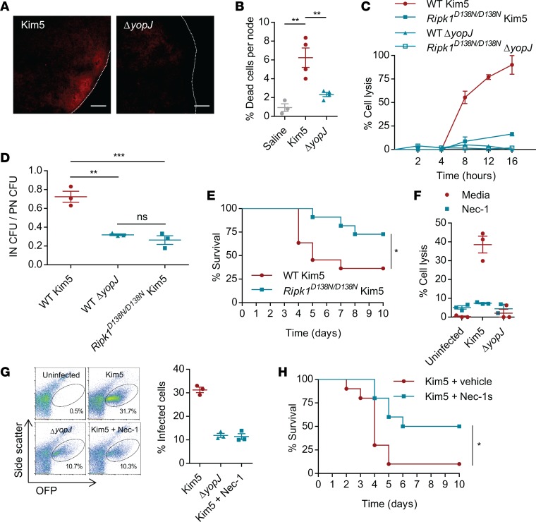
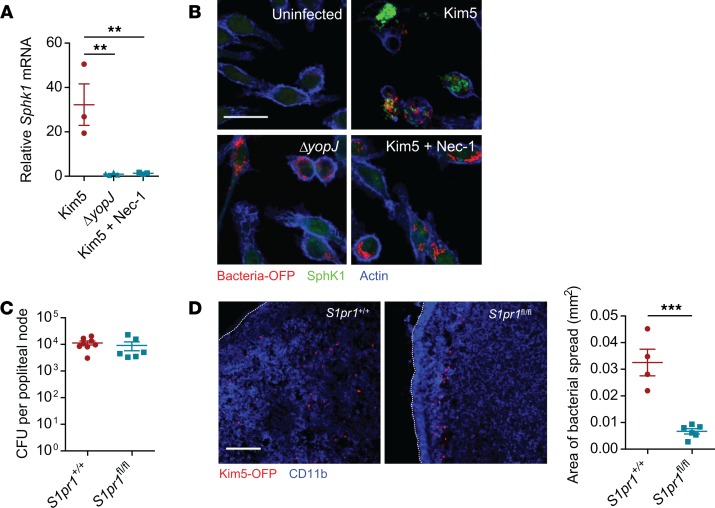
When draining lymph nodes become infected by Yersinia pestis (Y. pestis), a massive influx of phagocytic cells occurs, resulting in distended and necrotic structures known as buboes. The bubonic stage of the Y. pestis life cycle precedes septicemia, which is facilitated by trafficking of infected mononuclear phagocytes through these buboes. However, how Y. pestis convert these immunocytes recruited by host to contain the pathogen into vehicles for bacterial dispersal and the role of immune cell death in this context are unknown. We show that the lymphatic spread requires Yersinia outer protein J (YopJ), which triggers death of infected macrophages by downregulating a suppressor of receptor-interacting protein kinase 1-mediated (RIPK1-mediated) cell death programs. The YopJ-triggered cell death was identified as necroptotic, which released intracellular bacteria, allowing them to infect new neighboring cell targets. Dying macrophages also produced chemotactic sphingosine 1-phosphate, enhancing cell-to-cell contact, further promoting infection. This necroptosis-driven expansion of infected macrophages in buboes maximized the number of bacteria-bearing macrophages reaching secondary lymph nodes, leading to sepsis. In support, necrostatins confined bacteria within macrophages and protected mice from lethal infection. These findings define necrotization of buboes as a mechanism for bacterial spread and a potential target for therapeutic intervention.

Keywords: Bacterial infections; Immunology; Macrophages; Microbiology.


Conflict of interest statement

Conflict of interest: SNA is the cofounder and chief scientific officer for Mastezellen Bio Inc.

Figures

Figure 1
Figure 1. YopJ is a critical virulence factor promoting bacterial dispersal.
(A) Survival of mice challenged with bacteria instilled into a single rear footpad. Data are combined from 2 independent experiments, n = 9–10. (B) Bacterial numbers (CFU) in the blood, 48 hours after footpad infection with Kim5 or ΔyopJ strain (n = 5). (C) Bacterial numbers in spleen, 72 hours after infection (h.p.i.) (n = 8–9). (D and E) Bacterial numbers in iliac nodes (INs) (D) and popliteal nodes (PNs) (E) 24 h.p.i. (n = 5–7). Data are representative of 3 independent experiments. (F) Immunofluorescence staining of PN cross-sections 24 hours after footpad infection with OFP-labeled Kim5 or ΔyopJ bacteria. The images are representative of 2 independent experiments. Scale bar: 25 μm. The graph shows bacteria-containing pockets counted in 4 fields (n = 4) (G) Quantification of OFP+ cells in PNs 24 h.p.i. by flow cytometry. Data are combined from 2 independent experiments (distinguished by open and closed symbols), each with 4 mice per group. Significance for Kaplan-Meier curves was determined by the log-rank test. Other data were analyzed via unpaired 2-tailed Student’s t test. *P < 0.05, **P < 0.01, ***P < 0.001.
Figure 2
Figure 2. YopJ-triggered macrophage death promotes infection of neighboring cells.
(A) Time-lapse images showing intracellular OFP+ bacteria (red) and their release from J774A.1 macrophages (magnification 200×). Time 00:00 (hh:mm) indicates 4 hours after initial infection. Also see Supplemental Videos 1 and 2. (B) Number of intracellular bacteria per well at 1.5 hours and 4 h.p.i. of 2 × 10⁴ J774A.1 cells for 30 min. (n = 3). Input indicates CFU count at 0 min. from the bacterial suspension added to each well. (C) Cytolysis visualized by the entry of propidium iodide (red) into J774A.1 macrophages (magnification 200×). Time 00:00 (hh:mm) indicates 4 hours after initial infection. The arrow in each panel indicates the initially infected cell. Ratio of infected/uninfected cells, 1:10. Also see Supplemental Video 3. (D) Percent cell lysis representing death of initially and newly infected cells combined at various time points after initial infection. Ratio of initially infected/uninfected cells, 1:10. Data are represented as the mean of duplicate for each time point. (E and F) Total OFP+ cells (E) and bacterial count (F) in PNs 9 hours after footpad injection with J774.1A cells bearing Kim5-OFP or ΔyopJ-OFP (n = 3). Data are representative of at least 2 independent experiments. Data were analyzed via unpaired 2-tailed Student’s t test. *P < 0.05.
Figure 3
Figure 3. Virulence of YopJ depends on RIPK1-mediated programed cell death.
(A) Representative images showing degree of necrosis in PNs revealed by propidium iodide staining 24 h.p.i. with Kim5 or ΔyopJ strains (n = 2). Scale bar 100 μm. (B) Quantification of dead cells in the PNs by flow cytometry (n = 3–4). Data are representative of 2 independent experiments. (C) Lysis of BMDMs isolated from WT or RipK1D138N/D138N mice following in vitro infection with Kim5 or ΔyopJ (MOI 10). Data are representative of 3 independent experiments. (D) Bacterial counts in INs relative to bacterial counts in PNs 24 hours after footpad infection (n = 3). (E) Survival of WT and RipK1D138N/D138N mice following footpad challenge with Kim5 bacteria. Data are combined from 2 independent experiments, each with 5–6 mice per group. (F) Lysis of J774A.1 macrophages at 8 h.p.i. during Kim5 or ΔyopJ infections in the presence or absence of Necrostatin-1 (Nec-1, 30 μM) (n = 3). (G) Flow cytometry plots and percentage graph showing infection of neighboring macrophages from Kim5-OFP or ΔyopJ-OFP infected macrophages in the presence or absence of Nec-1, 10 hours after initial infection (n = 3). Data are representative of 2 independent experiments. (H) Survival of Kim5-infected mice with or without Nec-1s treatment. Data are combined from 2 independent experiments, n = 5 in each experiment. Significance for Kaplan-Meier curves was determined by the log-rank test. Other data were analyzed via 1-way ANOVA. *P < 0.05, **P < 0.01, ***P < 0.001.
Figure 4
Figure 4. YopJ-triggered cell death is necroptotic and is activated after a distinct lag period.
(A) RIPK1 immunoprecipitated from WT (B6) or RIPK1 mutant (D138N) BMDMs 3 h.p.i. and the presence of RIPK1, Caspase-8, RIPK3, and FLIPL detected by immunoblotting. Total lysates were also assayed for these proteins and β-actin. p30 and p20 indicate cleavage products of corresponding proteins. (B) Immunoblot analysis of lysates prepared from Kim5-infected BMDMs at indicated time points and probed for MLKL, phospho-MLKL, and β-actin. Intensity measurements relative to actin were shown under corresponding bands. (C) Immunoblot analysis of lysates prepared from uninfected macrophages or macrophages collected at indicated time points after infection with Kim5 or ΔyopJ strain. Immunoblots were probed with anti-FLIPS/L and anti–β-actin. Band intensities relative to actin were shown for the short-exposure panel. (D) Fold change of FLIP mRNA 2 h.p.i. relative to expression in uninfected macrophages (n = 3). (E) Lysis of WT or Ripk1D138N/D138N BMDMs 4 h.p.i. with Y. pestis Kim5 or ΔyopJ strain in presence or absence of 10 μM SAHA, a FLIP inhibitor (n = 3–4). Data are representative of 2 independent experiments. Data were analyzed via unpaired 2-tailed Student’s t test. *P < 0.05, **P < 0.01. See complete unedited blots in the supplemental material.
Figure 5
Figure 5. Necroptosis of infected macrophages alters local S1P gradients promoting intranodal bacterial spread.
(A) Fold change of Sphk1 mRNA in J774A.1 macrophages at 8 h.p.i. relative to uninfected controls (n = 3). (B) Immunofluorescence staining for SphK1 (green) in uninfected, Kim5-OFP–infected or in ΔyopJ-infected macrophages. Scale bar: 25 μm. (C) Bacterial numbers in PNs from mice whose mononuclear phagocytes were S1PR1-sufficient (Cx3cr1-Cre S1pr1+/+) or -deficient (Cx3cr1-Cre S1pr1fl/fl) 24 hours following footpad infection with Kim5 Y. pestis (n = 6). (D) Immunofluorescence staining of PNs of Cx3cr1-Cre S1pr1+/+ or Cx3cr1-Cre S1pr1fl/fl mice, 24 hours after footpad infection with Kim5-OFP bacteria. Scale bar: 50 μm. Graph shows area of bacterial spread measured from the PN images (n = 4–6). Data were analyzed via unpaired 2-tailed Student’s t test or 1-way ANOVA. Data are representative of at least 2 independent experiments. **P < 0.01, ***P < 0.001.

References

    1. Junt T, Scandella E, Ludewig B. Form follows function: lymphoid tissue microarchitecture in antimicrobial immune defence. Nat Rev Immunol. 2008;8(10):764–775. doi: 10.1038/nri2414. - DOI - PubMed
    1. Bar-Haim E, Gat O, Markel G, Cohen H, Shafferman A, Velan B. Interrelationship between dendritic cell trafficking and Francisella tularensis dissemination following airway infection. PLoS Pathog. 2008;4(11):e1000211. doi: 10.1371/journal.ppat.1000211. - DOI - PMC - PubMed
    1. St John AL, Abraham SN. Salmonella disrupts lymph node architecture by TLR4-mediated suppression of homeostatic chemokines. Nat Med. 2009;15(11):1259–1265. doi: 10.1038/nm.2036. - DOI - PMC - PubMed
    1. St John AL, et al. S1P-Dependent trafficking of intracellular yersinia pestis through lymph nodes establishes Buboes and systemic infection. Immunity. 2014;41(3):440–450. doi: 10.1016/j.immuni.2014.07.013. - DOI - PMC - PubMed
    1. Perry RD, Fetherston JD. Yersinia pestis--etiologic agent of plague. Clin Microbiol Rev. 1997;10(1):35–66. - PMC - PubMed

Publication types